pH/redox dual-sensitive nanoparticles based on the PCL/PEG triblock copolymer for enhanced intracellular doxorubicin release

Yan Caoa, Junqiang Zhaoa, Yumin Zhangb, Jianfeng Liub, Jinjian Liub, Anjie Dongac and Liandong Deng*a
aDepartment of Polymer Science and Technology and Key Laboratory of Systems Bioengineering of the Ministry of Education, School of Chemical Engineering and Technology, Tianjin University, Tianjin 300072, China. E-mail: dengliandong@aliyun.com; Fax: +86 22 27890706; Tel: +86 22 27890707
bTianjin Key Laboratory of Radiation Medicine and Molecular Nuclear Medicine, Institute of Radiation Medicine, Chinese Academy of Medical Science and Peking Union Medical College, Tianjin, 300192, China
cCollaborative Innovation Center of Chemical Science and Engineering (Tianjin), Tianjin 300072, China

Received 30th January 2015 , Accepted 12th March 2015

First published on 12th March 2015


Abstract

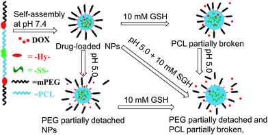
pH/redox dual-sensitive nanoparticles (NPs) based on a poly(ε-caprolactone) (PCL) and polyethylene glycol (PEG) triblock copolymer were developed and investigated aiming at improving the drug release property of PCL in cancer chemotherapy. A redox-sensitive disulfide bond and pH-sensitive benzoic-imine linkers were introduced to the backbone of the amphiphilic block copolymer termed SCHE, which could self-assemble into stable spherical NPs in aqueous solutions with an average size of about 100 nm. Doxorubicin (DOX) as a hydrophobic anticancer drug was loaded into the NPs by a dialysis method. The in vitro drug release results showed that the accumulative release amount of DOX at pH 5.0 with 10 mM glutathione (GSH) was accelerated apparently by more than twice compared to that at pH 7.4 without GSH. After a pre-treatment with GSH, the intracellular fluorescence intensity of Hela cells was enhanced compared to those without the pretreatment, which indicated faster DOX release in cells with higher GSH concentration. In an MTT assay, no obvious toxicity of blank NPs was found. In conclusion, SCHE NPs could serve as a novel colloidal drug delivery system in cancer chemotherapy and the introduced unstable linkers could enhance the drug release rate at tumor sites.


1. Introduction

In the past decade, amphiphilic block copolymeric nanoparticles (NPs), due to their stability and facility to be functionalized, have been extensively studied in the field of drug delivery, especially in anticancer drug delivery.1,2 Stimuli-triggered release of drugs is an efficient way to reduce the premature leakage of drugs to normal tissues and improve drug accumulation in targeted sites as well as reverse multidrug resistance in tumor therapy.3,4 To this end, smart nanocarriers sensitive to various external or internal stimuli such as light,5–7 temperature,8,9 enzyme,10,11 pH,12–14 reductive15,16 or oxidative stress17,18 have been developed. Among these sensitivities, pH-responsiveness and reduction-responsiveness are the most studied since tumor tissues have some characteristics over normal tissues such as lower pH (cancerous tissues pH 6.5–7.2, endosomes 5.0–6.5, lysosomes 4.5–5.0 whereas physiological pH 7.4) and high redox potential (the concentration of reducing glutathione (GSH) tripeptide in tumor cytosol and cell nuclei is 100–1000 times higher than that of body fluids).19

However, most of these smart stimuli-sensitive nanocarriers are based on non-biodegradable poly-olefins or their derivatives obtained by living polymerization such as atom transfer radical polymerization (ATRP) or reversible addition–fragmentation chain transfer (RAFT) polymerization.20,21 Biodegradable aliphatic polyesters do not arouse enough interest in this field due to their lack of modifiable groups.

Biodegradable aliphatic polyesters such as poly(ε-caprolactone) (PCL),22,23 polylactic acid (PLA)24 and poly(lactic-co-glycolic acid) (PLGA)25 are proper candidates for drug carriers due to their good biocompatibility, biodegradability and non-immunogenicity.26 They are approved by Food and Drug Administration (FDA) for clinical application and can be biodegraded by hydrolysis in physiological conditions.27 It should be noted, nevertheless, that these aliphatic polyesters, especially PCL, are far from being satisfactory with respect to drug release profiles.28 A critical challenge to be addressed is the slow and uncontrolled release of encapsulated drugs. The slow drug release profile is attributed to the delayed diffusion through the hydrophobic aliphatic polyesters core due to both hydrophobic interactions and slow hydrolysis of the ester linkages in backbones.29 To address this problem, strategies as co-assembled with other sensitive copolymers into micelles was adopted, which gained unsatisfactory drug release profiles.30 Redox-responsive PLA-based micelles with a disulfide linkage in the middle of triblock copolymers were synthesized.31 Modest improvement of DOX release rate was achieved in the presence of 10 mM dithiothreitol (DTT), which remained limited. To improve the performance of aliphatic polyesters as drug delivery materials, the drug release profiles should be further optimized.

In the present study, pH/redox dual-sensitive nanoparticles (NPs) based on poly(ε-caprolactone) (PCL) and polyethylene glycol (PEG) triblock copolymer were developed and investigated aiming at improving the drug release property of PCL in cancer chemotherapy. As shown in Scheme 1, a redox-sensitive disulfide bond (–SS–) in the middle of PCL segment and pH-sensitive benzoic-imine linkers (–Hy–) between hydrophilic and hydrophobic segments were introduced to the copolymer mPEG–Hy–PCL–SS–PCL–Hy–mPEG termed SCHE, which could self-assemble into stable drug-loaded NPs in physiological environment using doxorubicin (DOX) as model drug. In weak acid environment, PEG segments will detached from SCHE NPs. In the presence of GSH, the disulfide bonds in hydrophobic core will break into thiols and make the core less hydrophobic.32 Both the changes of core and hydrophilic/hydrophobic balance can prompt the release of DOX. In this way, the SCHE NPs keep stable and low leakage of DOX in system circulation whereas quickly release DOX when internalized by cancer cells. The pH/redox triggered destabilization of SCHE NPs and in vitro DOX release from DOX-loaded SCHE NPs were investigated. The intracellular DOX release in enhanced GSH concentration was compared with that of normal cancer cells. As drug carriers, the cytotoxicity of SCHE and DOX-loaded SCHE NPs was also evaluated.


image file: c5ra01833a-s1.tif
Scheme 1 Schematic illustration of the self-assembly of drug-loaded SCHE NPs as well as acid/redox-induced destabilization and enhanced drug release in stimulating environments.

2. Materials and methods

2.1 Materials

Methoxy poly(ethylene glycol) (mPEG, Mn = 2.0 kDa), ε-caprolactone (ε-CL) and stannous octoate were obtained from Sigma-Aldrich. ε-CL was dried over CaH2 for 48 h at room temperature and distilled under reduced pressure just before use. mPEG was dried by azeotropic distillation from toluene. Bis(2-hydroxyethyl)disulfide (HES, 97%, Alfa Aesar) was dried over CaH2 and distilled under reduced pressure prior to use. p-formylbenzoic acid, hydrazine monohydrate (98%), N,N′-carbonyldiimidazole (CDI, 97%), N,N′-dicyclohexylcarbodiimide (DCC, 99%) and 4-dimethylaminopyridine (DMAP, 99%) were obtained from Aladdin and used as received. Doxorubicin hydrochloride (DOX·HCl) was purchased from Wuhan Hezhong Biochemical in manufacturing co., Ltd (China, Wuhan). All other reagents were analytical grade and water-removed before use.

2.2 Characterization

1H NMR spectra were recorded on a Varian INOVA 500 MHz spectrometer (Varian Inc., Palo Alto, USA) using deuterated chloroform (CDCl3) or deuterium dimethyl sulfoxide (CD3SOCD3) as solvent and tetramethylsilane (TMS) as the internal standard. The molecular weight (Mw, Mn) and molecular weight polydispersity index (Mw/Mn) of polymers were determined by a Waters 1515 gel permeation chromatography (GPC, Waters company, Milford, USA) equipped with refractive index detector, using PLgel MIXED C (MW200-3M), PLgel C (MW500-20K) and PLgel C (MW4-400K) column. Tetrahydrofuran (THF) was used as the eluent at a flow rate of 1.0 mL min−1 at 25 °C and polystyrene was used as the standard for calibration. Fourier transform infrared spectroscopy measurement (FT-IR) was executed on a BIO-RAD FT-IR 3000 spectrometer (BIO-RAD Company, Hercules, USA) with KBr pellets at room temperature. The size and size distribution of NPs were monitored by dynamic laser scattering (DLS) measurements on a Brookhaven BI-200SM (Brookhaven Instruments Co., Holtsville, USA) at λ = 532 nm with a fixed detector angle of 90° at room temperature. Each measurement was performed in triplicate. Morphologies of NPs were observed by transmission electron microscope (TEM) on a JEOL JEM-1011 transmission electron microscope with an accelerating voltage of 100 kV. The samples were prepared by dripping a drop of NPs solution (1.0 mg mL−1) onto a 400 mesh copper grid coated with carbon. Then the samples were air-dried at 25 °C before measurement. The fluorescence images of cells were obtained with an inverted fluorescence microscope (IFM) (DMI6000B, Leica, Wetzlar, Germany).

2.3 Synthesis of methoxy poly(ethylene glycol) benzaldhyde (PEG–CHO)

PEG–CHO was synthesized as previously described.33 Typically, mPEG (8 g, 4 mmol) was dissolved in 150 mL of dried dichloromethane. Then p-formylbenzoic acid (6 g, 40 mmol), DCC (8.2 g, 40 mmol) and DMAP (1.2 g, 10 mmol) were added. The solution was kept at room temperature under stirring for 24 h. The resulting mixture was filtered and the filtrate was concentrated by vacuum rotary evaporation. The concentrated mixture was dissolved in 80 mL of isopropanol and recrystallized at 0 °C. The product mPEG–CHO was collected by filtration and then washed with isopropanol and diethyl ether in turn. Yield: 6.6 g, 78%. 1H NMR (500 MHz, CDCl3): δ ppm: 10.10 (–CHO), 8.21–8.22 (aromatic proton), 7.95–7.96 (aromatic proton), 3.64 (–CH2–CH2–O–), 3.34 (–CH3).

2.4 Synthesis of PCL–SS–PCL

PCL–SS–PCL was synthesized by ring-opening polymerization (ROP) of ε-CL using HES as initiator and stannous octoate as catalyst. Typically, under an argon atmosphere, HES (154 mg, 1 mmol), stannous octoate (70 mg, 0.172 mmol) and ε-CL (4.28 g, 37.5 mmol) were added into a dried Schlenk flask. Then the resulting mixture was deoxygenated by four freeze–pump–thaw cycles. The reaction was allowed to proceed under magnetic stirring at 100 °C for 20 h. The rude product was cooled to room temperature and dissolved in dichloromethane. PCL–SS–PCL was gained by precipitated from cold diethyl ether and dried in vacuum at room temperature for 24 h. Yield: 4.1 g, 92%. 1H NMR (500 MHz, CDCl3): δ ppm: 4.33 (–CH2–SS–CH2–), 2.92 (–CH2–CH2–SS–CH2–CH2–), 4.06, 1.38, 2.31, 1.64 (–CH2– in ε-caprolactone repeat units).

2.5 Synthesis of NH2–NH–PCL–SS–PCL–NH–NH2

The amination of PCL–SS–PCL was conducted by activating PCL–SS–PCL with CDI followed by reacting with hydrazine hydrate. Typically, PCL–SS–PCL (2.1 g, 0.5 mmol) and CDI (1.62 g, 10 mmol) were dissolved in dried dichloromethane. The reaction was kept at room temperature with stirring for 24 h under nitrogen atmosphere. Then, dichloromethane was partly removed by vacuum rotary evaporation. The concentrated mixture was precipitated in a large excess amount of cold diethyl ether twice. CDI–PCL–SS–PCL–CDI was collected as white solid and dried in vacuum overnight. Yield: 2.0 g, 87%. 1H NMR (500 MHz, CDCl3): δ ppm: 7.11 (imidazole proton), 7.46 (imidazole proton), 8.20 (imidazole proton), 4.33 (–[C with combining low line][H with combining low line]2–SS–[C with combining low line][H with combining low line]2–), 2.92 (–[C with combining low line][H with combining low line]2–CH2–SS–CH2[C with combining low line][H with combining low line]2–), 4.06, 2.31, 1.64, 1.38 (–CH2– in ε-caprolactone repeat units).

CDI–PCL–SS–PCL–CDI (1.1 g, 0.25 mmol) and hydrazine hydrate (250 mg, 5 mmol) was mixed with dried methyl alcohol and the reaction was carried out by reflux for 12 h. The resulting solution was dialyzed against water in a dialysis bag (molecular weight cutoff (MWCO) = 3500 Da) for 24 h to remove methyl alcohol and unreacted hydrazine hydrate. NH2–NH–PCL–SS–PCL–NH–NH2 was collected by lyophilization. Yield: 0.9 g, 90%. 1H NMR (500 MHz, CDCl3): δ ppm: 8.05 (–OOC–NH–), 4.08, 2.31, 1.66, 1.38 (–CH2– in ε-caprolactone repeat units), 4.33 (–CH2–SS–CH2–), 2.93 (–[C with combining low line][H with combining low line]2–CH2–SS–CH2[C with combining low line][H with combining low line]2–), 1.83 (–NH–[N with combining low line][H with combining low line]2).

2.6 Preparation of SCHE

SCHE was prepared by coupling reaction of PEG–CHO and NH2–NH–PCL–SS–PCL–NH–NH2. The whole synthesis route is shown in Scheme 2. NH2–NH–PCL–SS–PCL–NH–NH2 (0.5 g, 0.125 mmol) and PEG–CHO (0.64 g, 0.3 mmol) was dissolved in anhydrous dichloromethane. The reaction proceeded for 10 h, and then dichloromethane was removed by vacuum rotary evaporation. The rude product was dissolved in THF and dialyzed against slightly alkaline water. SCHE was collected by lyophilization. Yield: 0.9 g, 95%. 1H NMR (500 MHz, CDCl3): δ ppm: 4.33, 2.92 (–CH2–CH2–SS–CH2–CH2–), 4.06, 2.31, 1.63, 1.39 (–CH2– in ε-caprolactone repeat units), 3.65, 3.38 (repeat units of mPEG), 8.22–8.23, 7.95–7.97 (aromatic proton).
image file: c5ra01833a-s2.tif
Scheme 2 Synthesis route of the mPEG–Hy–PCL–SS–PCL–Hy–mPEG. (i) ROP: stannous octoate, 100 °C, 20 h; (ii) activating reaction: dichloromethane, room temperature, 24 h; (iii) amination reaction: methanol, 70 °C, 12 h; (iv) esterification: dichloromethane DCC/DMAP, room temperature, 24 h; (v) coupling reaction: dichloromethane, room temperature, 24 h.

2.7 Preparation and characterization of SCHE NPs

SCHE NPs were prepared by nanoprecipitation method. 10 mg of SCHE was dissolved in 2 mL of THF and dropwise added into 10 mL of phosphate buffer (PB, 10 mM, pH 7.4) under stirring at 25 °C. THF was removed by evaporation at room temperature under stirring.

The critical aggregation concentration (CAC) of SCHE in water was estimated by steady-state fluorescent-probe methodology using pyrene as a probe on a Varian fluorescence spectrophotometer at room temperature.34 The concentrations of SCHE ranged from 1 × 10⁻² to 5 × 10² mg L−1 and the final concentration of pyrene was fixed at 6.0 × 10⁻⁷ M. The mixture was sonicated and then equilibrated for at least 24 h in the dark at 25 °C. Fluorescence spectra at various SCHE concentrations were recorded at an emission wavelength of 380 nm, with the excitation and emission bandwidths set at 5 nm. The ratio of the peak intensity at 337 nm over 333 nm (I337/I333) of the excitation spectra were recorded and plotted versus polymer concentration. The inflection point of the curve at low and high concentration regions was determined as CAC.

2.8 Stimuli-triggered destabilization of SCHE NPs

The stability and stimuli-triggered destabilization of SCHE NPs was evaluated by the size changes of NPs.35 The size changes of SCHE NPs in response to pH, 10 mM GSH or both pH and 10 mM GSH were monitored with a DLS. SCHE NPs dispersions (1.0 mg mL−1, pH 7.4) were prepared by nanoprecipitation as mentioned above and equally divided into three groups. For groups of pH 5.0, the pH values were adjusted by dilute hydrochloric acid solution. The size changes of SCHE NPs in 10 mM GSH at pH 7.4 or pH 5.0 were also investigated. The samples were incubated at 37 °C with shaking at 250 rpm. The size changes of SCHE NPs were measured at 4 h and 20 h.

2.9 DOX loading and in vitro DOX release

DOX-loaded NPs of SCHE-4k were prepared by dialysis method. DOX (2 mg) and SCHE-4k (20 mg) were dissolved in dimethyl formamide (DMF, 2 mL). Then the solution was added dropwise into 20 mL of PB (pH 7.4, 10 mM) at 25 °C under stirring. The solution was stirred for another 4 h. Then the DOX-loaded SCHE NPs were dialyzed (MWCO = 3500) against PB (pH 7.4, 10 mM) for 24 h. The dispersion was filtered and lyophilized. The whole procedure was carried out in the dark. For determination of drug loading content (DLC) and drug loading efficiency (DLE), a standard curve method was adopted. The lyophilized powder of DOX-loaded SCHE NPs was dissolved in DMSO and analyzed with UV-vis spectroscopy at 480 nm. DLC and DLE were calculated according to the following formula:
DLC (wt%) = (weight of loaded drug/weight of drug-loaded NPs) × 100%

DLE (%) = (weight of loaded drug/weight of drug in feed) × 100%

The in vitro DOX release profiles from DOX-loaded SCHE NPs were investigated at pH 7.4, 6.5 and 5.0 with or without 10 mM GSH. 5 mL of the DOX-loaded NPs dispersion (1 mg mL−1) was sealed in a dialysis tube (MWCO = 3500 Da) then immersed in 10 mL of different dialysis medium and shaken (80 rpm) at 37 °C. At predetermined time intervals, 4 mL of the dialysis medium was taken out for UV-vis spectroscopy measurement and the same volume of corresponding release media was added. All release experiments were performed in triplicate in the dark and the samples were protected in nitrogen to prevent oxidization of GSH.

2.10 Cellular uptake studies

The cellular uptake of DOX and DOX-loaded SCHE-4k NPs were observed using an inverted fluorescence microscope (IFM) (DMI6000B, Leica, Wetzlar, Germany) in Hela cell line. The cells were seeded in 24-well plates at a density of 2 × 10⁵ cells per well in 2 mL of complete Dulbecco's modified Eagles's medium (DMEM) and cultured at 37 °C in a 5% CO2 atmosphere for 24 h. The cells for the GSH pretreated groups were then treated with GSH (used for cell culture) for 2 h to enhance the GSH concentration. After the pretreatment, no obvious difference of cell viability was observed. After being washed with PBS, the cells were incubated with DOX-loaded NPs or free DOX·HCl at a final DOX concentration of 10 mg L−1 in complete DMEM at 37 °C for additional 2 h. Cells without any pretreatment were evaluated for comparison. Then, the culture medium was removed and cells were washed with PBS thrice. Finally, the cells were fixed with 4% formaldehyde for 30 min at room temperature, and the cell nuclei were stained with 4′,6-diamidino-2-phenylindole (DAPI, blue) for 20 min. Images of cells were obtained using an IFM at 2 h and 4 h.

For flow cytometric analysis, Hela cells were placed into 24-well plates (2 × 10⁵ cell per well) and cultured in complete DMEM for 24 h and then pretreated with GSH for 2 h. Then cells were washed with PBS and cultured with DOX-loaded NPs or free DOX·HCl at a final DOX concentration of 10 mg L−1 in complete DMEM at 37 °C for additional 2 h. Cells without any pretreatment were evaluated for comparison. Then, the culture medium was removed and cells were washed with PBS thrice. The analysis was examined by flow cytometer on an FACS caliber (BD Biosciences US).

2.11 Cell culture and cytotoxicity assay

The human cervical carcinoma cell lines (Hela) and mouse breast cancer cell lines (4T1) were propagated in complete Dulbecco's modified Eagles's medium (DMEM) supplemented with 10% fetal bovine serum (FBS) and 100 μg mL−1 streptomycin at 37 °C in 5% CO2 atmosphere.

The cytotoxicity of SCHE-4k and the anti-tumor activity of DOX-loaded NPs were measured using the MTT assay. Hela and 4T1 cells were seeded in 96-well plates at a density of 7000 cells per well in 200 μL complete DMEM medium and cultured for 24 h. Culture medium was then removed. For the cytotoxicity study of SCHE-4k, fresh medium containing increasing concentration of SCHE-4k was added to each well. While the anti-tumor activity of DOX-loaded SCHE NPs was studied by comparing with free DOX at different concentrations from 0.1 to 20 μg mL−1. At each concentration, the cytotoxicity was tested in 8 wells. After 24 h incubation, 20 μL of MTT solution (5 mg mL−1 in phosphate-buffered saline) was added to each well. 4 h later, the medium containing MTT was removed and 150 μL of dimethyl sulfoxide was added to dissolve the formazan crystals. The optical density of the solution was measured at 570 nm on a microplate reader (Varioskan Flash, Thermo Fisher Scientific, South Logan, UT, USA).

3. Results and discussion

3.1 Synthesis and characterization of SCHE

As shown in Scheme 2, SCHE copolymers were prepared by coupling reaction of mPEG functionalized with p-formylbenzoic acid and PCL–SS–PCL modified with hydrazine hydrate. The PCL segment was designed with different molecular weights of 2 kDa, 4 kDa and 6 kDa, respectively. FT-IR, 1H NMR and GPC were used to confirm the structures and compositions.

Methoxy poly(ethylene glycol) benzaldehyde (mPEG–CHO) was obtained by esterification reaction. In the 1H NMR spectrum, as shown in Fig. 1A, the esterification efficiency calculated by comparing the integrals of peak at 8.2 ppm with peak at about 3.4 ppm was about 98.5%.


image file: c5ra01833a-f1.tif
Fig. 1 1H NMR spectra (500 MHz) with CDCl3 as the solvent of mPEG–CHO (A), NH2–NH–PCL–SS–PCL–NH–NH2 (B) and SCHE (C).

The ring-opening polymerization of ε-CL initiated by HES with Sn(Oct.)2 as catalyst gave PCL–SS–PCL. The degree of polymerization was calculated by the integrals of peaks at 4.06 ppm (–CH2–OOC–, a) belonging to ε-CL and 2.92 ppm (–CH2–SS–CH2, g) belonging to HES (Fig. S1). Gained PCL–SS–PCL was then activated with CDI. According to the 1H NMR spectrum shown in Fig. S2, the activation rate was near 100%. After the activation of PCL–SS–PCL with hydrazine hydrate, peaks at about 7.1 ppm (m), 7.4 ppm (n) and 8.2 ppm (p) belonging to the imidazole group were missing, which demonstrated the success of reaction (Fig. 1B). To further confirm the existence of hydrazine at the end of the polymer chain, 1H NMR spectrum of the polymer was tested in deuterium dimethyl sulfoxide and then in the mixture of deuterium oxide and deuterium dimethyl sulfoxide. Peak of liable protons appeared at about 8.0 ppm and after the addition of D2O, the peak was missing, which confirmed the existence of hydrazide group (Fig. S3).

SCHE was obtained by the coupling reaction of mPEG–CHO and NH2–NH–PCL–SS–PCL–NH–NH2. The coupling efficiency calculated using the integrals of peak at 3.4 ppm (–CH3, i) and 4.0 ppm (–CH2OCO–, a) was about 90% (Fig. 1C). Infrared spectra of polymers are shown in Fig. 2. In Fig. 2A, the peak at about 1110 cm−1 was the typical stretching vibration absorption of ether bond (C–O–C), which belonged to the mPEG segment. Two new peaks at about 1730 cm−1 and 1700 cm−1 were found, which belonged to the ester bond generated from the esterification reaction and the aldehyde group. In Fig. 2B, the strong peak at about 1730 cm−1 belonged to the ester bond, which was affiliated to PCL segment. In Fig. 2C, typical peaks of both PCL and mPEG appeared after the coupling reaction of mPEG–CHO and NH2–NH–PCL–SS–PCL–NH–NH2. A new specific band appearing at about 1630 cm−1 further confirmed the formation of the benzoic-imine linkers in SCHE. After the coupling reaction, the aldehyde peak at 1700 cm−1 was missing. GPC spectrum further proved the successful coupling of PEG–CHO and NH2–NH–PCL–SS–PCL–NH–NH2 by the increase of molecular weight after the coupling reaction (Fig. 3). Detailed information about SCHE is shown in Table 1.


image file: c5ra01833a-f2.tif
Fig. 2 FT-IR spectra of mPEG–CHO (A), NH2–NH–PCL–SS–PCL–NH–NH2 (B), and SCHE (C).

image file: c5ra01833a-f3.tif
Fig. 3 GPC elution chromatograms of mPEG–CHO (A), NH2–NH–PCL–SS–PCL–NH–NH2 (B), SCHE-4k (C).
Table 1 Characteristics of SCHE block copolymers
Block copolymera Mnb Mnc Mnd Mw/Mnd CAC (mg L−1)
a 2k, 4k, 6k represent the designed Mn of PCL–SS–PCL block in the middle of the chain.b Designed Mn of SCHE.c Determined by 1H NMR.d Determined by GPC (THF as the eluent, 1.0 mL min−1, 30 °C, polystyrene standards).
SCHE-2k 6000 6019 7236 1.38 10.54
SCHE-4k 8000 8100 9484 1.02 8.49
SCHE-6k 10[thin space (1/6-em)]000 10[thin space (1/6-em)]350 11[thin space (1/6-em)]448 1.52 7.28


3.2 Preparation and characterization of SCHE NPs

Amphiphilic block copolymer SCHE could self-assemble into NPs in aqueous solution due to the hydrophobic interaction of PCL segments and the hydrophilic interaction of mPEG chains. SCHE NPs were prepared by nanoprecipitation method and the CAC of SCHE was evaluated by pyrene fluorescence probe method. It has been well known that high sensitivity of a vibrational band contained in fluorescence spectra of pyrene solutions depends on the environmental polarity of pyrene. As shown in Fig. 4, the ratio of the peak intensity at 337 nm over 333 nm (I337/I333) of the excitation spectra was recorded and plotted versus polymer concentration. The inflection point of the curve at low and high concentration regions was determined as the CAC. The CAC of SCHE is about 10 mg L−1 (Table 1). As expected, the CAC value of SCHE is liable to decrease with increasing hydrophobic composition. The low CAC values mean the stability of NPs assembled from SCHE upon dilution at high dilution rate in blood.
image file: c5ra01833a-f4.tif
Fig. 4 Fluorescence excitation spectra of pyrene in PB (10 mM) at different SCHE-4k concentrations (A) and critical aggregation concentration (CAC) of SCHE-4k nanoparticles (B).

The morphology and size distribution of NPs were monitored by TEM and DLS, respectively. As shown in Fig. 5, the average hydrodynamic diameters of the blank NPs in aqueous dispersions were about 100 nm. Detailed particle sizes and polydispersity index (PDI) of size are shown in Table 2. The TEM image showed spherical morphology of SCHE NPs with a diameter roughly comparable to that gained by the DLS.


image file: c5ra01833a-f5.tif
Fig. 5 Size distribution of NPs (1.0 mg mL−1) and their morphology measured by DLS (A) and TEM (B), respectively.
Table 2 Characteristics and drug loading property of SCHE NPs
Block copolymera Sizeb (nm) PDIb DLCc (%) DLE (%)
a 2k, 4k, 6k represent the designed Mn of PCL–SS–PCL block in the middle of the chain.b Particle size and PDI measured by DLS.c Determined by UV-vis absorbance measurement.
SCHE-2k 118 0.318 2.9 58
SCHE-4k 93 0.216 3.7 74
SCHE-6k 96 0.232 4.2 84


3.3 Stimuli-triggered destabilization of SCHE NPs

The carriers' ability to keep stable in systemic circulation and destabilize upon stimuli at tumor sites is benefit to reduce the systemic toxicity and increase the drug concentration in the target. The stability of SCHE NPs was evaluated by the size changes of NPs. As shown in Fig. 6, SCHE NPs kept stable until 20 h in PB (pH 7.4, 10 mM) at 37 °C. In acid environment, the particle size tended to be larger and the original unimodal peak was liable to divide into double. The sizes of SCHE NPs increased from the original 115 nm to about 180 nm at 4 h and 210 nm at 20 h with a wide range (Fig. 6A). This could be attributed to the detachment of PEG from SCHE NPs by the breakage of benzoic-imine linkers at acid pH and the NPs changed into smaller fragments. Some of the fragments aggregated to form larger ones.
image file: c5ra01833a-f6.tif
Fig. 6 Size changes of SCHE-4k NPs in response to pH (A), 10 mM GSH (B) and pH + 10 mM GSH (C) measured by the DLS.

When 10 mM GSH was added, at both pH 7.4 or pH 5.0, the size changes had similar tendency to that of acid stimulus (Fig. 6B and C). For the group of pH 5.0 + 10 mM GSH, the size distribution at 20 h is broader than the other two groups. Disulfide bonds could be broken into thiols, which are hydrophilic to some extent.32 Under redox stimulus, the disulfide bonds in the middle of copolymer chains were broken into thiols, which might improve the hydrophily of the core, and thus expand the core to some degree. Both the acid stimulus and the existence of GSH contribute to the destabilization of SCHE NPs.

3.4 In vitro DOX release from SCHE NPs

DOX was selected as a hydrophobic model drug to investigate the drug release profiles of SCHE NPs under various stimulus conditions. The DOX-loaded NPs were prepared by dialysis method. The DLC and DLE are listed in Table 2.

The drug release properties were investigated using dialysis method at pH 7.4, 6.5 and 5.0 with or without 10 mM GSH. As shown in Fig. 7, in all the circumstances, no notable burst release behaviors were observed. This could be attributed to the metastable state of the DOX-loaded nanoparticles under stimulating conditions. The hydrophobic antitumor drug DOX is released from SCHE NPs mainly by diffusion which should conquer the hydrophobic interaction with the hydrophobic core. As shown in Fig. 7A, at pH 7.4, when the benzoic-imine linker was stable, the cumulative release of DOX was about 16% at 21.5 h. As pH value decreased, the benzoic-imine linker became unstable and the accumulative release rates of DOX were 20% and 42% at pH 6.5 and pH 5.0, respectively. The cumulative release of DOX at pH 6.5 was a little faster than that of pH 7.4 whereas the release rate of DOX greatly increased at pH 5.0. This could be attributed to the detachment of mPEG chains at different levels.31 At faintly acid environment, benzoic-imine linkers break slightly and with the decrease of pH value, the break percentage will increase. Thus the release rate of DOX is promoted differently. Besides, at acid environment, the solubility of DOX will be increased, which is conductive to the diffusion as well as the release of DOX.


image file: c5ra01833a-f7.tif
Fig. 7 In vitro DOX release from SCHE NPs at different pH values (A) and at different pH values with 10 mM GSH (B).

The influence of redox sensitivity to the drug release rate was also studied. As shown in Fig. 7B, at 21.5 h, the accumulative release rate of DOX in10 mM GSH were about 36%, 48% and 53% at pH 7.4, pH 6.5 and pH 5.0, respectively. Compared to Fig. 7A, the influence of GSH to the drug release rate was obvious. As discussed above, the disulfide bonds in the middle of SCHE could break into thiols under a redox condition. The change of NPs' hydrophobic cores also benefits the release of DOX. By comparing the accumulative release rates of DOX at pH 5.0 without GSH, at pH 7.4 with 10 mM GSH and at pH 5.0 with 10 mM GSH, it is apparent that the pH/redox dual sensitivities of SCHE are more efficient than single sensitivity as respect to improving the intracellular drug release in cancer cells.

3.5. Cellular uptake studies

IFM was used to investigate the cellular uptake of DOX-loaded NPs and the intracellular drug release. To observe the influence of GSH on intracellular redox-sensitive DOX release more obviously, Hela cells were pretreated with GSH for 2 h to enhance the intracellular GSH concentration.

As shown in Fig. S4 and 8, after incubation with free DOX for 2 h and 4 h, the fluorescence was mainly found in nuclei (Fig. S4A and B and 8A and B) whereas the fluorescence was found both in nuclei and cytoplasm after incubation with DOX-loaded NPs (Fig. S4C and D and 8C and D). The fluorescence intensities in nuclei of DOX-loaded NPs groups were weaker than those of free DOX groups. This is because DOX loaded in NPs could enter the nuclei only when it is released from NPs whereas free DOX can enter cells directly by diffusion and then enter nuclei easily.


image file: c5ra01833a-f8.tif
Fig. 8 Fluorescence microscopy images of Hela cells incubated for 4 h with free DOX (A and B) or DOX-loaded NPs (C and D). (A) and (C) were groups without the GSH pretreatment, (B) and (D) were groups with the GSH pretreatment. from left to right DOX (red), DAPI (blue) and merge of the two images.

Intracellular fluorescence with or without a GSH pretreatment was compared. For the groups of free DOX, with or without the GSH pretreatment, the fluorescence intensities were nearly the same. For the groups of DOX-loaded NPs, the GSH pretreated group showed enhanced intracellular fluorescence compared to the group without the pretreatment. The pretreatment of Hela cells with 10 mM GSH did not cause any detectable effect on cell viability, which was in accordance with literature.30,36 Besides, DOX loaded in NPs shows weaker fluorescence compared with free DOX at the same DOX concentration, which is caused by the self-quenching effect of DOX. Thus, the enhanced intracellular fluorescence of cells with the GSH pretreatment and incubated with DOX-loaded NPs could be attributed to the promoted intracellular DOX release, which benefits from the redox-sensitive breakage of disulfide bonds. The enhanced release of DOX into cytoplasm makes it convenient for DOX molecules to enter into nuclei and exert their effect. The flow cytometric analyses also showed enhanced fluorescence intensity after the cells were cultured for 4 h (Fig. 9) and 2 h (Fig. S5).


image file: c5ra01833a-f9.tif
Fig. 9 Flow cytometric results of Hela cells incubated with free DOX·HCl or DOX-loaded NPs for 4 h. The cells were not pretreated (A) or pretreated (B) with 10 mM GSH.

3.6 Cytotoxicity of blank NPs and anti-tumor activity of DOX-loaded NPs

The cytotoxicity of blank SCHE NPs and anti-tumor activity of DOX-loaded NPs was tested against Hela and 4T1 cell lines by MTT assays. As shown in Fig. 10, the blank SCHE NPs showed >90% cell viability against both Hela and 4T1 cells after incubation for 24 h even at the concentration of 500 μg mL−1. These results demonstrate that SCHE has good biocompatibility.
image file: c5ra01833a-f10.tif
Fig. 10 Cytotoxicity of blank SCHE-4k NPs against Hela cells and 4T1 cells.

To test the in vitro anti-tumor activity of SCHE-4k NPs encapsulated DOX, MTT assays toward the Hela and 4T1 cells were performed. Hela and 4T1 cells were incubated with free DOX and DOX-loaded SCHE NPs for 24 h and the DOX concentrations ranged from 0.1 to 20 μg mL−1. As shown in Fig. 11, the DOX-loaded NPs showed similar toxicity as compared to free DOX. The cell viability decreased with the increase of the DOX concentration. When the concentration reached 20 μg mL−1, the cell viability was below 30%. For DOX-loaded NPs, the half inhibitory concentration (IC50) against both Hela cells and 4T1 cells were about 1 μg mL−1. And the IC50 for free DOX against Hela cells and 4T1 cells were 0.6 and 1 μg mL−1, respectively.


image file: c5ra01833a-f11.tif
Fig. 11 Cytotoxicity of DOX-loaded NPs or free DOX against Hela cells (A) and 4T1 cells (B).

4. Conclusion

In this study, we successfully designed and synthesized a pH and redox dual-sensitive block copolymer SCHE based on PCL and PEG by introducing labile linkers aiming at improving drug release profiles of PCL in the field of anticancer drug delivery. The introduced sensitive linkers endowed SCHE NPs the ability to keep stable in physiological conditions whereas destabilize in pH and/or redox conditions. At physiological environment, SCHE NPs released DOX slowly and the final released DOX amount was low. At pH 5.0 and 10 mM GSH, the accumulative DOX release when reaching a platform was improved by more than three times as compared to that in physiological conditions due to the pH/redox dual-triggered destabilization of SCHE NPs. Enhanced intracellular DOX release was observed when the intracellular GSH concentration was improved. This proves the redox sensitivity is benefited to the intracellular DOX release. The introduced pH and redox dual-sensitive linkers efficiently improved the drug release profiles of carriers based on PCL. In summary, as a drug carrier for cancer therapy, the pH and redox dual-sensitive SCHE NPs possessed favorable properties such as good biocompatibility, low drug leakage during circulation, enhanced drug release in response to the acid and redox environment of cancer cells, which are features of efficient vehicles for drug delivery.

Acknowledgements

This work was supported by a grant from the National Natural Science Foundation of China (Number 81371667, 51203189) and the Specialized Research Fund for the Doctoral Program of Higher Education (20120032110013).

References

  1. C. Deng, Y. Jiang, R. Cheng, F. Meng and Z. Zhong, Nano Today, 2012, 7, 467–480 CrossRef CAS PubMed.
  2. E. Locatelli and M. Comes Franchini, J. Nanopart. Res., 2012, 14, 1–17 CrossRef.
  3. S. Ganta, H. Devalapally, A. Shahiwala and M. Amiji, J. Controlled Release, 2008, 126, 187–204 CrossRef CAS PubMed.
  4. Q. Yin, J. Shen, Z. Zhang, H. Yu and Y. Li, Adv. Drug Delivery Rev., 2013, 65, 1699–1715 CrossRef CAS PubMed.
  5. Z. Feng, L. Lin, Z. Yan and Y. Yu, Macromol. Rapid Commun., 2010, 31, 640–644 CrossRef CAS PubMed.
  6. X. Wang, G. Jiang, X. Li, B. Tang, Z. Wei and C. Mai, Polym. Chem., 2013, 4, 4574–4577 RSC.
  7. H. Lin, W. Xiao, S.-Y. Qin, S.-X. Cheng and X.-Z. Zhang, Polym. Chem., 2014, 5, 4437–4440 Search PubMed.
  8. A. Klaikherd, C. Nagamani and S. Thayumanavan, J. Am. Chem. Soc., 2009, 131, 4830–4838 CrossRef CAS PubMed.
  9. X. J. Loh, J. del Barrio, P. P. C. Toh, T.-C. Lee, D. Jiao, U. Rauwald, E. A. Appel and O. A. Scherman, Biomacromolecules, 2011, 13, 84–91 CrossRef PubMed.
  10. Y. Yang, D. Pan, K. Luo, L. Li and Z. Gu, Biomaterials, 2013, 34, 8430–8443 CrossRef CAS PubMed.
  11. C. Zhang, D. Pan, K. Luo, N. Li, C. Guo, X. Zheng and Z. Gu, Polym. Chem., 2014, 5, 5227–5235 RSC.
  12. S. Yu, C. He, J. Ding, Y. Cheng, W. Song, X. Zhuang and X. Chen, Soft Matter, 2013, 9, 2637–2645 RSC.
  13. W. Chen, P. Zhong, F. Meng, R. Cheng, C. Deng, J. Feijen and Z. Zhong, J. Controlled Release, 2013, 169, 171–179 CrossRef CAS PubMed.
  14. L. Li, Z. Bai and P. A. Levkin, Biomaterials, 2013, 34, 8504–8510 CrossRef CAS PubMed.
  15. Y. Sun, W. Zou, S. Bian, Y. Huang, Y. Tan, J. Liang, Y. Fan and X. Zhang, Biomaterials, 2013, 34, 6818–6828 CrossRef CAS PubMed.
  16. C. Cui, Y.-N. Xue, M. Wu, Y. Zhang, P. Yu, L. Liu, R.-X. Zhuo and S.-W. Huang, Biomaterials, 2013, 34, 3858–3869 CrossRef CAS PubMed.
  17. W. Chen, Y. Zou, J. Jia, F. Meng, R. Cheng, C. Deng, J. Feijen and Z. Zhong, Macromolecules, 2013, 46, 699–707 CrossRef CAS.
  18. K. E. Broaders, S. Grandhe and J. M. J. Fréchet, J. Am. Chem. Soc., 2010, 133, 756–758 CrossRef PubMed.
  19. D. Maciel, P. Figueira, S. Xiao, D. Hu, X. Shi, J. Rodrigues, H. Tomás and Y. Li, Biomacromolecules, 2013, 14, 3140–3146 CrossRef CAS PubMed.
  20. R. Tang, W. Ji, D. Panus, R. N. Palumbo and C. Wang, J. Controlled Release, 2011, 151, 18–27 CrossRef CAS PubMed.
  21. Y. Gu, Y. Zhong, F. Meng, R. Cheng, C. Deng and Z. Zhong, Biomacromolecules, 2013, 14, 2772–2780 CrossRef CAS PubMed.
  22. R. Feng, W. Zhu, Z. Song, L. Zhao and G. Zhai, J. Nanopart. Res., 2013, 15, 1–12 Search PubMed.
  23. O. Issarachot, J. Suksiriworapong, M. Takano, R. Yumoto and V. Junyaprasert, J. Nanopart. Res., 2014, 16, 1–15 CrossRef.
  24. S.-Y. Lee, S. Kim, J. Y. Tyler, K. Park and J.-X. Cheng, Biomaterials, 2013, 34, 552–561 CrossRef CAS PubMed.
  25. K. Zhang, X. Tang, J. Zhang, W. Lu, X. Lin, Y. Zhang, B. Tian, H. Yang and H. He, J. Controlled Release, 2014, 183, 77–86 CrossRef CAS PubMed.
  26. G. G. Pitt, M. M. Gratzl, G. L. Kimmel, J. Surles and A. Sohindler, Biomaterials, 1981, 2, 215–220 CrossRef.
  27. H. K. Makadia and S. J. Siegel, Polymers, 2011, 3, 1377–1397 CrossRef CAS PubMed.
  28. M. N. Ganivada, V. Rao, N. H. Dinda, P. Kumar, J. Das Sarma and R. Shunmugam, Macromolecules, 2014, 47, 2703–2711 CrossRef CAS.
  29. P. Olsen, J. Undin, K. Odelius and A.-C. Albertsson, Polym. Chem., 2014, 5, 3847–3854 RSC.
  30. X. L. Wu, J. H. Kim, H. Koo, S. M. Bae, H. Shin, M. S. Kim, B.-H. Lee, R.-W. Park, I.-S. Kim, K. Choi, I. C. Kwon, K. Kim and D. S. Lee, Bioconjugate Chem., 2010, 21, 208–213 CrossRef CAS PubMed.
  31. A. Cunningham and J. K. Oh, Macromol. Rapid Commun., 2013, 34, 163–168 CrossRef CAS PubMed.
  32. J. Aldana, Y. A. Wang and X. Peng, J. Am. Chem. Soc., 2001, 123, 8844–8850 CrossRef CAS PubMed.
  33. J. Gu, W.-P. Cheng, J. Liu, S.-Y. Lo, D. Smith, X. Qu and Z. Yang, Biomacromolecules, 2007, 9, 255–262 CrossRef PubMed.
  34. J. Zhao, J. Liu, S. Xu, J. Zhou, S. Han, L. Deng, J. Zhang, J. Liu, A. Meng and A. Dong, ACS Appl. Mater. Interfaces, 2013, 5, 13216–13226 CAS.
  35. K. Miao, W. Shao, H. Liu and Y. Zhao, Polym. Chem., 2014, 5, 1191–1201 RSC.
  36. J. Liu, Y. Pang, W. Huang, Z. Zhu, X. Zhu, Y. Zhou and D. Yan, Biomacromolecules, 2011, 12, 2407–2415 CrossRef CAS PubMed.

Footnote

Electronic supplementary information (ESI) available. See DOI: 10.1039/c5ra01833a

This journal is © The Royal Society of Chemistry 2015
Click here to see how this site uses Cookies. View our privacy policy here.