Computational investigation of the ligand field effect to improve the photoacoustic properties of organometallic carbonyl clusters

Arijit Bag and Pradip Kr. Ghorai*
Indian Institute of Science Education and Research Kolkata, Mohanpur-741246, West Bengal, India. E-mail: pradip@iiserkol.ac.in

Received 29th January 2015 , Accepted 17th March 2015

First published on 17th March 2015


Abstract

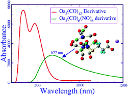
Water soluble organometallic carbonyl clusters are biocompatible, stable and reliable high-contrast photoacoustic contrast agents (PACAs). However, they have limited applications and efficacy due to their absorption in the visible region, which consists of wavelengths that have poor penetration depth inside tissue. In this article, we present the molecular level understanding of these compounds and investigate an alternative way to improve their photoacoustic properties. We discovered that organometallic nitrosyl carbonyl compounds, such as [Os3(CO)6(NO)4(μ-NO)(μ-S(CH2)2COO)]Na+, which shows high absorption in the near infrared region (λmax = 755 nm), are more suitable photoacoustic contrast agents than other carbonyl clusters that have been reported in the literature to date. As the metal nitrosyl bond is easily oxidisable, these compounds may also be used to study the reactive oxygen species in a living cell. We introduce a theoretical model to calculate the relative toxicity of a compound in terms of electric dipole moment (μ) and reactivity index (ω). Therefore, we computed the μ and ω values for all of the clusters. It was shown that the modeled nitrosyl carbonyl compound is much less toxic than the reported carbonyl clusters.


1 Introduction

Photoacoustic spectroscopy1,2 is one of the most important fields of research today, due to its spatial resolution and deeper tissue penetration capability in live cell imaging.3–8 Nowadays, exogenous contrast agents9 are used for most of the cell imaging techniques.7,8,10,11 For photoacoustic imaging, several classes of contrast agent have been tried, among which organic dyes,12,13 nanoparticles,14–18 nanodyes19 and organometallic cluster compounds20 are of the greatest interest. All of these contrast agents have many advantages over the others and also have some drawbacks. Very recently, organometallic cluster compounds, M3(CO)12 (where M = Fe, Ru and Os), of group 8 elements have been used as PACAs.20 These compounds have drawn a huge amount of interest, due to their biocompatibility, solubility, stability and negligible cytotoxicity.21–23 These compounds have also been used to synthesize effective anti-angiogenic agents that may be used for cancer treatment.24 They are also the common synthetic precursors to other organometallic complexes.25–27

These compounds are surface-active catalysts and have been shown to catalyze the Fischer–Tropsch28,29 and water-gas shift30 reactions. But the application of these compounds as PACAs is limited, because their optical absorbance is in the visible region.20 Photoacoustic tomography is used for live cell imaging to detect life-threatening diseases like cancer, tumors etc. This method is more accurate than other imaging techniques, such as MRI, CT, USG, etc. A very good PACA should exhibit significant absorbance within the wavelength range of 700 nm–1000 nm, as the absorbance in this region by living cells is much less. In this article, we report the molecular level understanding of these compounds and discover new compounds that exhibit absorbance at a longer wavelength (λ) through effective ligand substitution.

2 Theory

The linearized photoacoustic energy equation12,31 for a light absorptive material is defined as
 
image file: c5ra01757b-t1.tif(1)
where τ is a small perturbation in the temperature, and ζ, ρ, C, β, δ and γ are the thermal conductivity, density of the absorbent, heat capacity at constant volume, thermal expansivity, density perturbation and specific heat ratio, respectively. [script letter H](t) is the amount of heat generated due to photoacoustic interactions. Photoacoustic interactions in a material, when exposed to light, can be written as a function of the absorption coefficient (μa) and time integrated flux density, fluence (Φ),32 as follows
 
[script letter H](x,t) = μa(x)Φ(x,t,μa) (2)
where x is the position vector and t is the absorption time. It is known that the optical penetration depth is inversely proportional to the absorption coefficient (μa) and hence proportional to the wavelength λ of the incident radiation, as follows33
 
image file: c5ra01757b-t2.tif(3)
where k is the molar extinction coefficient. From eqn (2) and (3), we can say that a photoacoustic signal [script letter H](x,t) is inversely proposal to λ. If λ varies within the tissue transparency window of 700 nm to 1000 nm, a great contrast with the background signal is expected. Thus, for better contrast, one must use radiation of longer λ. It is also possible to increase the signal strength by increasing the Φ (see eqn (2)) within the biomedical application limits.34 By reducing the pulse duration (tp), Φ can also be increased provided image file: c5ra01757b-t3.tif.12 Thus, compounds that have a very small oscillator strength value may also be used successfully as PACAs by using a high frequency laser pulse.

To calculate relative toxicity (γ), we used the absolute hardness (η),35 absolute electronegativity (χ) and reactivity index (ω) of a compound.36,37 These three properties are calculated from the following relations

 
image file: c5ra01757b-t4.tif(4)
 
image file: c5ra01757b-t5.tif(5)
 
image file: c5ra01757b-t6.tif(6)
where EHOMO is the highest occupied molecular orbital energy and ELUMO is the lowest unoccupied molecular orbital energy. Toxicity (Ctox) and reactivity index (ω) are related as follows37
 
Ctox = k1 + k2ω (7)
where k1 and k2 are the constants. For a large value of ω, image file: c5ra01757b-t7.tif and hence
 
Ctoxω (8)
Toxicity also depends on the partition coefficient (Po/w) of the compound in 1-octanol and water. Considering Deneer’s equation38 we find that toxicity is related to Po/w as follows
 
Ctox ∝ (Po/w)−0.36 (9)
Thus, we can write
 
Ctoxω(Po/w)−0.36 (10)
Bayat and Movaffagh39 showed that the partition coefficient may be expressed in terms of the solvation free energy of the compound in two different solvents, as below
 
image file: c5ra01757b-t8.tif(11)
where ΔGsol,w and ΔGsol,oct are the solvation free energies of the compound in water and in 1-octanol, T is the temperature and R is the universal gas constant. The solvation free energy in water is the measure of the dipole–dipole interactions between the solute and solvent. Thus, we may consider that ΔGsol,w is proportional to the dipole moment (μ) of the solute i.e. dipole moment of the compound. If the test compound and reference compound are of the same type, then ΔGsol,oct is constant. We may consider that Po/w ∝ e, where k is the constant for a particular temperature. Using eqn (10), we get
 
Ctoxωe (12)
If we now assign the subscripts ref and comp to the reference compound and the compound under investigation, respectively, then the γ of similar compounds can be written as
 
image file: c5ra01757b-t9.tif(13)
k may also be considered as a scaling factor. For simplicity, we assume k = 1. Hence, the relative toxicity of similar compounds is as below
 
image file: c5ra01757b-t10.tif(14)
Thus, γref is 1.0. If γ of a compound is greater than 1.0, then that compound is more toxic than the reference one.

3 Computational details

All of the calculations were performed by using the Gaussian 09 package.40 The structures were optimized without any symmetry constraints. All of the minimum energy structures were confirmed by the harmonic vibrational frequency without any imaginary modes. The convergence thresholds were set to 0.000015 Hartrees per Bohr for the forces, 0.00006 Å for the displacement and 106 Hartrees for the energy change. All calculations were performed with density functional theory (DFT) using the unrestricted Becke’s three parameter hybrid exchange functional41 combined with Lee–Yang–Parr non-local correlation functional,42 abbreviated as B3LYP. We have used the LanL2DZ basis set43,44 along with the corresponding Los Alamos relativistic effective core potentials45 provided by the Gaussian 09 package. We have performed time dependent density functional theory (TDDFT) calculations for the UV-visible spectra of the chosen compounds. It has been reported that, for large main group element clusters, the B3LYP/LanL2DZ method is sufficient.46–51

4 Results and discussion

4.1 Metal carbonyl clusters

First, we carried out the geometry optimization of the three trimetal carbonyl clusters, Fe3(CO)12, Ru3(CO)12 and Os3(CO)12 in the gas phase. Fig. 1 represents the optimized structures and Table 1 lists the metal–metal (M–M) bond distances of these carbonyl clusters. In the Fe cluster, two metal atoms are connected through two bridging CO molecules. The bond distance between the two Fe atoms is 2.59 Å, which is very close to the experimental value of 2.57 Å. The length of the two other metal–metal bonds, i.e. bonds between bridging and non-bridging metals, is 2.68 Å, as compared to the experimental value of 2.70 Å. In the case of the Ru cluster, the bond between bridging atoms is 2.86 Å long, which is again in agreement with the experimental value of 2.85 Å. The length of the bonds between the bridging and non-bridging metals is 2.89 Å, as compared to the experimental values of 2.85 Å and 2.86 Å. For both the Fe and Ru clusters, the bonds between bridging and non-bridging metals are longer than the bridging M–M bond distances, which is expected because bridging stabilises the bond. On the other hand, in the case of the Os cluster, all three metal–metal bonds are of nearly the same length and there is no bridging carbonyl. The M–M bond distances are 2.89 Å, 2.89 Å and 2.89 Å, and are in very good agreement with the experimental values of 2.87 Å, 2.88 Å and 2.88 Å, respectively.
image file: c5ra01757b-f1.tif
Fig. 1 Optimized geometries of M3(CO)12 (M = Fe, Ru and Os) metal carbonyl clusters.
Table 1 Absorption maxima and other properties of Fe3(CO)12, Ru3(CO)12 and Os3(CO)12 clusters
Compound Calculated absorbance peak (nm) Experimental52,53 absorbance peak (nm) M–M bond distance (Å) (calculated) M–M bond distance (Å) (experimental) Charge on metal (au)
a Indicates metal atoms (or bond between two atoms) that are connected by a bridging ligand.
Fe3(CO)12 565 602 (ref. 52) 2.59a 2.57a (ref. 55) −0.940a
301 310 (ref. 52) 2.68 2.70 −0.940a
2.68 2.70 −0.653
Ru3(CO)12 390 390 (ref. 52), 392 (ref. 53) 2.86a 2.85 (ref. 56) 0.059a
230 238 (ref. 52) 2.89 2.85 0.059a
2.89 2.86 0.216
Os3(CO)12 386.71, 386.55 385 (ref. 52 and 53) 2.89 2.87 (ref. 56) 0.171
320.93, 320.87 330 (ref. 52 and 53) 2.89 2.88 0.171
254.43, 254.43 240 (ref. 52 and 54) 2.89 2.88 0.171


By using these optimized structures, we have computed the absorption spectra for all three clusters. The calculated values of the absorption peaks, which are listed in Table 1, were in very good agreement with the experimental findings. The computed value for the Fe cluster at 565 nm had a deviation of 37 nm from the experimental value, but all other deviations were less than 10 nm. In the osmium cluster, we found that all three absorption peaks actually resulted from the overlap of two nearly degenerate transitions, which probably arose due to the higher order of symmetry. Thus, for the osmium cluster, the absorbance intensity is higher than those of the Fe and Ru clusters. A comparison of the absorption spectra is presented in Fig. 2. The natures of all three plots are similar, they differ only in their absorption peaks being at different frequencies.


image file: c5ra01757b-f2.tif
Fig. 2 UV-visible spectra of different M3(CO)12 clusters (where M = Os, Ru and Fe).

4.2 Metal carbonyl cluster derivatives

From the metal carbonyl cluster studies, it is not clear why the osmium cluster exhibits better contrast as a PACA.20 Thus, we computed the UV-visible absorption spectra of all of the carbonyl cluster derivatives to explain the experimental results reported by Kong et al.20 Fig. 3 presents three optimized geometries of carbonyl cluster derivatives of Fe, Ru and Os. We used a thioacetate derivative of the Fe, Ru and Os clusters [M3(CO)10(μ-H)(μ-S(CH2)2COO)Na+] as these derivatives were used in the experiment by Kong et al. The optimized structures are shown in Fig. 3. Due to organic ligand substitution, the geometries of all three clusters have changed significantly.
image file: c5ra01757b-f3.tif
Fig. 3 Optimized geometries of metal carbonyl cluster derivatives.

From Table 2, it can be seen that in both the Fe and Ru clusters the metal–metal bond between the two bridged metals increased in length by 0.07 Å, but the bonds between the bridging and non-bridging metals remained almost the same, as the deviation was less than 0.01 Å. A dramatic change was observed for the osmium cluster: the symmetry was lost. Sulfide and hydride ions formed metal–ligand–metal (M–L–M) bridges. As a result, the bond between the two bridging Os molecules was decreased in length by 0.29 Å and the two other bond lengths were decreased by 0.21 Å and 0.20 Å, respectively. Fig. 4 presents the UV-visible spectra of these compounds. The absorption peaks of the Fe and Ru derivatives underwent a blue shift, whereas the Os derivative exhibited a red shift. From Table 2, it is clear from the maximum value of the absorption peak that the Os derivative is a better PACA than the Fe and Ru derivatives. The significant difference in the UV-visible spectra was due to their different structural transformations, which were observed due to organic ligand substitution. In the cluster-only form, i.e. in M3(CO)12, we observed two bridging CO molecules (M–L–M bridging) for both Fe and Ru but not for Os. Interestingly, in its derivative form, the Os cluster also exhibited a M–L–M structure like Fe and Ru. Thus, the red shift of the Os derivative was only due to the change in its geometry. We also observed that, for the Fe and Ru derivatives, only one absorption peak was obtained instead of two for the unsubstituted carbonyl clusters. For the Os derivative, there were two peaks, but they became broad.

Table 2 Absorption maxima of the Fe3(CO)12, Ru3(CO)12 and Os3(CO)12 cluster derivatives
Compound Calculated absorbance peak (in nm) Oscillator strength (calculated) HOMO–LUMO energy gap (au) M–M bond distance (Å) (calculated) Charge on metal (au)
a Indicates metal atoms (or bond between two atoms) which are connected by ligand bridge.
Fe3(CO)12 derivative 300 0.14 0.12101 2.66a −0.856a
2.69 −0.855a
2.69 −0.782
Ru3(CO)12 derivative 296 0.045 0.12975 2.92a −0.094a
2.88 −0.093a
2.88 0.190
Os3(CO)12 derivative 473 0.060 0.05965 2.61a 0.132a
349 0.075   2.70 0.180a
2.69 0.725


image file: c5ra01757b-f4.tif
Fig. 4 UV-visible spectra of different metal carbonyl cluster derivatives (M3(CO)10(μ-H)(μ-S(CH2)2COO)Na+, where M = Os, Ru and Fe).

Charge density analysis (see Table 1) showed that in the cluster-only form, all of the Fe atoms were negatively charged, the Ru atoms had a very small positive charge, and the Os atoms were positively charged. In the Fe and Ru clusters, the bridging atoms were more negative or less positive than the non-bridging atom, and the bond length between the M–M bridging atoms was less than that of the non-bridging bonds. This implies that, due to bridging, ligand-to-metal charge transfer occurred. In the Os cluster, all three metals had the same positive charge, which indicated the absence of ligand-to-metal charge transfer. Due to the more positive charge on Os and larger value of the crystal field splitting energy, it was more stable than the Fe and Ru clusters.

With regard to the Os cluster, a recent experiment did not observe any significant difference in its absorbance due to the formation of its salt derivative. We observed one absorption peak at 386 nm with an oscillator strength of 0.05 for the pure cluster, whereas for the derivative (see Fig. 4) there were two distinct peaks of nearly equal intensity, one was at 475 nm with an oscillator strength of 0.06 and another was at 340 nm with an oscillator strength of 0.08. Thus, due to salt formation, and hence a change in the ligand field, the optical properties of the compound changed significantly. If we consider the overlap between the two absorption maxima, we find a maximum at 407.5 nm, which is in very good agreement with the experimentally observed value of 410 nm.20

Analyzing the excited states of the M3(CO)12 clusters and their derivatives, we observed degeneracy only for the lowest unoccupied molecular orbital (LUMO) of the Os3(CO)12 cluster, which does not contain any bridging ligand. However, due to bridging in the derivative form, the degeneracy is lost. This loss of degeneracy was responsible for the decrease in absorbance. The twofold degeneracy indicates electronic transitions from one metal to two other equivalent metal centers. However, due to the presence of the ligand in the bridge, the two metal atoms differ from the third. In this situation, the absence of degeneracy indicates that the electron transition occurs from the bridging metals to the non-bridging metal.

4.3 Effect of the organic chain length

To model ways of improving the properties of this compound as a PACA, first we changed the organic chain length, keeping the functional group fixed. From Table 3, we can see that for C = 3 (C is the number of carbon atoms present in the organic salt) the photoacoustic activity is at its maximum. From the charge distribution of the residue, we find that for C = 3 the charge on the S atom is at its maximum (−0.286) and very large in comparison with those for C = 2, 4 and 5. The same charge on S for C = 2, 4 and 5 yields the same absorption maxima positions of the corresponding derivatives, irrespective of the salt.
Table 3 Influence of organic chain length on the optical absorption of the Os3(CO)12 derivative
Number of carbon atoms in the residue Charge on sulphur atom Lowest energy absorption peak (nm) Oscillator strength (calculated)
2 −0.069 290 0.05
3 −0.286 475 0.06
4 −0.072 287 0.07
5 −0.070 294 0.07


4.4 Effect of NO substitution

As we did not observe a significant improvement in photoacoustic properties by substituting the organic chain, we substituted the strong field CO ligand with a weak field NO ligand. For an osmium nitrosyl carbonyl cluster, such as Os3(CO)10(NO)2, we found that the effects of introducing bridging NO agrees very well with the experimental findings.57 As discussed before, we did not observe any bridging CO in the Os3(CO)12 cluster. The different metal–ligand bonding is due to the crystal field splitting. In the presence of a strong field CO ligand, the crystal field splitting stabilization energy is high and hence strong metal–metal bond formation is favorable. Thus, only terminal metal–ligand binding occurs in the case of the Os3(CO)12 cluster. The presence of a weak field ligand weakens the M–M bond and leads to the formation of a M–L–M bridge. Metal–metal bond analysis of the two optimized geometries Os3(CO)12 and Os3(CO)10(NO)2 supported this argument. All of the M–M bond distances in Os3(CO)12 were 2.89 Å, whereas in Os3(CO)10(NO)2 the bond distances were 2.96, 2.96 and 3.26 Å. The elongation of one of the Os–Os bonds, where two bridging ligands were connected, was a reflection of the weak M–M bonding. This change in the geometry and ligand field around the metals also caused a red-shift54 and blue-shift (from 386 nm → 339 nm) in the UV-visible spectrum. The metal–metal bond distances in the Os3(CO)11(NO) cluster were 2.88, 2.90 and 2.90 Å. Although the substitution of one CO by one NO did not affect the metal–metal bond length significantly, a huge red-shift in the UV-visible spectrum (386 nm → 494 nm, a change of 108 nm) was observed. Due to the substitution of one CO by NO, the HOMO–LUMO energy gap decreased from 0.14336 a.u. to 0.10673 a.u., i.e. by 109 nm, which is exactly the same as the spectral shift of 108 nm. Thus, the spectral shift was only due to the change in the ligand field. For this compound, the lowest energy transition was a metal–metal d–d transition. Thus, for the osmium cluster derivative, to improve its photoacoustic properties we have to substitute CO with a weak field ligand.

Following this, we computed the lower energy absorption spectra of all of the possible osmium nitrosyl carbonyl compounds. Interestingly, there was no linear relationship between λmax (wavelength for maximum absorbance) and the number of NO molecules. We discovered that, although the Os3(NO)12 compound had the highest λmax of 939 nm (see Table 4), its oscillator strength was very low (0.006). Thus, considering both its high λmax value and acceptable value for oscillator strength, Os3(CO)6(NO)6 may be the best PACA (λmax = 657 nm and an oscillator strength of 0.03). Applying the same organic salt substitution to this compound led to a better PACA (λmax = 755 nm and an oscillator strength of 0.0134).

Table 4 Absorption peak of the Os3(CO)12 cluster due to the substitution of CO by a NO ligand
Composition Absorbance peak (in nm) Oscillator strength
Os3(CO)12 386 0.091
Os3(CO)11(NO) 494 0.046
Os3(CO)10(NO)2 339 0.025
Os3(CO)9(NO)3 361 0.014
Os3(CO)8(NO)4 337 0.028
Os3(CO)7(NO)5 429 0.006
Os3(CO)6(NO)6 654 0.028
Os3(CO)5(NO)7 545 0.006
Os3(CO)4(NO)8 540 0.034
Os3(CO)3(NO)9 674 0.006
Os3(CO)2(NO)10 655 0.016
Os3(CO)1(NO)11 764 0.004
Os3(NO)12 939 0.005


Since we have found that the Os3(CO)6(NO)4(μ-NO)(μ-S(CH2)2COO)Na+ may be the best PACA, we further studied this compound extensively. To investigate the effect of the solvent on this compound, we included water as a solvent. For this particular study, we used the IEFPCM model and B3PW91 functional with the same basis set. The optimized structure is given in Fig. 5. The metal–metal bond distances were 3.22 Å between the bridging metals and 3.06 Å and 2.97 Å between the bridging and non-bridging metals.


image file: c5ra01757b-f5.tif
Fig. 5 Optimized structure of Os3(CO)6(NO)4(μ-NO)(μ-S(CH2)2COO)Na+.

IR and UV-visible spectra of Os3(CO)6(NO)4(μ-NO)(μ-S(CH2)2COO)Na+ are shown in Fig. 6 and 7, respectively. It was observed that our modeled compound showed similar efficiency as a PACA in solution phase as well. Absorption above 800 nm was the same in both the gas phase and solution phase. Between 500 nm and 800 nm, it had a better efficiency in the solution phase. Thus, the use of this compound as a PACA is viable. We found that it is a very strongly polar compound with a dipole moment of 14.29 debye. Thus, it is expected that this compound would be highly water soluble and hence less cytotoxic. A comparison of the relative toxicities of three cluster derivatives and this nitro-substituted compound is presented in Table 5. The osmium carbonyl cluster was taken as the reference compound. We know that cytotoxicity depends on the reactivity index and log[thin space (1/6-em)]Po/w.58,59 As discussed before, a compound of high dipole moment (μ) should have a low value of log[thin space (1/6-em)]Po/w and low toxicity.58 On the other hand, a high reactivity index value (ω) corresponds to high toxicity. Here, we have computed the relative toxicity, as mentioned in the theory section. Using these parameters, we found that Ru3(CO)6(NO)4(μ-NO)(μ-S(CH2)2COO)Na+ has six times (6.227) higher toxicity than Os3(CO)10(μ-H)(μ-S(CH2)2COO)Na+. Our modeled compound, Os3(CO)6(NO)4(μ-NO)(μ-S(CH2)2COO)Na+, is considerably less toxic than the osmium and iron cluster derivatives.


image file: c5ra01757b-f6.tif
Fig. 6 IR spectrum of Os3(CO)6(NO)4(μ-NO)(μ-S(CH2)2COO)Na+.

image file: c5ra01757b-f7.tif
Fig. 7 UV-visible spectra of Os3(CO)6(NO)4(μ-NO)(μ-S(CH2)2COO)Na+.
Table 5 Reactivity descriptors and relative toxicity (with respect to osmium cluster derivatives) of three metal carbonyl cluster derivatives and our modeled compound
Compounds Absolute hardness (η) Absolute electro-negativity (χ) Reactivity index (ω) Dipole moment (μ) Relative toxicity (γ)
Fe3(CO)10 salt 0.060 0.1779 0.2628 11.954 0.014
Ru3(CO)10 salt 0.050 0.1760 0.3094 5.988 6.227
Os3(CO)10 salt 0.071 0.1753 0.2161 7.458 1.00
Os3(CO)6(NO)5 salt 0.024 0.1876 0.7325 14.292 0.004


4.5 Free energy (ΔG) calculation for NO substitution

We have used modeling to produce an improved PACA by substituting CO by a NO ligand. The synthesis of these compounds may not be trivial. So far, mono- and di-substituted compounds have already been synthesized. Thus, using a free energy calculation, we investigated whether other substitutions are possible or not. The computed ΔG values are listed in Table 6. The ΔG values for the first two substitutions (i.e. for Os3(CO)11(NO) and Os3(CO)10(NO)2) are negative. But overall the free energy change for the tri-substituted derivative is positive, as is that for the penta-substituted derivative. The total free energy change values for the other derivatives are negative. From this calculation, we conclude that tri- and penta-substituted derivatives may not be prepared, but the rest can be prepared.
Table 6 Calculation of the free energy change values for the substitution reaction of CO by NO ligands in Os3(CO)12
Composition Free energy change (ΔG) (in a.u.) from the previous derivative Free energy change (ΔG) (in a.u.) from the initial compound
Os3(CO)11(NO) −0.3742 −0.3742
Os3(CO)10(NO)2 −0.0463 −0.4205
Os3(CO)9(NO)3 0.4498 0.0293
Os3(CO)8(NO)4 −0.3892 −0.3599
Os3(CO)7(NO)5 0.4405 0.0806
Os3(CO)6(NO)6 −0.3738 −0.2932
Os3(CO)5(NO)7 0.0038 −0.2894
Os3(CO)4(NO)8 −0.4471 −0.7365
Os3(CO)3(NO)9 0.0035 −0.7329
Os3(CO)2(NO)10 0.0835 −0.6495
Os3(CO)1(NO)11 −0.0002 −0.6497
Os3(NO)12 0.0169 −0.6328


4.6 Drug potentiality

As organometallic compounds are used as potential drugs for several fatal diseases,24,60 we have tested our modeled compound, Os3(CO)6(NO)4(μ-NO)(μ-S(CH2)2COO)Na+, for a few select diseases using the ChemoSophia in silico testing program.61 This compound was found to have drug potentiality for AIDS, cancer and as a painkiller. The results of the biological activity tests are given in Table 7. The compound was also found to have relatively low ecotoxicity. This supports our previously discussed relative toxicity calculation result for this compound. It is also possible for this compound to be metabolized by CYP450-2D6 and CYP450-3A4.
Table 7 Drug potentiality of our modeled compound: an in silico test using the ChemoSophia61 package
Biological activity Success probability (%)
HIV1-protease inhibitory activity 66.8
Anti-inflammatory activity (ks2-p38-MAP-kinase inhibitors) 69.4
Antioxidant activity 33.8
Antitumor anti-mitotic activity 27.5
Antitumor dihydrofolate reductase inhibitory activity 25.9
Antitumor topoisomerase-II inhibitory activity 99.6
Ecotoxicity 8.7
Metabolism at CYP450-2D6 54.9
Metabolism at CYP450-3A4 78.9


5 Conclusions

We studied the UV-visible spectra of organometallic carbonyl clusters, as these compounds have very recently been used as photoacoustic contrast agents. We found that Os3(CO)6(NO)4(μ-NO)(μ-S(CH2)2COO)Na+ is a better PACA, with high absorption from 500 nm to 1000 nm. Absorption by this compound even extended up to 2000 nm. Thus, it may be used in IR detection. This compound is paramagnetic, hence it may also be used as an MRI contrast agent. The absolute hardness (η) of this compound is 0.024 and its reactivity index (ω) is 0.7325, which suggests that it may be used to study reactive oxygen species in the living cell. Free energy calculations show that this compound can be easily synthesized. In silico activity testing of this compound shows very good activity towards HIV (66.8%) and cancer (99.6%). Other biological activities are given in Table 7. From these studies, we can say that our modeled compound Os3(CO)6(NO)4(μ-NO)(μ-S(CH2)2COO)Na+ may serve as an eco-friendly (as its ecotoxicity is only 8.7%), biocompatible (as it is non-cytotoxic), water soluble PACA capable of being detected in deeper tissue (absorption from 700 nm to 1000 nm), and may also function as an effective drug for HIV, cancer and inflammation.

Acknowledgements

A. Bag acknowledges IISER Kolkata for funding and providing research facilities. PKG would like to thank CSIR grant no. 01(2558)/12/EMR-II for financial support.

References

  1. A. G. Bell, Am. J. Sci., 1880, 20, 305–324 CrossRef.
  2. A. C. Tam, Rev. Mod. Phys., 1986, 58, 381–431 CrossRef CAS.
  3. L. V. Wang, Med. Phys., 2008, 35, 5758–5767 CrossRef.
  4. N. V. Thakor, J. Cereb. Blood Flow Metab., 2012, 32, 936–937 CrossRef PubMed.
  5. X. Wang, Y. Pang, G. Ku, X. Xie, G. Stocia and L. V. Wang, Nat. Biotechnol., 2003, 21, 803–806 CrossRef CAS PubMed.
  6. X. Wang, Y. Pang, G. Ku, G. Stocia and L. V. Wang, Opt. Lett., 2003, 28, 1739–1741 CrossRef CAS.
  7. D. J. Stephens and V. J. Allan, Science, 2003, 300, 82–86 CrossRef CAS PubMed.
  8. J. K. Jaiswal, E. R. Goldman, H. Mattoussi and S. M. Simon, Nat. Methods, 2004, 1, 73–78 CrossRef PubMed.
  9. V. Tsytsarev, C. Bernardelli and K. I. Maslov, J. Neurosci. Neuroeng., 2012, 1, 180–192 CrossRef PubMed.
  10. R. B. Sekar and A. Periasamy, J. Cell Biol., 2003, 160(5), 629–633 CrossRef CAS PubMed.
  11. X. Michalet, F. F. Pinaud, L. A. Bentolila, J. M. Tsay, S. Doose, J. J. Li, G. Sundaresan, A. M. Wu, S. S. Gambhir and S. Weiss, Science, 2005, 307, 538–544 CrossRef CAS PubMed.
  12. K. Firouzi, E. Stride and N. Saffari, J. Acoust. Soc. Am., 2013, 133(6), 3853–3862 CrossRef CAS PubMed.
  13. A. Hellebust and R. Richards-Kortum, Nanomedicine, 2012, 7(3), 429–445 CrossRef CAS PubMed.
  14. A. Agarwal, S. W. Huang, M. OD́onnell, K. C. Dey, M. Dey, N. Kotov and S. Ashkenazi, Appl. Phys. Lett., 2007, 102, 064107 Search PubMed.
  15. K. Pu, A. J. Shuhendler, J. V. Jokerst, J. Mei, S. S. Gambhir, Z. Bao and J. Rao, Nat. Nanotechnol., 2014, 9, 233–239 CrossRef CAS PubMed; J. V. Jokerst, A. J. Cole, D. V. de Sompel and S. S. Gambhir, ACS Nano, 2012, 6, 10366–10377 CrossRef PubMed.
  16. Y. S. Chen, W. Frey, S. Kim, P. Kruizinga, K. Homan and S. Emelianov, Nano Lett., 2011, 11, 348–354 CrossRef CAS PubMed.
  17. M. Pramanik, M. Swierczewska, D. Green, B. Sitharaman and L. V. Wang, J. Biomed. Opt., 2009, 14, 034018 CrossRef PubMed.
  18. X. Liang, Z. Deng, L. Jing, X. Li, Z. Dai, C. Li and M. Huang, Chem. Commun., 2013, 49, 11029–11031 RSC.
  19. C. J. Flynn, E. B. E. Oh, S. M. McCullough, R. W. Call, C. L. Donley, R. Lopez and J. F. Cahoon, J. Phys. Chem. C, 2014, 118, 14177–14184 CAS.
  20. K. V. Kong, L. Lun-De, Z. Lam, N. V. Thakor, W. K. Leong and M. Olivo, Chem. Commun., 2014, 50, 2601–2603 RSC.
  21. K. V. Kong, W. K. Leong and L. H. Lim, Chem. Res. Toxicol., 2009, 22, 1116–1122 CrossRef CAS PubMed.
  22. K. V. Kong, W. K. Leong, S. P. Ng, T. H. Nguyen and L. H. Lim, ChemMedChem, 2008, 3, 1269–1275 CrossRef CAS PubMed.
  23. K. V. Kong, W. Chew, L. H. K. Lim, W. Y. Fan and W. K. Leong, Bioconjugate Chem., 2007, 18, 1370–1374 CrossRef CAS PubMed.
  24. A. A. Nazarov, M. Baquie, P. N. Sliwinska, O. Zava, J. R. van Beijnum, M. Groessl, D. M. Chisholm, Z. Ahmadi, J. S. McIndoe, A. W. Griffion, H. van den Bergh and P. J. Dyson, Sci. Rep., 2013, 3, 1485 Search PubMed.
  25. M. G. Richmond, Coord. Chem. Rev., 2004, 248, 881–901 CrossRef CAS PubMed.
  26. N. E. Leadbeater, J. Organomet. Chem., 1999, 573, 211–216 CrossRef CAS.
  27. Y. M. Wuu, J. G. Bentsen, C. G. Brinkley and M. S. Wrighton, Inorg. Chem., 1987, 26, 530–540 CrossRef CAS.
  28. M. Deeba and B. C. Gates, J. Catal., 1981, 67, 303–307 CrossRef CAS.
  29. F. Hugues, A. Dalmon, P. Bussiere, A. K. Smith, M. J. Basset and D. Olivier, J. Phys. Chem., 1982, 86, 5136–5144 CrossRef CAS.
  30. R. Ganzerla, M. Lenarda, F. Pinna and M. Graziani, J. Organomet. Chem., 1981, 208, C43–C45 CrossRef CAS.
  31. H. X. Chen, I. E. McGrath, A. C. Beveridge and G. J. Diebold, Proc. – IEEE Ultrason. Symp., 1997, 1, 719–725 Search PubMed.
  32. B. T. Cox and P. C. Beard, J. Acoust. Soc. Am., 2005, 117, 3616–3627 CrossRef CAS PubMed.
  33. http://pveducation.org/pvcdrom/pn-junction/absorption-coefficient.
  34. M. Xu and L. V. Wang, Rev. Sci. Instrum., 2006, 77, 041101 CrossRef PubMed.
  35. R. G. Parr and R. G. Pearson, J. Am. Chem. Soc., 1983, 105, 7512–7516 CrossRef CAS.
  36. M. Ishihara, M. Kawase, G. Westman, K. Samuelsson, N. Motohashi and H. Sakagami, Anticancer Res., 2007, 27, 4053–4058 CAS.
  37. J. Padmanabhan, R. Parthasarathi, V. Subramanian and P. K. Chattaraj, J. Phys. Chem. A, 2006, 110, 2739–2745 CrossRef CAS PubMed.
  38. J. W. Deneer, W. Sinnige and J. L. M. Hermens, Aquat. Toxicol., 1988, 12, 185–192 CrossRef CAS.
  39. Z. Bayat and J. Movaffagh, Russ. J. Phys. Chem. A, 2010, 84, 2293–2299 CrossRef CAS.
  40. M. J. Frisch, G. W. Trucks, H. B. Schlegel, G. E. Scuseria, M. A. Robb, J. R. Cheeseman, G. Scalmani, V. Barone, B. Mennucci, G. A. Petersson, H. Nakatsuji, M. Caricato, X. Li, H. P. Hratchian, A. F. Izmaylov, J. Bloino, G. Zheng, J. L. Sonnenberg, M. Hada, M. Ehara, K. Toyota, R. Fukuda, J. Hasegawa, M. Ishida, T. Nakajima, Y. Honda, O. Kitao, H. Nakai, T. Vreven, J. A. Montgomery Jr, J. E. Peralta, F. Ogliaro, M. Bearpark, J. J. Heyd, E. Brothers, K. N. Kudin, V. N. Staroverov, R. Kobayashi, J. Normand, K. Raghavachari, A. Rendell, J. C. Burant, S. S. Iyengar, J. Tomasi, M. Cossi, N. Rega, J. M. Millam, M. Klene, J. E. Knox, J. B. Cross, V. Bakken, C. Adamo, J. Jaramillo, R. Gomperts, R. E. Stratmann, O. Yazyev, A. J. Austin, R. Cammi, C. Pomelli, J. W. Ochterski, R. L. Martin, K. Morokuma, V. G. Zakrzewski, G. A. Voth, P. Salvador, J. J. Dannenberg, S. Dapprich, A. D. Daniels, O. Farkas, J. B. Foresman, J. V. Ortiz, J. Cioslowski and D. J. Fox, Gaussian 09, Revision D.01, Gaussian, Inc., Wallingford CT, 2009 Search PubMed.
  41. A. D. Becke, Phys. Rev. A, 1988, 38, 3098–3100 CrossRef CAS.
  42. C. Lee, W. Yang and R. G. Parr, Phys. Rev. B: Condens. Matter Mater. Phys., 1988, 37, 785–789 CrossRef CAS.
  43. P. J. Hay and W. R. Wadt, J. Chem. Phys., 1985, 82, 270–283 CrossRef CAS PubMed.
  44. W. R. Wadt and P. J. Hay, J. Chem. Phys., 1985, 82, 284–298 CrossRef CAS PubMed.
  45. P. J. Hay and W. R. Wadt, J. Chem. Phys., 1985, 82, 299–310 CrossRef CAS PubMed.
  46. S. Chiodo, N. Russo and E. Sicilia, J. Chem. Phys., 2006, 125, 104107 CrossRef CAS PubMed.
  47. J. F. Alvino, T. Bennett, D. Anderson, B. Donoeva, D. Ovoshchnikov, R. H. Adnan, D. Appadoo, V. Golovko, G. Andersson and G. F. Metha, RSC Adv., 2013, 3, 22140–22149 RSC.
  48. T. Bennett, R. H. Adnan, J. F. Alvino, V. Golovko, G. G. Andersson and G. F. Metha, Inorg. Chem., 2014, 53, 4340–4349 CrossRef CAS PubMed.
  49. H. Wang, Y. Xie, R. B. King and I. H. F. Schaefer III, J. Am. Chem. Soc., 2006, 128, 11376–11384 CrossRef CAS PubMed.
  50. T. Andruniow, M. Jaworska, P. Lodowski, M. Z. Zgierski, R. Dreos, L. Randaccio and P. M. Kozlowski, J. Chem. Phys., 2008, 129, 085101 CrossRef PubMed.
  51. M. R. Silva-Junior, M. Schreiber, S. P. A. Sauer and W. Thiel, J. Chem. Phys., 2008, 129, 104103 CrossRef PubMed.
  52. D. R. Tyler, R. A. Levenson and H. B. Gray, J. Am. Chem. Soc., 1978, 100, 7888–7893 CrossRef CAS.
  53. F. W. Vergeer, P. Matousek, M. Twrie, P. J. Costa, M. J. Calhorda and F. Hartl, Chem.–Eur. J., 2004, 10, 3451–3460 CrossRef CAS PubMed.
  54. J. P. Lomont, A. J. Shearer, S. C. Nguyen and C. B. Harris, Organometallics, 2013, 32, 2178–2186 CrossRef CAS.
  55. Y. Kou, H. L. Wang, M. Te, T. Tanaka and M. Nomura, J. Catal., 1993, 141, 660–670 CrossRef CAS.
  56. M. J. Calhorda, P. J. Costa, F. Hartl and F. W. Vergeer, C. R. Chim., 2005, 8, 1477–1486 CrossRef CAS PubMed.
  57. J. R. Norton, J. P. Collman, G. Dolcetti and W. T. Robinson, Inorg. Chem., 1972, 11(2), 382–388 CrossRef CAS.
  58. D. Asturiol and A. Worth, The use of chemical reactivity Assays in toxicity prediction, E. Farmi 2749, 21027 Ispra(VA), Italy.
  59. M. Ishihara, M. Kawase, G. Westman, K. Samuelsson, N. Motohashi and H. Sakagami, Anticancer Res., 2007, 27, 4053–4058 CAS.
  60. G. Gasser, I. Ott and N. Metzler-Nolte, J. Med. Chem., 2011, 54, 3–25 CrossRef CAS PubMed.
  61. http://www.chemosophia.com.

This journal is © The Royal Society of Chemistry 2015
Click here to see how this site uses Cookies. View our privacy policy here.