Amino-functionalized magnetic magnesium silicate double-shelled hollow microspheres for enhanced removal of lead ions

Bingfang Zouab, Ke Chena, Yongqiang Wang*a, Chunyu Niua and Shaomin Zhoua
aKey Laboratory for Special Functional Materials of the Ministry of Education, Henan University, Kaifeng 475004, P. R. China. E-mail: wangyq@henu.edu.cn; Fax: +86 3712 3881358; Tel: +86 3712 3881358
bSchool of Physics and Electronics, Henan University, Kaifeng 475004, P. R. China

Received 23rd January 2015 , Accepted 23rd February 2015

First published on 23rd February 2015


Abstract

Magnesium silicate hollow microspheres with porous structure were synthesized using silica microspheres as chemical template, which exhibited excellent adsorption properties to heavy metal ions in water in our previous report. Herein, both an iron magnetic core and amino-groups were put forward to functionalize the magnesium silicate hollow microspheres for a fast recoverable and highly efficient absorbent. In the synthesis process, double-shelled hollow microspheres with iron oxide (Fe3O4) inner shell and magnesium silicate outer shell (DS-Fe3O4/MS) were realized via hydrothermal treatment of Fe3O4@SiO2 double-shelled hollow microspheres in ammonia solution containing magnesium ions, and the amino-group was introduced into the porous surface of the magnesium silicate shell by a refluxing method to increase the active adsorption sites. The experimental results show that the amino-functionalized magnetic magnesium silicate double-shelled hollow microspheres (DS-Fe3O4/MS-AG) exhibited a high adsorption capacity of 315.5 mg g−1, and the high adsorption capacity was proposed to be due to synergic adsorption from the magnesium silicate shell (ion-exchange adsorption), surface-modified amino-groups (complexation adsorption) and the carboxyl groups on the inner iron oxide hollow microspheres (electrostatic adsorption). Additionally, the used DS-Fe3O4/MS-AG hollow microspheres could be easily regenerated through immersing them in a solution containing magnesium ions.
1. Introduction

Heavy metal pollutants (such as Pb2+, Cd2+ and Hg2+) in aquatic environments have brought a severe threat to public health and ecological systems.1 Considerable efforts have been made to develop effective treatment techniques for removing heavy metal ions from water; including electrochemical methods, adsorption, chemical coagulation, photo degradation, and biodegradation.2 Among these methods, adsorption is probably the most common method in practical use, for the adsorbent could be used repeatedly, meanwhile the heavy metal ions could be recovered through a desorption process. Unquestionably, the development of the adsorption technique relies heavily on research progress of adsorbent materials, thus development of heavy metal adsorbents with high adsorption capacity, fast adsorption desorption kinetics, and easy separation and regeneration is in great demand.

The rapid advance of nanoscience and nanotechnology has brought new opportunities for water treatment.2 Due to unique physical and chemical properties such as large specific surface areas, high adsorption capacity and fast adsorption rate, nanomaterials have shown their tremendous potential for capture of inorganic or organic pollutants in water. Up to now, various kinds of nanomaterials, especially micro/nanostructured material like metal oxide,3–5 silicate,6–10 and surface modified mesoporous silica,11–13 have been focused on as the adsorbents in waste-water treatment, and proven to be promising for environmental remediation. Among them, silicate nanomaterials were attracted many researchers' interest for their stable and environment-friendly features. Our group reported a serial silicate nanomaterial through using silica particles as chemical template,14–16 like magnesium silicate hollow microspheres assembled by nanosheets, which could be used as excellent and environment-friendly absorbents of heavy metal ions through ions exchange.16 Although this kind of silicate adsorbent exhibited good adsorption performance, it is very difficult to recycle them due to their dispersive properties, which are often encountered in other micro/nanostructured absorbents. Therefore, magnetic-based silicate adsorbents have been extensively studied due to their special magnetic properties. These magnetic adsorbents can be easily separated from aqueous systems by an external magnet, then lower operational costs for absorbent separations and recycling of the adsorbent materials could be easily achieved.17–19

Moreover, higher adsorption capacity are still required and pursued at all time when using absorbent to remove pollutants.20 It is well-known that higher specific surface area means more adsorption active sites, thus absorbents with porous or hierarchical, even hollow structures was continually developed. For example, urchin-like or flower-like iron oxide, especial with hollow interior, were successively synthesized to improve their high specific surface area as efficient adsorbents.21–23 However, the specific surface area couldn't be improved infinitely, thus composite adsorbents were becoming attractive recently, for all the compositions could contribute to the adsorption of pollutants, and this complex structure could also achieved higher adsorption capacity, for example, amino-groups or thiol-groups was often used to functionalize mesoporous silica materials;12,24 graphene oxide with carboxyl groups were used to modify other absorbents like iron oxides and silicates for improved adsorption capability.25–28 Therefore, new structures or composites are still needed to be explored and developed.

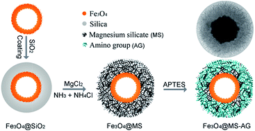
Here in this paper, a kind of composite adsorbent with magnetic core and amino-modified surface was developed as illustrated in Scheme 1. Fe3O4 hollow microspheres coated with silica were used as chemical template, for the silica shell were transformed into porous magnesium shell with high specific area under hydrothermal treatment, which both exhibited fast magnetic separability and high adsorption capacity. Furthermore, the large specific surface area of porous magnesium silicate shell was further utilized to be functionalized with amino-groups, which could increase the adsorption sites for higher adsorption capacities. Therefore, the special designed composite structure was expected to both exhibit high adsorption capacity and fast recoverability when used as adsorbent of Pb2+ ions in wastewater.


image file: c5ra01373a-s1.tif
Scheme 1 The synthesis of amino-functionalized magnetic magnesium silicate double-shelled hollow microspheres.

2. Experimental section

2.1 Double-shelled Fe3O4/SiO2 hollow microspheres

The magnetic Fe3O4 hollow microspheres with diameter about 250 nm were prepared through a hydrothermal reaction.29 The as-obtained Fe3O4 (0.1 g) microspheres were then dispersed in water–ammonia–ethanol mixture (10 mL/8 mL/180 mL) by sonication for 30 min, then a solution of TEOS in ethanol was slowly dropped into the above solution under a mechanical stirrer. After the reaction was performed for 120 min, the product was collected with a magnet, and washed with ethanol and water for several times.30 Finally, the product was dried in vacuum for 6 h to obtain the Fe3O4/SiO2 microspheres.

2.2 Double-shelled Fe3O4/magnesium silicate hollow microspheres (DS-Fe3O4/MS)

The above DS-Fe3O4/SiO2 microspheres (0.075 g) were dispersed in 40 mL H2O, then MgCl2 (0.15 g), NH4Cl (1.5 g) and NH3·H2O (1.5 mL) were added into the above solution in turn. The above mixture was sonicated for 30 min, and then was transferred into a Teflon-lined stainless-steel autoclave (80 mL) and then sealed to heat at 140 °C. After a 12 h reaction period, the autoclave was cooled to room temperature. The precipitate was collected by the help of a magnet, and then dried in vacuum oven for 12 h.

2.3 Double-shelled Fe3O4/MS hollow microspheres functionalized with amino-group (DS-Fe3O4/MS-AG)

Amino-functionalized double-shelled Fe3O4/MS hollow microspheres were prepared by surface modification of Fe3O4/MS hollow microspheres using (3-aminopropyl) trimethoxysilane (APTMS). Double-shelled Fe3O4/MS hollow microspheres (0.05 g), toluene (50 mL) and APTMS (5 mL) were added to a 250 mL three-necked flask. After the mixture was ultrasonically dispersed for 30 min, it was refluxed at 100 °C with continuous stirring for 6 h under a nitrogen flow (30 mL min−1). The final product was collected by the help of a magnet, followed by washing with ethanol several times and drying at 60 °C in vacuum oven for 12 h.

2.4 Characterization

The products were analyzed by X-ray diffraction (XRD), in a 2θ range from 10° to 80°, using CuKα radiation (Philips X'pert diffractometer). The morphology of the prepared products was studied by field emission scanning electron microscopy (FESEM, Sirion 200 FEG) and field emission transmission electron microscopy (FETEM, JEOL-2010, 200 kV). The powders were dispersed in ethanol using ultrasonic vibration. The samples for microscopy studies were prepared by deposition of dispersions of the powder in ethanol directly on the SEM stubs or holey carbon grid for TEM examination. Infrared (IR) spectra were recorded in the wave numbers ranging from 4000 to 400 cm−1 with a Nicolet model 759 Fourier transform infrared (FT-IR) spectrometer using a KBr wafer. Magnetic measurements were performed with a superconducting quantum interference device (SQUID) magnetometer (Quantum Design, MPMS XL). The surface area of the samples was determined by nitrogen adsorption (Micrometrics ASAP 2020).

2.5 Pb2+ ions removal experiments

Pb(NO3)2 was used as the source of Pb2+ ions. Pb2+ ions solution (20 mL) with different concentrations were firstly prepared, and then the above as-prepared absorbent (20 mg) was added to test the adsorption performance. The mixtures were shaken at a speed of 100 rpm for 6 hours to establish adsorption equilibrium at room temperature, and then the adsorbent was separated from the mixture by a magnet. To determine Pb2+ ions removal by the adsorbent, the Pb2+ concentration in the remaining solution was measured by an inductively coupled plasma atomic emission spectrophotometer (ICP-AES). The adsorption rate experiments were carried out inside 100 mL bottle by using DS-Fe3O4/MS-AG hollow microspheres (50 mg) as adsorbent, and the samples were collected at desired time intervals and separated by a magnet immediately. The Pb2+-loaded amino-functionalized Fe3O4/MS hollow microspheres were regenerated through immersing in magnesium chloride solution for 6 h. After that, the absorbent was washed with distilled water for cyclic adsorption, and the adsorption process were measured by similar steps.

3. Results and discussion

3.1. Morphology and phase characterizations of Fe3O4, DS-Fe3O4/MS, DS-Fe3O4/MS-AG hollow microspheres

The superparamagnetic Fe3O4 hollow microspheres were prepared according to our previous report shown in Fig. 1a, the hollow microspheres had a diameter of 250 nm in Fig 1b. These as-prepared Fe3O4 hollow microspheres are highly water dispersible for abundant carboxyl surface groups which made the outer SiO2 coating easily. As shown in Fig 1b, monodispersed double-shelled Fe3O4/SiO2 (DS-Fe3O4/SiO2) hollow microspheres were synthesized successfully by sol–gel method using tetra-ethoxysilane (TEOS) as the silica source, a thick SiO2 layer about 100 nm could be seen around Fe3O4 hollow microspheres. As marked by the dotted line in Fig. 1b, the hollow structure of magnetic core still could be seen clearly. After hydrothermal treatment in alkaline solution containing magnesium ions, the SiO2 shell is gradually dissolved and reacted with Mg2+ to generated magnesium silicates, then magnetic magnesium silicate double-shelled hollow microspheres (DS-Fe3O4/MS) were obtained in Fig. 1c. Clear core shell structure were observed from SEM image, and the core was surrounded by lamellae-assembled shell, and the magnesium silicate grew in the form of a lamellar structure which were origin of its talc structure. The thin lamellae wrapped together and formed porous shell which was subsequently functionalized with amino-groups for increasing adsorption sites. After functionalized with amino-groups, the product (DS-Fe3O4/MS-AG) show no obviously change seen from Fig. 1d. The DS-Fe3O4/MS-AG still kept core shell structure with rough shell in Fig. 1e, further magnified image in Fig. 1f show that the shell kept thin lamella structure. Since the magnesium silicate was transformed from silica shell in the above experiment, thicker magnesium silicate shell could be obtained easily when Fe3O4/SiO2 hollow microspheres with thicker silica shell was used, and the corresponding results could be seen in Fig. S1.
image file: c5ra01373a-f1.tif
Fig. 1 (a) SEM image of Fe3O4 hollow microspheres, (b) TEM image of DS-Fe3O4/SiO2 hollow microspheres, (c) SEM image of DS-Fe3O4/MS hollow microspheres and (d) SEM image of DS-Fe3O4/MS-AG hollow microspheres, (e) and (f) TEM images of DS-Fe3O4/MS-AG hollow microspheres.

Powder XRD was used to monitor the phase structure of the products during the synthesis process (Fig. 2). In the XRD pattern of the obtained Fe3O4 sample (curve a), all the diffraction peaks could be easily indexed to the standard Fe3O4 reflection (JCPDS card no.75-1609). Curve b is a typical XRD pattern of the Fe3O4/SiO2 hollow microspheres, which show almost all the same features as those shown in curve a. No diffraction peaks corresponding to SiO2 were observed except the broad peaks around 23°, which was due to the fact that the prepared SiO2 shell is amorphous. In comparison to the XRD patterns of the Fe3O4 hollow microspheres, four additional peaks like (020), (200), (332) and (335) were vaguely observed in curve c as shown by the arrows, which represent the generation of magnesium silicate hydroxide hydrate (Mg3Si4O10(OH)2, JCPDS no. 03-0174) and can be indexed through careful comparison with pure magnesium silicate in Fig. 2d.16 Meanwhile no broad peak around 23° was detected, showing the complete transformation of silica layer into magnesium silicate shell around Fe3O4 hollow microspheres. Therefore, the gradual phase changes of the products at all stages proved the successfully synthesis of magnetic magnesium silicate composite material. After amino-functionalization, the final products showed a similar XRD pattern with curve c (not shown here), however, it could be identified by other techniques like IR spectrum.


image file: c5ra01373a-f2.tif
Fig. 2 X-ray diffraction (XRD) patterns of (a) Fe3O4 hollow microspheres, (b) DS-Fe3O4/SiO2 hollow microspheres, (c) DS-Fe3O4/MS hollow microspheres and (d) pure magnesium silicate hollow microspheres.

3.2. Characterizations of amino-groups functionalized DS-Fe3O4/MS (DS-Fe3O4/MS-AG) hollow microspheres

The surface area and pore size distribution of DS-Fe3O4/MS and DS-Fe3O4/MS-AG hollow microspheres were tested by nitrogen adsorption–desorption. As seen in Fig. S2, both curve a and b belong to type IV isotherm with a type H3 hysteresis loop, which indicated the as-obtained product was mesoporous material. For the introduction of magnetic cores, the BET surface area of DS-Fe3O4/MS hollow microspheres are calculated to be 438 m2 g−1, which is a little smaller than that (521 m2 g−1) of our previously reported magnesium silicate hollow microspheres. Although they were further functionalized with amino-groups, the final DS-Fe3O4/MS-AG hollow microspheres are still as high as 402 m2 g−1. Such a high surface area is mainly attributed to the hollow structures and porous shell. There is a sharp pore distribution with an average diameter of 3.97 nm in DS-Fe3O4/MS, which are most likely from the void space of magnesium silicate self-assembly lamellae structures, and the pore became smaller in size with center peak at 3.76 nm which may be ascribed that small amount of silica generated during the surface functionalization. The high surface area and porous structure of the final DS-Fe3O4/MS-AG are beneficial for adsorption of heavy metal ions.
image file: c5ra01373a-f3.tif
Fig. 3 IR spectra of DS-Fe3O4/MS hollow microspheres (a) before and (b) after amino-functionalization, and the color of (c) DS-Fe3O4/MS hollow microspheres and (e) DS-Fe3O4/MS-AG hollow microspheres aqueous solution after adding ninhydrin, and the solution color of (d) DS-Fe3O4/MS hollow microspheres and (f) DS-Fe3O4/MS-AG hollow microspheres aqueous solution after magnetic collection.

FTIR spectra of DS-Fe3O4/MS and DS-Fe3O4/MS-AG hollow microspheres are further characterized in Fig. 3. For these two samples, most absorption peaks was similar, like the peaks at 1023 cm−1 and 461 cm−1, which could be ascribed to Si–O–Si bonds. However, two other weak peaks including 2922 cm−1 and 1382 cm−1 associated with the stretching vibration of methylene groups were also observed form curve b,31 which indirectly proved the APTES was attached onto the surface of DS-Fe3O4/MS shell. For further verifying the existence of amino-groups, ninhydrin test was used in our experiment. The principle of this test is that once the surface amine group reacts with ninhydrin, the solution will become purple color for the generated purple colored molecules. As shown in the inset of Fig. 3c and d, when ninhydrin were added into the solution contained DS-Fe3O4/MS hollow microspheres, no color was observed even the product was collected to the side of vial under external magnet. However, after DS-Fe3O4/MS-AG hollow microspheres were functionalized with amino-groups, the color of the solution became dark in Fig. 3e, and purple color could be observed clearly after magnetic attraction, which is the typical color of ninhydrin (Fig. 3f), indicating the existence of amino-groups on the surface of DS-Fe3O4/MS-AG hollow microspheres. These results directly proved the successful shell functionalization with amino-groups.

The magnetic hysteresis loops of three products including Fe3O4, DS-Fe3O4/MS and DS-Fe3O4/MS-AG hollow microspheres were characterized in the applied field sweeping from −20 KOe to 20 KOe in Fig. 4. All of them show superparamagnetic behaviors at room temperature, and the magnetic saturation (Ms) values are 78.0, 10.9, and 8.4 emu g−1 for Fe3O4, DS-Fe3O4/MS and DS-Fe3O4/MS-AG hollow microspheres, respectively. Compared to the Ms value of Fe3O4, both DS-Fe3O4/MS and DS-Fe3O4/MS-AG hollow microspheres are quite lower, which may be due to the decrease in the density of Fe3O4 in the as-obtained microspheres after external shell was introduced like magnesium silicate. The Ms value of DS-Fe3O4/MS-AG hollow microspheres was a litter lower than that of DS-Fe3O4/MS hollow microspheres, which may be ascribed to the formation of small amount of silica during the functionalization of amino-groups. Although the magnetic saturation of DS-Fe3O4/MS-AG hollow microspheres was low, they still demonstrated high magnetic responsivity and could be completely accumulated to the wall of bottle within 2 min under an external magnetic field. After removing the magnet, these magnetic composite microspheres will be redispersed in water again by slight agitation. Such an excellent magnetic property allows them to be separated from solution and redispersed into solution easily in practical application.


image file: c5ra01373a-f4.tif
Fig. 4 Room temperature magnetization curves of the products (a) Fe3O4 hollow microspheres, (b) DS-Fe3O4/MS hollow microspheres, and (c) DS-Fe3O4/MS-AG hollow microspheres.

3.3 Adsorption performance tests of DS-Fe3O4/MS-AG hollow microspheres

Amino-groups were commonly used to functionalize mesoporous silica materials and demonstrated outstanding ability to remove a wide variety of heavy metal ions such as Cu(II), Pb(II), and Cd(II) from aqueous solutions owing to the strong metal complex capability of amino-groups.31–35 Here the porous shell of the as-obtained Fe3O4/MS hollow microspheres, which showed large specific surface area, were fully utilized in our experiment and functionalized with the amino-groups to increase the adsorptive sites.

In the as-obtained DS-Fe3O4/MS-AG hollow microspheres, on the one hand, metal ions like Pb2+ could enter into the skeleton of material through ion-exchange silicate materials which were reported by many researchers and our group. On the other hand, after functionalized with amino-groups, the porous shell was fully utilized, and the adsorption sites were increased. Besides, the carboxyl groups from the PAAS attached on the Fe3O4 hollow microsphere also contributed to the adsorption performance. Thus, there are three kinds of adsorption active sites in the as-prepared DS-Fe3O4/MS-AG hollow microspheres including ion-exchange adsorption, complexation adsorption and electrostatic adsorption. The detailed relationship between the removal ability and the concentration of the lead ions solution was firstly illustrated by an adsorption isotherm.

The Fig. 5 shows the adsorption isotherm of lead ions for the as-prepared DS-Fe3O4/MS-AG hollow microspheres. The Langmuir and Freundlich isotherm models are applied to simulate lead ions adsorption. The Langmuir model is expressed as:

 
Qe = bQmCe/(1 + bCe) (1)


image file: c5ra01373a-f5.tif
Fig. 5 The Pb2+ adsorption isotherm of DS-Fe3O4/MS-AG hollow microspheres. (The inset was the Ce/Qe versus Ce plot.)

The Freundlich isotherm model can be expressed by the following formula:

 
Qe = kCe1/n (2)
where Ce is the equilibrium concentration of lead ions in the supernatant (mg L−1); Qe is the amount of lead ions adsorbed on per weight of absorbent (mg g−1) after adsorption equilibrium; Qm represents the maximum adsorption capacity of lead ions on per weight of absorbent (mg g−1); and b is the Langmuir adsorption constant (L mg−1). The Freundlich constant k is correlated to the relative adsorption capacity of the adsorbent (mg g−1), and 1/n is the adsorption intensity.

The experimental data were simulated with the Langmuir and Freundlich isotherm models respectively. The relative parameters calculated from the two models were listed in Table S1, where the correlation coefficient of the Langmuir model (R2 = 0.99) was higher than that of the Freundlich model (R2 = 0.78), indicating that the adsorption data are better fitted by the Langmuir model. The calculated values of Qm are 315.5 mg g−1 for DS-Fe3O4/MS-AG hollow microspheres. Obviously, the as-obtained magnetic composite silicate microsphere was much higher than many other reported magnetic nanomaterials like metal oxides, mesoporous silica and carbonaceous materials.22,32,36,37 To investigate the origin of the adsorption sites in DS-Fe3O4/MS-AG hollow microspheres, the synthesized Fe3O4, and DS-Fe3O4/MS hollow microspheres in our experiment were further tested as absorbent of Pb2+ as shown in Fig. S3. Firstly, the adsorption capacity was calculated to be 261.8 mg g−1 for DS-Fe3O4/MS hollow microspheres in Fig. S3a, thus there was a differential value about 53.7 mg g−1 in adsorption capacities between DS-Fe3O4/MS and DS-Fe3O4/MS-AG hollow microspheres, which could be attributed to the surface modified amino-groups on DS-Fe3O4/MS hollow microspheres. Secondly, as for the DS-Fe3O4/MS hollow microspheres, the mass percentage of magnesium silicate shell could be roughly estimated to be 86% according to the Ms values including Fe3O4 and DS-Fe3O4/MS hollow microspheres in Fig. 4, and then the adsorption value of magnesium silicate shell could be calculated to be 258 mg g−1 (Qm of pure magnesium silicate was about 300 mg g−1 in our previous report16). Therefore the differential value in adsorption capacities between DS-Fe3O4/MS and magnesium silicate shell was about 3.8 mg g−1, which may be ascribed to the adsorption sites of magnetic core, considering that the adsorption capacity of the polyacrylate modified Fe3O4 hollow microspheres was about 24.6 mg g−1 in Fig. S3b. Based on the above analysis, in the as-obtained DS-Fe3O4/MS-AG hollow microspheres, there are three kinds of adsorption sites including magnesium silicate shell, surface modified amino-groups and the surface attached carboxyl groups in Fe3O4 hollow microspheres,29 therefore the high adsorption capacity was proposed to be synergic adsorption from the magnesium silicate shell (ion-exchange adsorption),17 surface-modified amino-groups (complexation adsorption)31 and the surface attached carboxyl groups on Fe3O4 hollow microspheres (electrostatic adsorption).29

The rate of removal process was an important consideration for industrial water treatment, which directly influenced the efficiency. During the experiment, the as-prepared DS-Fe3O4/MS-AG hollow microspheres (50 mg) were used as adsorbent, and added into the Pb2+ solution (50 mL) with concentration (200 mg L−1). As shown in Fig. 6, there are two different stages in the Pb2+ removal process. At initial stage, the DS-Fe3O4/MS-AG hollow microspheres demonstrated a fast removal performance, and nearly 70% of Pb2+ ions were removed in 5 minutes. However, the removal rate of Pb2+ ions became slow in the following stage, and the adsorption equilibrium was gradually established after almost 6 h. This kind of adsorption process could be proposed that the initial fast removal was attributed to ions exchange at the absorbent surface layer, and the amino-groups complexation with Pb2+ ions on the surface was also included. The ions exchange took place quickly for two reasons, one is that the Pb2+ ions could contact with large surface area of adsorbent, the other is the absorbent was assembled by a lot of nanoscaled and active lamella. After the Mg2+ ions in the surface layer were exchanged with Pb2+ ions, the subsequent ions exchange became slowly. The reason may be that the new adsorption sites would only be provided through the slow diffusion and exchange with Mg2+ ions in the interior of the absorbent, thus the Pb2+ removal from water became slow. Although the adsorption equilibrium needed a relative long time, most of Pb2+ ions could be removed in only 60 min. A large adsorption capacity and rapid uptake rate signifies the advantages of DS-Fe3O4/MS-AG hollow microspheres as an adsorbent of Pb2+ ions in water treatment.


image file: c5ra01373a-f6.tif
Fig. 6 Relationship between the residual concentration of Pb2+ (Ct) at different time during the adsorption process.

Obviously, in our experiment, the obtained product could be easily recovered by external magnet, which improve the efficiency significantly in the recovery process. It was also found that the adsorbent could be used as absorbent of Pb2+ ions again after the following ion exchange in Mg2+ solution with high concentration, thus the DS-Fe3O4/MS-AG hollow microspheres could be regenerated through post-treatment as shown in Fig. 7a. Since the reusability of absorbents related closely to the cost, the adsorption–desorption cycles was further investigated. As shown in Fig. 7b, the adsorption capacity of the regenerated absorbent decreased, but the DS-Fe3O4/MS-AG hollow microspheres still demonstrated over 85% of the initial value after six cycles. We proposed that the decrease of adsorption capacity may come from two aspects, one is that the residual Pb2+ in the adsorbent increased gradually which is difficult to be replaced by Mg2+ again, and the other is the lamella structure of adsorbent maybe collapse during the multistep ions exchange process.14 Additionally, it was noticed that magnesium ions would be continually released in the treated water in every recycling adsorption process according to the ion exchange mechanism, however, magnesium ions are a common ion in drinking water, and the safe concentration of magnesium ions is far too higher than the concentration of lead.17 Based on the results above, the as-prepared DS-Fe3O4/MS-AG hollow microspheres exhibit excellent adsorption performance, and are also safe and environment-friendly, thus they could be used as a prospective magnetically recoverable adsorbent.


image file: c5ra01373a-f7.tif
Fig. 7 (a) The schematic recycling process of DS-Fe3O4/MS-AG hollow microspheres and (b) the adsorption efficiency of Pb2+ at different cycles.

4. Conclusions

The magnetic separable DS-Fe3O4/MS-AG hollow microspheres were facile synthesized as high efficient absorbent of Pb2+ ions by hydrothermal method. The introduction of iron magnetic core made the absorbent fast recoverable, and amino-group surface modification increased the active adsorption sites for higher adsorption efficiency. The experimental results show that the amino-functionalized magnetic magnesium silicate double-shelled hollow microspheres (DS-Fe3O4/MS-AG) exhibited high adsorption capacity with 315.5 mg g−1, and the high adsorption capacity were proposed to be synergic adsorption from the magnesium silicate shell (ion-exchange adsorption), surface-modified amino-groups (complexation adsorption) and the surface attached carboxyl groups on Fe3O4 hollow microspheres (electrostatic adsorption).

Acknowledgements

This work was supported by the Natural Science Foundation of China (no. 51102077, no. 51372070 and no. 21203055), the Excellent Youth Scientific Research Foundation of Henan University (0000A40409) and Changjiang Scholars and Innovative Research Team in University (no. PCS IRT1126).

Notes and references

  1. C. J. Vorosmarty, P. B. McIntyre, M. O. Gessner, D. Dudgeon, A. Prusevich, P. Green, S. Glidden, S. E. Bunn, C. A. Sullivan, C. R. Liermann and P. M. Davies, Nature, 2010, 467, 555 CrossRef CAS.
  2. L. Zhang and M. Fang, Nano Today, 2010, 5, 128 CrossRef CAS.
  3. Y. Wang, G. Wang, H. Wang, W. Cai, C. Liang and L. Zhang, Nanotechnology, 2009, 20, 155604 CrossRef PubMed.
  4. X. Wang, W. Cai, Y. Lin, G. Wang and C. Liang, J. Mater. Chem., 2010, 20, 8582 RSC.
  5. G. Liu, Q. Deng, H. Wang, S. Kang, Y. Yang, D. H. Ng, W. Cai and G. Wang, Chem.–Eur. J., 2012, 18, 13418 CrossRef CAS.
  6. J. Qu, C. Cao, Y. Hong, C. Chen, P. Zhu, W. Song and Z. Wu, J. Mater. Chem., 2012, 22, 3562 RSC.
  7. R. Jin, Y. Yang, Y. Xing, L. Chen, S. Song and R. Jin, ACS Nano, 2014, 8, 3664 CrossRef CAS.
  8. Y. Zhuang, Y. Yang, G. Xiang and X. Wang, J. Phys. Chem. C, 2009, 113, 10441 CAS.
  9. J. Qu, W. Li, C. Cao, X. Yin, L. Zhao, J. Bai, Z. Qin and W. Song, J. Mater. Chem., 2012, 22, 17222 RSC.
  10. J. Zheng, B. H. Wu, Z. Y. Jiang, Q. Kuang, X. L. Fang, Z. X. Xie, R. B. Huang and L. S. Zheng, Chem.–Asian J., 2010, 5, 1439 CAS.
  11. A. Walcarius and L. Mercier, J. Mater. Chem., 2010, 20, 4478 RSC.
  12. L. Mercier and T. J. Pinnavaia, Adv. Mater., 1997, 9, 500 CrossRef CAS.
  13. J. Dong, Z. H. Xu and F. Wang, Appl. Surf. Sci., 2008, 254, 3522 CrossRef CAS.
  14. Y. Q. Wang, C. J. Tang, Q. A. Deng, C. H. Liang, D. Ng, F. L. Kwong, H. Q. Wang, W. P. Cai, L. D. Zhang and G. Z. Wang, Langmuir, 2010, 26, 14830 CrossRef CAS.
  15. Y. Q. Wang, G. Z. Wang, H. Q. Wang, W. P. Cai and L. D. Zhang, Chem. Commun., 2008, 6555 RSC.
  16. Y. Q. Wang, G. Z. Wang, H. Q. Wang, C. H. Liang, W. P. Cai and L. D. Zhang, Chem.–Eur. J., 2010, 16, 3497 CrossRef CAS.
  17. C. Cao, F. Wei, J. Qu and W. Song, Chem.–Eur. J., 2013, 19, 1558 CrossRef CAS.
  18. S. Zhang, W. Xu, M. Zeng, J. Li, J. Li, J. Xu and X. Wang, J. Mater. Chem. A, 2013, 1, 11691 CAS.
  19. Q. Ou, L. Zhou, S. Zhao, H. Geng, J. Hao, Y. Xu, H. Chen and X. Chen, Chem. Eng. J., 2012, 180, 121 CrossRef CAS.
  20. J. Hu, L. Zhong, W. Song and L. Wan, Adv. Mater., 2008, 20, 2977 CrossRef CAS.
  21. Y. F. Lin and J. L. Chen, RSC Adv., 2013, 3, 15344 RSC.
  22. B. Wang, H. Wu, L. Yu, R. Xu, T. Lim and X. W. Lou, Adv. Mater., 2012, 24, 1111 CrossRef CAS.
  23. F. Mou, J. Guan, Z. Xiao, Z. Sun, W. Shi and X. Fan, J. Mater. Chem., 2011, 21, 5414 RSC.
  24. X. Feng, G. E. Fryxell, L. Q. Wang, A. Y. Kim, J. Liu and K. M. Kemner, Science, 1997, 276, 923 CrossRef CAS.
  25. C. Gui, Q. Wang, S. Hao, J. Qu, P. Huang, C. Cao, W. Song and Z. Yu, ACS Appl. Mater. Interfaces, 2014, 6, 14653 CAS.
  26. Y. Jung, H. I. Lee, J. H. Kim, M. H. Yun, J. Hwang, D. H. Ahn, J. N. Park, J. H. Boo, K. S. Choi and J. M. Kim, J. Mater. Chem., 2010, 20, 4663 RSC.
  27. X. Wu, L. Wang, C. Chen, A. Xu and X. Wang, J. Mater. Chem., 2011, 21, 17353 RSC.
  28. Y. Lin, W. Cai, X. Tian, X. Liu, G. Wang and C. Liang, J. Mater. Chem., 2011, 21, 991 RSC.
  29. B. Zou, Y. Liu and Y. Wang, RSC Adv., 2013, 3, 23327 RSC.
  30. Y. Q. Wang, K. Wang, B. F. Zou, T. Gao, X. L. Zhang, Z. L. Du and S. M. Zhou, J. Mater. Chem. C, 2013, 1, 2441 RSC.
  31. J. Wang, S. Zheng, Y. Shao, J. Liu, Z. Xu and D. Zhu, J. Colloid Interface Sci., 2010, 349, 293 CrossRef CAS.
  32. I. F. Nata, G. W. Salim and C. K. Lee, J. Hazard. Mater., 2010, 183, 853 CrossRef CAS.
  33. H. S. Li, X. Y. Miao, Y. X. Hao, H. Y. Zhao, X. Y. Sun and L. J. Wang, J. Colloid Interface Sci., 2008, 318, 309 CrossRef.
  34. Y. Hao, C. Man and Z. Hu, J. Hazard. Mater., 2010, 184, 392 CrossRef CAS.
  35. S. Huang and D. Chen, J. Hazard. Mater., 2009, 163, 174 CrossRef CAS.
  36. H. Wang, Q. Chen, J. Chen, B. Yu and X. Hu, Nanoscale, 2011, 3, 4600 RSC.
  37. I. F. Nata, G. W. Salim and C. K. Lee, J. Hazard. Mater., 2010, 183, 853 CrossRef CAS.

Footnote

Electronic supplementary information (ESI) available: SEM and TEM images of Fe3O4@MS hollow microspheres; N2 adsorption–desorption isotherm of DS-Fe3O4/MS and DS-Fe3O4/MS-AG microspheres. See DOI: 10.1039/c5ra01373a

This journal is © The Royal Society of Chemistry 2015
Click here to see how this site uses Cookies. View our privacy policy here.