Acid–base equilibria and coordination chemistry of the 5,10,15,20-tetraalkyl-porphyrins: implications for metalloporphyrin synthesis

Dao Tkhe Nama, Yulia B. Ivanovab, Svetlana G. Puhovskayaa, Mikalai M. Kruk*c and Sergei A. Syrbua
aIvanovo State University of Chemistry and Technology, Sheremetevsky str. 7, 153000, Ivanovo, Russian Federation. E-mail: svetlana.puhovskaya@mail.ru; syrbu@isuct.ru; Fax: +7 4932 417995; Tel: +7 4932 329241
bInstitute of Solution Chemistry of Russian Academy of Sciences, Academicheskaya str. 1, 153045, Ivanovo, Russian Federation. E-mail: jjiv@yandex.ru; Fax: +7 4932 371277; Tel: +7 9109 893245
cBelarusian State Technological University, Physics Department, Sverdlov Str., 13a, 220006, Minsk, Belarus. E-mail: m.kruk@belstu.by; krukmikalai@yahoo.com; Fax: +375 17 226 14 32; Tel: +375 17 399 49 60

Received 22nd January 2015 , Accepted 6th March 2015

First published on 9th March 2015


Abstract

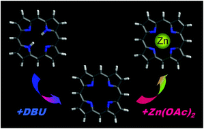
A spectrophotometric study of the acid–base equilibria and complexation with metal ions has been carried out for 5,10,15,20-tetrakis(trifluoromethyl)porphine and 5,10,15,20-tetrakis(iso-buthyl)porphine in acetonitrile solutions. Protonation and deprotonation of these porphyrins was found to be stepwise with sequential formation of mono- and doubly protonated/deprotonated species. The overall basicity and acidity constants have been determined. The porphyrin complexation with Zn2+ and Cu2+ ions was studied and the rates constants of the macrocycle metallation have been determined. The structure–property relationship derived from the metal chelation studies and the prospects for the use of the above systems for the design of highly sensitive sensors for metal ions were discussed in detail.


Introduction

Interest in the studies of the coordination and physical chemistry of tetrapyrrolic compounds, and, first of all, of the representatives of the porphyrin family, is due to the exceptionally important biological role of the metallocomplexes of these compounds which they play in life. The diversity of the functionalities relates to the diversity of their molecular structures, ability to form complexes with different metal ions, possibility to form five- and six-coordinated axially ligated complexes as well as the ability to tune the oxidation state of the chelated metal ion according to microenvironment properties.1–9 The success of the porphyrin applications is directly related to the optimisation of the synthetic procedures, as well as to the detailed understanding of the structure–property relationships in the physico-chemical properties of these compounds. Selective modifications of the porphyrin macrocycle, especially those of non-symmetric type, frequently leading to the pronounced out-of-plane deformations of the tetrapyrrolic macrocycle, are considered to be the tool to understand and mimic the diversity of the biological functions of the tetrapyrroles.10–12

The results of the literature data analysis indicate that porphyrin macrocycle chelation with metal ions is able to proceed with two mechanisms: the molecular and ionic ones.2,6,13–16 In the former case the complexation takes place between the metal salts and the porphyrin in the free base form (molecular form), whereas in the latter case deprotonated form of porphyrin undergoes the complexation. The first reaction is the generally accepted way to obtain the metalloporphyrins and has been studied in detail repeatedly,2,6,17,18 while the macrocycle complexation with ionic mechanism was expected to be somewhat exotic and being far from the practical interest. However, our recent studies have demonstrated that the efficient metal chelation takes place also with doubly protonated forms of several porphyrin derivatives,15,19–21 we have revealed that the rate of the porphyrin complexation with Zn2+ ions dramatically increases upon the addition to the solution of the deprotonating agent such as 1,8-diazabicyclo-[5,4,0]-undec-7-en (DBU), acting as “proton sponge” and ultimately leading to the full deprotonation of the porphyrin macrocycle core. Our findings are in line with few early communications on the ionic mechanism of complexation, where the possibility to obtain the metalloporphyrins has been reported for the core H-bonded and monodeprotonated porphyrins.22–24

Based on the above results, the extension of the metal complexation studies, involving the compounds with different substitution pattern of the porphyrin macrocycle, seems to be of interest. These studies aim to establish the structure–function relationship for the complexation of doubly deprotonated porphyrin macrocycles with different metal salts. The problem of metal ions determination in solutions is studied extensively during last two decades.25–31 The reported to date conventional optical (spectrophotometric and luminescent) methods of the metal ion detection with porphyrin sensors approach to the detection limit of 1 × 10⁻⁹ mol L−1.31 Increased complexation rate for ionic mechanism allows to extend the detection limit at least down to 5 × 10⁻¹¹ ÷ 1 × 10⁻¹⁰ mol L−1 (according to difference in the porphyrin to metal salt ratio required for the metal ion chelating15,21). In this communication we report on the results of the studies of the acid–base equilibria and the complexation with Cu(OAc)2 and Zn(OAc)2 salts for two meso-alkyl-substituted porphyrins. The kinetic parameters related to the metal ion complexation by porphyrin macrocycle are also reported and discussed.

Results and discussion

Free base porphyrin complexation

Molecular structures of studied 5,10,15,20-tetrakis-(trifluoromethyl)porphine (1) and 5,10,15,20-tetrakis(iso-buthyl)-porphine (2) are shown on the Scheme 1. It is accepted, that complexation of the free base porphyrins with divalent metal ions in nonaqueous solutions takes place according eqn (1):
 
H2P + [MX2(Solv.)n−2] → MP + 2HX + (n − 2)Solv., (1)
where the H2P is the porphyrin, M is the chelated metal ion, X is the salt anion, Solv is the solvent molecule serving as ligand.2,6,8

image file: c5ra01323b-s1.tif
Scheme 1

The kinetic parameters for the Cu-complexes formation of porphyrins 1 and 2 in the acetic acid are summarized in Table 1. Corresponding spectral changes measured during the spectrophotometric titrations are given at Fig. 1 and 2. Acetic acid has been chosen for the titration procedure since it forms relatively labile solvated complexes of transition metals, which can be easily broken in the presence of the porphyrin macrocycles to form metallocomplexes: thus, for example, 2,3,7,8,12,13,17,18-octamethylporphyrin complexation rate constant with Cu2+ was found to be about 105 times higher compared to that measured in pyridine.32,33

Table 1 Kinetic parameters of the Cu-complex formation for porphyrins 1 and 2 in acetic acid at 298 K
  CCuAcO2, mol L−1 keff × 104, s−1 kv × 104, (L mol−1)0.5 s−1 Ea, kJ mol−1 ΔS, J mol−1 K−1
1 1.81 × 10⁻³ 1.77 ± 0.10 41.5 ± 2.3 79 ± 2 −34 ± 6
2 3.62 × 10⁻⁴ 85.8 ± 0.9 4511 ± 135 75 ± 1 −7 ± 3


image file: c5ra01323b-f1.tif
Fig. 1 Ground state absorption spectra changes in the course of the titration of porphyrin 1 with Cu(OAc)2 in the acetic acid at 298 K. Cp = 5.20 × 10⁻⁵ mol L−1; CCuAc2 = 1.81 × 10⁻³ mol L−1. Arrows on this and following figures indicate the direction of the spectral changes upon increase in the titrant concentration.

image file: c5ra01323b-f2.tif
Fig. 2 Ground state absorption spectra changes in the course of the titration of porphyrin 2 with Cu(OAc)2 in the acetic acid at 298 K. Cp = 1.79 × 10⁻⁵ mol L−1; CCuAc2 = 3.62 × 10⁻⁴ mol L−1.

The obtained data on kinetic parameters of Cu–porphyrins formation indicate that electronic effects play the important role in the complexation. Replace of the electron donating substituents in porphyrin 1 for the electron withdrawing ones in the porphyrin 2 leads to almost 100-fold increase in the efficient rate constant keff of the complexation. At the same time, the energy of activation remains practically unchanged. Thus, four iso-butyl groups in the meso-positions of macrocycle in porphyrin 2 bring to the substantial changes of the electronic density of the tertiary nitrogens of pyrrole rings compared to that for porphyrin 1. As a result, the basicity of porphyrin 2 is higher. According to bimolecular one stage mechanism of reaction (1), increase in basicity leads to more efficient interaction of porphyrin ligand with metal cation in the transition state, and, consequently, leads to substantial increase in the efficient rate constant of complexation.

Formation of mono- and doubly protonated porphyrin species

Porphyrins, is known, to be the amphoteric compounds, i.e. having both basic (N-bases) and acidic (weak NH-acids) properties.1,4,10,34 There is a relationship between the coordination and acid–base properties of porphyrins; namely, the protonation of porphyrin macrocycle is the competing reaction with respect to the coordination of metal ion. Increase in the basicity of the porphyrin 2 as compared to that of porphyrin 1 are supported by the data obtained with the spectrophotometric titrations of both studied porphyrins in acetonitrile solutions with perchloric acid (Fig. 3–6).
image file: c5ra01323b-f3.tif
Fig. 3 Ground state absorption spectra changes in the course of the titration of porphyrin 1 with HClO4 in acetonitrile at 298 K. Cp = 3.04 × 10⁻⁵ mol L−1; CHClO4 = 0 ÷ 0.576 mol L−1.

image file: c5ra01323b-f4.tif
Fig. 4 The titration curve at λ = 509 nm for porphyrin 1 with HClO4 in acetonitrile at 298 K.

image file: c5ra01323b-f5.tif
Fig. 5 Ground state absorption spectra changes in the course of the titration of porphyrin 2 with HClO4 in acetonitrile at 298 K. Cp = 1.10 × 10⁻⁴ mol L−1; CHClO4 = 0 ÷ 2 × 10⁻⁴ mol L−1.

image file: c5ra01323b-f6.tif
Fig. 6 The titration curve at λ = 629 nm for porphyrin 2 with HClO4 in acetonitrile at 298 K.

One can see that increase in the acid concentration leads to the formation of two series of spectral curves, with each of them has its own set of isosbestic points, indicating that protonation of porphyrins macrocycles takes place in two sages for both porphyrin 1 and porphyrin 2. First of these series of spectral curves corresponds to the free base ↔ monoprotonated species equilibrium, whereas the second one relates to the equilibrium between the mono- and doubly protonated porphyrin species. Presence of the deflection point at the titration curve for the porphyrin 1 (Fig. 4) enables the easy determining the acid concentration ranges where the porphyrin is in mono- and doubly protonated form. In case of the porphyrin 2 two basicity constants are much closer each to other and therefore the titration curve does not reveal any deflection point (Fig. 6).35 The formation of the doubly protonated species can be treated with the overall basicity constant Kb describing the two-step formation of doubly protonated species. This value can be calculated with eqn (2):

 
lg[thin space (1/6-em)]Kb = lg[thin space (1/6-em)]Ind − nlg[thin space (1/6-em)]CHClO4, (2)
where, Ind is the indicator of concentration ratio [H4P2+]/[H2P], CHClO4 is analytic value of the perchloric acid concentration HClO4 in solution (mol L−1), n = 2 is the number of the protons attached.

The overall basicity constant Kb calculated with eqn (2) amounts lg[thin space (1/6-em)]Kb = 3.58 and lg[thin space (1/6-em)]Kb = 9.04 at 298 K for porphyrins 1 and 2, respectively. These values support the increase in the basicity in going from the porphyrin 1 to porphyrin 2. No differences in the macrocycle planarity are expected to occur between two studied porphyrins based on the extensive crystallography data for the porphyrins with the same type of peripheral substitution,36 and the macrocycle is suggested to be planar in both cases. Thus, the increase in the basicity of porphyrin 2 for almost 5 orders of magnitude needs to be assigned to pure electronic effects of substitution.

Formation of mono- and doubly deprotonated porphyrin species

The acidic properties of studied porphyrins were evaluated with spectrophotometric titration in acetonitrile solution. The reactions between two organic compounds when one of them is used as the proton acceptor were reported.37 The advantage of organic bases such as DBU over inorganic bases is the high solubility in organic solvents, enabling the use them in nonaqueous solutions.

The titration results revealed the difference in the acidity between porphyrin 1 and porphyrin 2. Thus, even the maximum available DBU concentration have been applied to the solutions of porphyrin 2, its doubly protonated form has not been obtained, whereas at moderate DBU concentrations two sequential steps, corresponding to formation of mono- and doubly deprotonated species of porphyrin 1, were observed (Fig. 7). At the end of titration all the porphyrin 1 molecules have been converted into the doubly protonated species. The ground state absorption spectrum of the doubly deprotonated porphyrin 1: λmax, nm (lg[thin space (1/6-em)]ε): 431 (4.96), 598 (4.00), 717 (3.89).


image file: c5ra01323b-f7.tif
Fig. 7 Ground state absorption spectra changes in the course of the titration of porphyrin 1 with the DBU in acetonitrile at 298 K. Cp = 1.89 × 10⁻⁵ mol L−1; CDBU = 0 ÷ 7.9 × 10⁻³ mol L−1. Insert shows the corresponding titration curve measured at λ = 400 nm.

The overall acidity constant was calculated with eqn (3):

 
lg[thin space (1/6-em)]Ka = lg[thin space (1/6-em)]Ind + nlg[thin space (1/6-em)]CDBU, (3)
where, Ind is the indicator of concentration ratio [P2−]/[H2P], CHClO4 is analytic value of the DBU in solution (mol L−1), n = 2 is the number of the protons attached. The calculations of the current concentrations of deprotonated forms and free base species, taking into account the material balance equation Co = C(H2P) + C(P2−), show that all the porphyrin 1 molecules found themselves in the doubly deprotonated form at the DBU concentration ∼7 × 10⁻³ mol L−1. The overall acidity constant was found to be −lg[thin space (1/6-em)]Ka = 9.69.

Complexation of the doubly deprotonated porphyrin

The Zn-complex formation of the porphyrin 1 was studied in the acetonitrile solution containing the DBU amount (∼7 × 10⁻³ mol L−1, as was indicate above) required for the full conversion of the free base species into the doubly deprotonated ones. The spectral changes in the Soret band region measured during the titration of the free base porphyrin 1 are shown on Fig. 8 for reference purposes. The spectral changes observed in course of the titration of the doubly deprotonated porphyrin 1′ are shown on Fig. 9.
image file: c5ra01323b-f8.tif
Fig. 8 Ground state absorption spectra changes in the course of the titration of the free base porphyrin 1 with Zn(OAc)2 in acetonitrile at 298 K. Cp = 1.31 × 10⁻⁵ mol L−1; CZnAc2 = 0 ÷ 1.14 × 10⁻² mol L−1.

image file: c5ra01323b-f9.tif
Fig. 9 Ground state absorption spectra changes in the course of the titration of the doubly deprotonated porphyrin 1′ with Zn(OAc)2 in acetonitrile at 298 K. Cp = 1.67 × 10⁻⁵ mol L−1; CZnAc2 = 0 ÷ 2.83 × 10⁻⁴ mol L−1; CDBU = 6.98 × 10⁻³ mol L−1.

Table 2 summarizes the kinetic and thermodynamic parameters of the complexation. One can see that in going from the free base to the doubly deprotonated form, the rate of the complexation increases for more than 1000 times. The efficient rate constant of complexation reveals almost 200-fold increase.

Table 2 Kinetic parameters of the Zn-complex formation for the porphyrin 1 free base and its doubly deprotonated form 1′ in acetonitrile at 298 K
  CZnAcO2, mol L−1 keff × 104, s−1 kv × 103, (L mol−1) s−1 Ea, kJ mol−1 ΔS, J mol−1 K
1 1.14 × 10⁻² 6.9 ± 0.10 6.05 ± 0.08 52 ± 2 −175 ± 6
1′ 2.83 × 10⁻⁴ 115 ± 5 4006 ± 5 24 ± 1 −144 ± 5


At the same time, the activation energy value decreases twice compared to that measured for the complexation of the free base porphyrin 1, which is followed with entropy decrease ΔS ∼ 30 J mol−1 K. These features are likely to be due to the absence of the energy losses required for the deformation and rupture of the N–H bonds in the porphyrin core, as well as due to nonuniform charge distribution over the macrocycle, leading to polarization of the electronic cloud. As a result, the doubly deprotonated porphyrin 1′ may have higher solvation in the transition state which facilitates the metal ion chelation.

The Zn–porphyrin 1 molecules formed with two different procedures are identical each to other. Small long wavelength shift of the Soret band maximum (see Fig. 8 and 9) for the Zn-complex of the porphyrin 1 formed with titration of doubly deprotonated form compared to that measured for Zn-complex obtained with titration of the free base porphyrin 1 is likely to be due to the solvent properties changes (most likely, the polarity), since in the former case the substantial amount of DBU was added to the acetonitrile solution. Stabilization of the electronic states with large transition dipole moment in (more) polar environment ultimately leads to the long wavelength shift of the absorption band.

Conclusions

The presented results unambiguously indicate that DBU can be successfully used for the formation of the mono- and doubly deprotonated porphyrins species in nonaqueous solutions. Introduction of the strong electron withdrawing substituents in the meso-positions of the porphyrin macrocycle increase the polarization of the N–H bonds, making difficult the complete deprotonation of the macrocycle. Provided that the macrocycle has low or moderate basicity, the full conversion of the free base porphyrins into the doubly deprotonated species is observed.

A significant decrease (down to 50 times) in the concentration of the metal salt required for the metal chelation and instant proceeding of the reaction appear to be the most promising features of the ionic mechanism of the metal complexation with the porphyrin macrocycles. This approach also needs substantially lower porphyrin to salt ratio (1[thin space (1/6-em)]:[thin space (1/6-em)]5) as compared with conventional molecular approach, where it amounts up to 1[thin space (1/6-em)]:[thin space (1/6-em)]100 or even higher value.2,23,24 All these facts taken together allow us to suggest the ionic mechanism of porphyrin metallocomplexes formation as the promising tool for the everyday practical use.

We hope also that the presented results to be of interest for the understanding of the synthesis and functioning of the endogenous porphyrins, since the formation of the metallocomplexes of endogenous porphyrins in the human and mammalian bodies occur also in the “very soft” conditions compared to those used for the synthesis of porphyrin metallocomplexes by conventional molecular procedure in the laboratory practice.

Experimental

Synthesis

The studied compounds were prepared according to the described earlier synthetic procedures.38–40
5,10,15,20-Tetrakis-(trifluoromethyl)porphine (1). 5,10,15,20-Tetrakis-(trifluoromethyl)porphine (1) was purified by column chromatography (silica, eluent hexane–benzene 10[thin space (1/6-em)]:[thin space (1/6-em)]1) followed with recrystallization from the methylene chloride–methanol mixture. Elem.: calcd for C24H10N4F12: C, 49.50; H, 1.73; N, 9.62; found: C, 49.53; H, 1.64; N, 9.33. 1H NMR δ(ppm): 9.60 (s, 8H), −2.08 (s, 2H, NH); abs.: (CH2Cl2), λmax, nm (lg[thin space (1/6-em)]ε): 403 (5.08), 510 (3.97), 545 (3.97), 593 (3.67), 649 (4.00).
5,10,15,20-Tetrakis(iso-buthyl)porphine (2). 5,10,15,20-Tetrakis(iso-buthyl)porphine (2) was purified by column chromatography (Al2O3, type III by Brockman, eluent chloroform) followed with recrystallization from the chloroform–methanol mixture. Elem.: calcd for C36H46N4: C 80.86, H 8.67, N 10.48; found: C 80.64, H 8.50, N 10.59. 1H NMR δ(ppm): −2.65 (br, s, 2H, NH), 1.19 (d, 24H, CH3), 2.62–2.81 (m, 4H, CH), 4.86 (d, 8H, CH2), 9.45 (s, 8H, β-H). Abs.: (CH2Cl2), λmax, nm (lg[thin space (1/6-em)]ε): 417 (5.65), 519 (4.15), 553 (4.04), 598 (3.7), 658 (3.95).

General experimental methods and instrumentation

NMR spectra in CDCl3 solutions were acquired on commercial instrument Bruker Avance 500 MHz and chemical shifts (δ) are reported in parts per million (ppm) referenced to tetramethylsilane (TMS) or the internal (NMR) solvent signals. Mass spectra were run using a HP5989A apparatus (CI and EI, 70 eV ionisation energy) with Apollo 300 data system or a Thermo Finnigan LCQ Advantage apparatus (ESI). Ground state absorption spectra and spectrophotometric titration experiments were carried out with spectrophotometer Shimadzu UV-1800. The methods of the titration procedure and protocols of experimental data analysis were described in our previous papers.41,42 The relative uncertainty in determined basicity and acidity constants did not exceeded 5%.

Cu(OAc)2 and Zn(OAc)2 “for analysis” were purified by recrystallization with acetic acid followed with dehydration at 380–390 K according methods described.43 Acetic acid “for analysis” was dehydrated by freezing followed with fractional distillation. Water contain was determined with Fischer method and did not exceeded 0.03%.44 Dry acetonitrile (water contain no more then 0.03%) was used in the titration experiment. The 1,8-diazabicyclo-[5,4,0]-undec-7-en (DBU) was used as the deprotonating agent (pKa = 13.2 in acetonitrile).29 DBU and perchloric acid HClO4 were used as received without further purification.

Complexation titration procedure and data treatment

Complexation was studied with the spectrophotometric method in the thermostated cuvettes at temperatures 293–308 K. Temperature during the titration set was kept with precision ± 0.1 K. Metalloporphyrin formation has the first order with respect to the porphyrin ligand.2 Titration was carried out in the conditions of the 100-fold excess of metal salts M(OAc)2 concentration over that of the porphyrin. In such conditions the efficient rate constant of the complexation reaction keff was calculated according to eqn (4):
 
keff = (1/t)ln[(AoA)/(AA)], (4)
where, Ao, A, A are the solution absorbance (at defined wavelength) before titration, at time t and at the end of titration, respectively. Rate constants of the n + 1 order were calculated according to eqn (5):
 
kn+1 = keff/cn(M(OAc)2), (5)
where, n is the reaction order with salt, which is 0.5 for Cu(OAc)2 in acetic acid,45 and 1 for Zn(OAc)2 in acetonitrile.21 Activation energy Ea for the studied temperature range was calculated with Arrhenius equation (eqn (6)):
 
Ea = 19.1[(T1T2)/(T2T1)]lg(k2/k1) (6)

Entropy change for the formation of transition state ΔS was calculated with eqn (7):

 
ΔS = 19.1[thin space (1/6-em)]lg[thin space (1/6-em)]kv + Ea/T − 253 (7)

Acknowledgements

The synthetic part of the work was performed with support of the grant of the Russian Science Foundation (no. 14-23-00204). This work was supported by the Russian Foundation of the Fundamental Research, project no. 13-03-01343-a. Partial financial support to Prof. Mikalai Kruk from the State Program of Scientific Researches of the Republic of Belarus “Convergence”, project 3.2.02 “Synthesis of new tetrapyrroles containing supramolecular systems and their applications for the luminescent sensors design”, is also acknowledged.

Notes and references

  1. G. P. Gurinovich, A. N. Sevchenko and K. N. Solovyov, Spectroscopy of chlorophyll and related compounds, Publishing house Science and Technology, Minsk, 1968, p. 520.[Engl. transl.: Nat. Tech. Informat. Serv. US Dept. of Commerce, Springfield, Virginia, 1971, 506 p.] Search PubMed.
  2. B. D. Berezin, Coordination compounds of porphyrins and phthalocyanines, Wiley, New York, Toronto, 1981 Search PubMed.
  3. D. J. Davis, in The Porphyrins, ed. D. Dolphin, Academic Press, New-York, 1978, vol. 5, p. 127 Search PubMed.
  4. L. Latos-Grażyński, K. Rachlewicz and J. Wojaczyński, Coord. Chem. Rev., 1999, 190–192, 109 CrossRef.
  5. E. J. Baerends, G. Ricciardi, A. Rosa and S. J. A. van Gisbergen, Coord. Chem. Rev., 2002, 230, 5 CrossRef CAS.
  6. B. D. Berezin, J. Porphyrins Phthalocyanines, 2003, 7, 715 CrossRef CAS.
  7. I. Beletskaya, V. S. Tyurin, A. Y. Tsivadze, R. Guilard and C. Stern, Chem. Rev., 2009, 109, 1659 CrossRef CAS PubMed.
  8. M. G. H. Vicente, in The Porphyrin Handbook: Synthesis an Organic Chemistry, ed. K. M. Kadish, K. M. Smith and R. Guilard, Academic Press, New York, 2000, vol. 1, pp. 149–200 Search PubMed.
  9. M. O. Senge, in The Porphyrin Handbook: Synthesis an Organic Chemistry, ed. K. M. Kadish, K. M. Smith and R. Guilard, Academic Press, New York, 2000, vol. 1, pp. 239–348 Search PubMed.
  10. J. A. Shelnutt, in The Porphyrin Handbook: Theoretical and Physical Characterization, ed. K. M. Kadish, K. M. Smith and R. Guilard, Academic Press, New York, 2000, vol. 7, pp. 167–223 Search PubMed.
  11. W. R. Scheidt and Y. Lee, Struct. Bonding, 1987, 64, 1–70 CrossRef CAS.
  12. O. A. Golubchikov, S. G. Pukhovskaya and E. M. Kuvshinova, Russ. Chem. Rev., 2005, 74, 249 CrossRef CAS PubMed.
  13. V. B. Sheinin, O. R. Simonova and E. L. Ratkova, Macroheterocycles, 2008, 1, 72 CAS.
  14. T. Sato, K. Ebisawa, K. Sue, S. Ito, T. Saito and N. Itoh, Ind. Eng. Chem. Res., 2012, 51, 13908 CrossRef CAS.
  15. Y. B. Ivanova, D. T. Nam, S. A. Syrbu and M. M. Kruk, Russ. J. Gen. Chem., 2013, 83, 1155 CrossRef CAS.
  16. Y. Qi and Ji G. Pan, Bull. Korean Chem. Soc., 2014, 35, 3313 CrossRef.
  17. P. Hambright, in The Porphyrin Handbook: Synthetic Methodology, ed. K. M. Kadish, K. M. Smith and R. Guilard, Academic Press, New York, 2000, vol. 3, pp. 129–210 Search PubMed.
  18. P. Hambright, Russ. Chem. Rev., 1977, 46(7), 1207 CAS.
  19. Y. B. Ivanova, S. G. Pukhovskaya, A. S. Semeykin and S. A. Surby, Russ. J. Gen. Chem., 2013, 83, 1406 CrossRef CAS.
  20. Y. B. Ivanova, N. V. Chizhova, S. G. Pukhovskaya and N. Z. Mamardashvili, Russ. J. Gen. Chem., 2014, 84, 848 CrossRef.
  21. Y. B. Ivanova, N. V. Chizhova and M. M. Kruk, Russ. J. Gen. Chem., 2013, 83, 558 CrossRef CAS.
  22. P. A. Stuzhin and O. G. Khelevina, Coord. Chem. Rev., 1996, 147, 41 CrossRef CAS.
  23. P. A. Stuzhin, O. G. Khelevina, M. N. Ryabova and B. D. Berezin, J. Appl. Spectrosc., 1990, 52, 70 CrossRef.
  24. B. D. Berezin, O. G. Khelevina and P. A. Stuzhin, Russ. J. Phys. Chem., 1985, 59, 1295 Search PubMed.
  25. X. B. Zhang, C. C. Guo, Z. Z. Li, G. L. Shen and R. Q. Yu, Anal. Chem., 2002, 74, 821 CrossRef CAS.
  26. H. Y. Luo, X. B. Zhang, J. H. Jiang, C. Y. Li, J. Peng, G. L. Shen and R. Q. Yu, Anal. Sci., 2007, 23, 551 CrossRef CAS.
  27. L. Lvova, P. Galloni, B. Floris, I. Lundström, R. Paolesse and C. Di Natale, Sensors, 2013, 13, 5841 CrossRef CAS PubMed.
  28. V. K. Gupta, A. K. Jain, G. Maheshwari, H. Lang and Z. Ishtaiwi, Sens. Actuators, B, 2006, 117, 99 CrossRef CAS PubMed.
  29. A. Kaljurand, L. Kütt, T. Sooväli, V. Rodima, I. Mäemets, I. Leito and A. Koppel, J. Org. Chem., 2005, 70, 1019 CrossRef PubMed.
  30. D. Vlascici, E. Fagadar, E. M. Pica, V. Cosma, O. Bizerea, G. Mihailescu and L. Olenic, Sensors, 2008, 8, 4995 CrossRef CAS PubMed.
  31. C. L. He, F. L. Ren, X. B. Zhang and Z. X. Han, Talanta, 2006, 70, 364 CrossRef CAS PubMed.
  32. S. G. Pukhovskaya, V. A. Efimovich, A. S. Semeikin and O. A. Golubchikov, Russ. J. Inorg. Chem., 2010, 55, 1494 CrossRef CAS.
  33. S. G. Pukhovskaya, V. A. Efimovich, A. S. Semeikin, E. A. Kolodina and O. A. Golubchikov, Russ. J. Gen. Chem., 2012, 82, 476 CrossRef CAS.
  34. M. Kruk, A. Starukhin and W. Maes, Macroheterocycles, 2011, 4, 69 CrossRef CAS.
  35. I. Y. Bernstein and Y. L. Kaminsky, Spectrophotometric Analysis in Organic Chemistry, Khimiya, Leningrad, 1986, p. 202, (in Russian) Search PubMed.
  36. M. O. Senge, in The Porphyrin Handbook, ed. K. M. Kadish, K. M. Smith and R. Guilard, Academic Press, San Diego, 2000, vol. 10, pp. 1–218 Search PubMed.
  37. A. F. Pozharskii, Russ. Chem. Rev., 1998, 67, 1 CrossRef PubMed.
  38. Y. Xie, J. P. Hill, R. Charvet and K. Ariga, J. Nanosci. Nanotechnol., 2007, 7, 2969 CrossRef CAS PubMed.
  39. J. G. Goll, K. T. Moore, A. Ghosh and M. J. Therien, J. Am. Chem. Soc., 1996, 118, 8344 CrossRef CAS.
  40. A. M. d'A Rocha Gonsalves, J. M. T. B. Varejao and M. M. Pereira, J. Heterocycl. Chem., 1991, 28, 630 Search PubMed.
  41. Y. B. Ivanova, V. B. Sheinin and N. Z. Mamardashvili, Russ. J. Gen. Chem., 2007, 77, 1458 CrossRef CAS.
  42. Y. B. Ivanova, Y. I. Churakhina and N. Z. Mamardashvili, Russ. J. Gen. Chem., 2008, 78, 673 CrossRef CAS.
  43. Y. V. Karyakin and I. I. Angelov, Chistye Khimicheskie Veschestva (Pure Chemical Reactants), Moscow, Khimiya, 1974, p. 407 Search PubMed.
  44. A. J. Gordon and R. A. Ford, The chemists's companion: a handbook of practical data, techniques, and references, A Wiley-Interscience publication, John Wiley and Sons, New-York, London, Sydney, Toronto, 1972, p. 541 Search PubMed.
  45. T. A. Ageeva, E. L. Ponomareva, O. A. Golubchikov and B. D. Berezin, Coord. Chem., 1987, 13, 886 CAS.

This journal is © The Royal Society of Chemistry 2015
Click here to see how this site uses Cookies. View our privacy policy here.