Two Schiff base ligands for distinguishing ZnII/CdII sensing—effect of substituent on fluorescent sensing†
Zhi-Peng Zhenga,
Qin Weia,
Wen-Xia Yina,
Lin-Tao Wana,
Xia Huanga,
Ying Yu*a and
Yue-Peng Cai*ab
aSchool of Chemistry and Environment, South China Normal University, Guangzhou Key Laboratory of Materials for Energy Conversion and Storage, Guangzhou 510006, P.R. China. E-mail: caiyp@scnu.edu.cn; Fax: +86-020-39310; Tel: +86-020-39310383
bState Key Laboratory of Structure Chemistry, Fuzhou 350002, Fujian, P.R. China
First published on 5th March 2015
Abstract
Two Schiff base ligands (HL1, HL2) were conveniently synthesised by one-step condensation between pyridine 2-ylmethanamine and 3-ethoxy-2-hydroxybenzenaldhyde (for HL1) or salicylaldehyde (for HL2) as fluorescent sensors for distinguishing sensing of Zn2+ or Cd2+. Both of the two fluorescent sensors present very weak emission at 463 nm (for HL1) or 453 nm (for HL2). For HL1, upon addition of Zn2+, the fluorescence intensity of HL1 enhanced and gradually red shifted to 493 nm with a green emission while addition of Cd2+ only induced enhancement of fluorescent intensity at 463 nm. For HL2, only addition of Zn2+ induced enhancement of fluorescence intensity, presenting a high Zn2+/Cd2+ selectivity. A Zn2+-induced red shift in fluorescent spectra of HL1 could be attributed to twisted intramolecular charge transfer (TICT) from the interaction between the Zn2+ ion and in situ formed ligand L1′ with the twisted structure in compound 1, which is absent in compound 2. The Zn2+/Cd2+ selectivity of fluorescent response for HL2 correlates with the Cd–HL2 and Zn–HL2 coordination bond distances. Obviously, introduction of ethoxyl groups onto the benzene ring as an electron-donating group facilitates the Zn-induced in situ dimerization of HL1 into new ligand L1′ with a twisted molecular structure, further resulting in a red shift of the fluorescence spectra.
Introduction
Fluorescent sensors for metal ions are some of the most sought-after detection methods for metal ions owing to their convenient use, high selectivity, potential use in living cells and discernible response with color variation under UV irradiation or with the naked eye.1 Whether in protein-bound or free form, Zn2+ is a type of abundant ion among the trace metal ions in the human body and is related to many crucial biological processes including nervous system function, enzyme regulation, gene expression, biological catalysis and Zn(II)-disorder-related diseases.2 Zn2+ is also environmentally important because increased levels of Zn2+ in water can cause environmental problems including poor soil microbial activity causing phytotoxic effects or smelly water.3 Some fluorescent sensors have been developed to investigate the distribution of mobile Zn2+ in living cells over the last decades which are mainly based on metal complexation and complexation-based photoinduced electron-transfer (PET), chelation-enhanced-fluorescence (CHEF) effects or internal charge-transfer (ICT) mechanisms.4 Twisted intramolecular charge transfer (TICT) based on a twisted molecular structure is among the types of ICT and is widely employed in fluorescent ion sensing due to its spectral shifts and color variation originating from locally emission (LE) band and TICT emission band.1c,5 Theoretically, introduction of electron donating groups (e.g. –OR, –R) and electron withdrawing groups (e.g. –NO2, –CN) onto the moiety of the organic sensor, may generate TICT upon coordination of the ligand with metal ions, based on which metal ions sensing could be achieved.5,6 On the other hand, the detection of Zn2+ is often related to Cd2+ because of their similar electron configurations and similar response to the same fluorescent sensor, making one of the biggest challenges in constructing Zn2+ fluorescent chemosensor.7 Moreover, because of cadmium's accumulation in the food chain and its great toxicity to human's body,8 it is desirable to develop some analytical methods for cadmium detection in the environment or living cells. Therefore, it is necessary to develop fluorescent sensor for distinguishing sensing of Zn2+ and Cd2+.Based on the above considerations, we designed and synthesized two Schiff base ligands HL1 and HL2 for distinguishing sensing of Zn2+ and Cd2+. Ethoxyl group as electron donating group was introduced onto HL1 endowing it with TICT property and subsequent spectral shift upon reaction with Zn2+. HL1 and HL2 were conveniently obtained through one-step condensation (Scheme 1) between pyridine 2-ylmethanamine and 3-ethoxy-2-hydroxybenzenaldhyde (for HL1) or salicylaldehyde (for HL2). Compared with other previously reported Zn2+ organic sensor,4 HL1 and HL2 present distinct advantages including its convenient synthesis and tridentate donor set of N2O capable of strongly chelating to Zn2+ or Cd2+ ions through forming five/six membered ring with common side, providing a binding site between metal ions and sensor. For HL2, together with the pyridine ring as electron withdrawing group,9 introduction of ethoxyl group as electron donating group onto the benzene ring facilitates the internal charge transfer of the sensor and further presents Zn-induced TICT and spectral red shift, which does not exist in the system of HL2 owing to absence of ethoxyl group. However, without ethoxyl group as hindrance effect, HL2 shows greater sensitivity towards sensing Zn2+ with a much lower detection limit (Scheme 2).
 | ||
| Scheme 2 Effect of substituent group on fluorescent sensing of Zn2+ and Cd2+ based on Schiff base complexes 1–4. | ||
Experimental
General procedures
All chemicals were of analytical reagent grade and were used as received without any further purification. Elemental analysis for C, H, and N were performed on a Perkin-Elmer 2400 analyzer. IR spectra were recorded with a Perkin-Elmer Fourier transform infrared spectrophotometer with samples prepared as KBr disks in the 4000–400 cm−1 range. UV-vis absorption spectra were recorded on a Shimadzu UV1800 UV-vis Spectrophotometer. Fluorescent spectra were recorded on a Hitachi F-2500 spectrometer with the excitation slit as 5 nm and emission slit as 5 nm.X-ray crystallography
Crystal data collections were performed at 298 K on a Bruker Smart Apex II diffractometer with graphite mono-chromated Mo Kα radiation (λ = 0.71073 Å) for four compounds 1–4. Absorption corrections were applied by using the multi-scan program SADABS.10 Structural solutions and full-matrix least-squares refinements based on F2 were performed with the SHELXS-97 (ref. 11) and SHELXL-97 (ref. 12) program packages, respectively. All the non-hydrogen atoms were refined anisotropically. The hydrogen atoms on organic motifs were placed at calculated positions. Details of the crystal parameters, data collections, and refinements for compounds 1–4 are summarized in Table S1.† Selected bond lengths and angles of compounds 1–4 are shown in Table S2.†Synthesis of HL1 and HL2
The Schiff-base ligand 2-ethoxy-6((pyridin-2-ylmethylimino)-methyl)phenol (HL1) was synthesized according to corresponding reference.13 2-((Pyridin-2-ylmethylimino)methyl)-phenol (HL2) was prepared according to related reference.14Synthesis of compounds 1–4
Results and discussion
Fluorescence/UV absorption spectra and titration of HL1
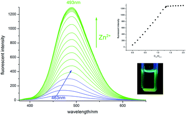
The fluorescence response of receptor HL1 towards Zn2+ was studied in 0.10 mM ethanol. Upon excitation at 350 nm, HL1 in ethanol presented dark blue emission at 463 nm which could be attributed to the intraligand π–π* transition.15 With stepwise addition of ZnCl2 solution in ethanol with concentration ranging from 0.1 equiv. to 2.0 equiv., fluorescent emission of HL1 gradually red-shifted to 493 nm with their fluorescent intensity at 493 nm enhanced linearly with the Zn2+ concentration and saturated when [Zn2+] reaches 1.33 equiv. (Fig. 1). Interaction between Zn2+ and HL1 was also studied by UV absorption spectra (Fig. S1†). Concomitant addition of ZnCl2 into HL1 in ethanol causes a new absorption peak at 380 nm with intensity increased while that at 220 nm, 264 nm, 334, 432 nm decreased with four isosbetic points at 249, 276, 303, 347 nm. The binding ratio between Zn2+ and ligand was estimated to be 4![[thin space (1/6-em)]](https://www.rsc.org/images/entities/char_2009.gif) :
:![[thin space (1/6-em)]](https://www.rsc.org/images/entities/char_2009.gif) 3 as confirmed by its job plot (Fig. S2†) and crystal structure. The limit of detection (LOD) was measured to be 1.11 × 10⁻⁶ M (R2 = 0.999) with a linearity range between 1 × 10⁻⁵ M and 9 × 10⁻⁵ M (LOD = 3σ/slope) (Fig. S3†).
3 as confirmed by its job plot (Fig. S2†) and crystal structure. The limit of detection (LOD) was measured to be 1.11 × 10⁻⁶ M (R2 = 0.999) with a linearity range between 1 × 10⁻⁵ M and 9 × 10⁻⁵ M (LOD = 3σ/slope) (Fig. S3†).
The selectivity of HL1 to Zn2+ was examined by fluorescence titration of HL1 with various metal ions (Fig. 2). The fluorescence intensity of HL1 was slightly quenched with some cations such as Ni2+, Cu2+, Co2+, and Fe3+, Mn2+. Other cations such as Na+, K+, Li+, Mg2+, Ba2+, Ca2+, Hg2+, Fe2+, Pb2+, Al3+ and Cr3+ did not cause any significant changes, showing selective CHEF & spectral red shift in the presence of Zn2+. Nevertheless, interaction of Cd2+ with HL1 also presents fluorescence spectral response which is relatively weaker compared with that of Zn2+. Concomitant addition of CdCl2 into HL1 in ethanol induced enhancement of the ligand-centered emission (Fig. 3) with binding ratio between Cd2+ and HL1 as 2![[thin space (1/6-em)]](https://www.rsc.org/images/entities/char_2009.gif) :
:![[thin space (1/6-em)]](https://www.rsc.org/images/entities/char_2009.gif) 1 confirmed by job plot (Fig. S4†) and crystallography with a limit of detection as 9.2 × 10⁻⁶ M (R2 = 0.999) with a linearity range between 2 × 10⁻⁵ to 1.8 × 10⁻⁴ M (LOD = 3σ/slope) (Fig. S5†). UV absorption titration also demonstrates interaction between Cd2+ and HL1 (Fig. S6†). The selectivity of HL1 on Zn2+ over Cd2+ was determined as 4.8 calculated from ratio of IZn/ICd (2 equiv.).
1 confirmed by job plot (Fig. S4†) and crystallography with a limit of detection as 9.2 × 10⁻⁶ M (R2 = 0.999) with a linearity range between 2 × 10⁻⁵ to 1.8 × 10⁻⁴ M (LOD = 3σ/slope) (Fig. S5†). UV absorption titration also demonstrates interaction between Cd2+ and HL1 (Fig. S6†). The selectivity of HL1 on Zn2+ over Cd2+ was determined as 4.8 calculated from ratio of IZn/ICd (2 equiv.).
To further evaluate selectivity of HL1 towards Zn2+, the interference of series of metal ions on detection of Zn2+ was examined. Seeing from Fig. 4, in the presence of various metal ions including Na+, K+, Ca2+, Mg2+, Fe3+, Hg2+, Mg2+ and Cd2+, the emission intensity of HL1–Zn remain hardly perturbed, while in the vicinity of other involved metals, it is slightly quenched but is still clearly detectable, indicating a high selectivity of HL1 on Zn2+.
Crystal structure of Zn4L1L1′Cl5 (1) and [Cd2(L1)Cl2-(DMSO)]2 (2) and proposed mechanism of HL1 sensing Zn2+/Cd2+
Reaction of HL1 with Zn2+ induced part of HL1 to undergo [3 + 2] cycloaddition16 generating a dimerized form of L1′ (Scheme S1†) and compound 1 was formed. Crystal analysis revealed that compound 1 crystallized in triclinic P![[1 with combining macron]](https://www.rsc.org/images/entities/char_0031_0304.gif) space group and each discrete tetranuclear unit comprised of four Zn2+ ions, one dimerized ligand L1′, one original Schiff base ligand L1 and five chlorides. As shown in Fig. 5, out of the four Zn ions, Zn1 was penta-coordinated to one pyridine nitrogen atom (N2), one amide nitrogen (N4), two hydroxyl oxygen atoms (O3, O4) all from L1′, while Zn2 was four-coordinated to one hydroxyl oxygen atom of L1′ and three chlorides (one μ2-bridged Cl2 and the other two terminal coordinated Cl1, Cl5). And Zn3 was penta-coordinated to one hydroxyl oxygen atom (O3) of L1′, two N atoms (N1, N2), one O atom (O1) of L1 and one μ2-bridged chlorides (Cl2), connecting L1 and L1′ together while Zn4 was quadra-coordinated to two nitrogen atoms (N5 N6) from L1′ and two terminal chlorides (Cl3, Cl4). Moreover, π⋯π stacking between benzene ring and pyridine ring helps to stabilize structure of compound 1 (Fig. 5b).
space group and each discrete tetranuclear unit comprised of four Zn2+ ions, one dimerized ligand L1′, one original Schiff base ligand L1 and five chlorides. As shown in Fig. 5, out of the four Zn ions, Zn1 was penta-coordinated to one pyridine nitrogen atom (N2), one amide nitrogen (N4), two hydroxyl oxygen atoms (O3, O4) all from L1′, while Zn2 was four-coordinated to one hydroxyl oxygen atom of L1′ and three chlorides (one μ2-bridged Cl2 and the other two terminal coordinated Cl1, Cl5). And Zn3 was penta-coordinated to one hydroxyl oxygen atom (O3) of L1′, two N atoms (N1, N2), one O atom (O1) of L1 and one μ2-bridged chlorides (Cl2), connecting L1 and L1′ together while Zn4 was quadra-coordinated to two nitrogen atoms (N5 N6) from L1′ and two terminal chlorides (Cl3, Cl4). Moreover, π⋯π stacking between benzene ring and pyridine ring helps to stabilize structure of compound 1 (Fig. 5b).
 | ||
| Fig. 5 (a) Molecular structure of compound Zn4(L1)(L1′)Cl5 (1) with partial atomic labels and (b) twisted conformation of the dimerized form of L1′ accounting for the TICT and spectral red-shift. | ||
When reacted with CdCl2, HL1 generated compound [Cd2(L1)Cl2(DMSO)]2 (2), which crystallized in triclinic, P![[1 with combining macron]](https://www.rsc.org/images/entities/char_0031_0304.gif) space group presenting a centrosymmetric discrete tetra-nuclear structure (Fig. 6). Its asymmetric unit contained two Cd2+ ions, one deprotonated (L1)− anion, three chlorides and one coordinated DMSO molecule. Cd1 was penta-coordinated to one pyridine nitrogen atom (N1), one azomethine amide nitrogen atom (N2), one hydroxyl oxygen atom (O1) and two chlorides (Cl1, Cl1, one μ2-bridged and the other terminal coordinated), exhibiting a distorted square pyramid coordination geometry. The other Cd2 ion is hexa-coordinated with two μ2-bridged chlorides (Cl2, Cl3), one hydroxyl oxygen atom (O1), one ethoxyl oxygen atom (O2) and one oxygen atom (O3) from the DMSO molecule, showing distorted octahedral coordination geometry. Moreover, by μ2-bridging of two chlorides (Cl3, Cl3a), two asymmetric units were connected to assemble a tetranuclear unit of compound 2.
space group presenting a centrosymmetric discrete tetra-nuclear structure (Fig. 6). Its asymmetric unit contained two Cd2+ ions, one deprotonated (L1)− anion, three chlorides and one coordinated DMSO molecule. Cd1 was penta-coordinated to one pyridine nitrogen atom (N1), one azomethine amide nitrogen atom (N2), one hydroxyl oxygen atom (O1) and two chlorides (Cl1, Cl1, one μ2-bridged and the other terminal coordinated), exhibiting a distorted square pyramid coordination geometry. The other Cd2 ion is hexa-coordinated with two μ2-bridged chlorides (Cl2, Cl3), one hydroxyl oxygen atom (O1), one ethoxyl oxygen atom (O2) and one oxygen atom (O3) from the DMSO molecule, showing distorted octahedral coordination geometry. Moreover, by μ2-bridging of two chlorides (Cl3, Cl3a), two asymmetric units were connected to assemble a tetranuclear unit of compound 2.
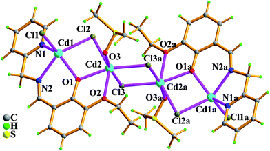 | ||
| Fig. 6 Molecular structure of tetranuclear compound [Cd2(L1)Cl2-(DMSO)]2 (2) with partial atomic labels. Asymmetry code: (a) 1 − x, 2 − y, 1 − z. | ||
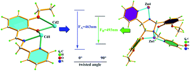
Emission spectra of compounds 1–2 in ethanol also accord with emission spectra generated from titration, further confirming that the products formed upon titration are compounds 1 and 2 (Fig. S7†). Hence, mechanism for Cd2+ and Zn2+ sensing of HL1 could be proposed referring to the structural analysis of compound 1 and 2. Compound 1 was obtained through reaction of ZnCl2 with HL1, in which HL1 as 1,3-dipole undergoes Zn-induced dimerization through [3 + 2] cycloaddition, generating a five-membered ring between two mole of HL1 (Scheme S1†). Viewing from the top of the five-membered ring, it is not difficult to find that L1′ was fixed to present a twisted molecular structure, in which the pyridine ring and benzene ring was almost twisted to be orthogonal. Such twisted structure induced a twisted-intramolecular charge transfer state upon excitation which narrowed down the energy gap between HOMO and LUMO, generating an emission peak of longer wavelength at 493 nm (Fig. 7).5a,17 Unlike compound 1, in compound 2, coordinated L1 presents a planar structure with the angle between the pyridine ring and benzene ring as nearly 0° (Fig. 7). Therefore, reaction of Cd2+ with HL1 presented locally excited (LE) emission at 463 nm.
Furthermore, fluorescent intensity enhancement of HL1 upon interaction with Zn2+ or Cd2+ should be ascribed to the CHFF (chelation enhanced fluorescence), inhabitation of C![[double bond, length as m-dash]](https://www.rsc.org/images/entities/char_e001.gif) N isomerization9,18 and weaken of PET9 (photo induced charge transfer) process in the ligand. On the other hand, different coordination structure of compounds 1 and 2 are probably related to the difference in radius between Cd2+ (0.96 Å) and Zn2+ (0.74 Å), which also affects the extend of the CHEF effect on the chromophores. Moreover, the greater heavy atom quenching effect of Cd2+ also conduces to weaker spectral response of HL1 on Cd2+ than that of Zn2+.
N isomerization9,18 and weaken of PET9 (photo induced charge transfer) process in the ligand. On the other hand, different coordination structure of compounds 1 and 2 are probably related to the difference in radius between Cd2+ (0.96 Å) and Zn2+ (0.74 Å), which also affects the extend of the CHEF effect on the chromophores. Moreover, the greater heavy atom quenching effect of Cd2+ also conduces to weaker spectral response of HL1 on Cd2+ than that of Zn2+.
Fluorescence/UV absorption spectra and titration of HL2
Twisted molecular structure of compound 1 could be ascribed to introduction of ethoxyl group which facilitates internal charge transfer of HL1. In order to explore the effect of ethoxyl group on its spectral response to Zn2+ and Cd2+, we choose HL2 absent of an ethoxyl group (Scheme 1) to examine its spectral response towards Zn2+/Cd2+ and found that HL2 has high selectivity on Zn2+ sensing with much more sensitivity.The fluorescent spectral response of HL2 toward Zn2+ was so sensitive compared to that of HL1 that the concentration of examined HL2 was diminished as 10 μM with the [Zn2+] ranging from 0–20 equiv. in ethanol. Unlike that of HL1 with an ethoxyl group, addition of ZnCl2 into HL2 in ethanol only induced enhancement of ligand-centered luminescence at 453 nm without any shift of emission peak and reached saturation when the ratio amounted to 1![[thin space (1/6-em)]](https://www.rsc.org/images/entities/char_2009.gif) :
:![[thin space (1/6-em)]](https://www.rsc.org/images/entities/char_2009.gif) 1 (Fig. 8). The UV absorption of HL2 also responds to titration of Zn2+ with a new absorption peak at 373 nm while that at 258, 319 nm decreased with four isosbetic points at 248, 269, 338, 291 nm (Fig. S8†). The binding ratio between Zn2+ and HL2 was determined to be 1
1 (Fig. 8). The UV absorption of HL2 also responds to titration of Zn2+ with a new absorption peak at 373 nm while that at 258, 319 nm decreased with four isosbetic points at 248, 269, 338, 291 nm (Fig. S8†). The binding ratio between Zn2+ and HL2 was determined to be 1![[thin space (1/6-em)]](https://www.rsc.org/images/entities/char_2009.gif) :
:![[thin space (1/6-em)]](https://www.rsc.org/images/entities/char_2009.gif) 1 which was further confirmed by both job plot (Fig. S9†) and crystal structure (Fig. 10). More sensitivity of HL2 on Zn2+ could be supported by the relatively low limit of detection as 7 × 10⁻⁸ M ranging (Fig. S10,† LOD = 3σ/slope, R2 = 0.997) with a linearity range between 1 × 10⁻⁶ M and 9 × 10⁻⁶ M.
1 which was further confirmed by both job plot (Fig. S9†) and crystal structure (Fig. 10). More sensitivity of HL2 on Zn2+ could be supported by the relatively low limit of detection as 7 × 10⁻⁸ M ranging (Fig. S10,† LOD = 3σ/slope, R2 = 0.997) with a linearity range between 1 × 10⁻⁶ M and 9 × 10⁻⁶ M.
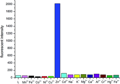
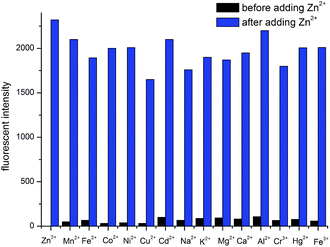
Likewise, selectivity of HL2 towards Zn2+ was examined by fluorescence of HL2 (Fig. 9) in the presence of equimolar metal ions including Mn2+, Fe2+, Co2+, Ni2+, Cu2+, Zn2+, Na+, K+, Mg2+, Ca2+, Al3+, Cr3+, Hg2+, and Fe3+ as their Cl− salts (Hg2+ as its NO3− salt). The results revealed that Mn2+, Fe2+, Co2+, Ni2+, Cu2+, Hg2+caused slight quenching of fluorescence of HL2 while Cd2+, Na+, K+, Mg2+, Ca2+, Al3+, Cr3+, and Fe3+ induced slight fluorescence enhancement, indicating that HL2 has high selectivity towards Zn2+. Specifically, the selectivity of HL2 on Zn2+ over Cd2+ was calculated as 30 using ratio of IZn/ICd, much higher than that of HL1. Furthermore, selectivity of HL2 on Zn2+ was studied by fluorescence respond of Zn2+ in the presence of other competing metal ions including Mn2+, Fe2+, Co2+, Ni2+, Cu2+, Zn2+, Cd2+, Na+, K+, Mg2+, Ca2+, Al3+, Cr3+, Hg2+, and Fe3+, results of which revealed that these metal ions have negligible disturbance on Zn–HL2 fluorescence, further indicating high selectivity of HL2 over Zn2+ (Fig. 10).
Crystal structure of (ZnL2Cl)2 (3) and [Cd4L26]·[CdCl4]·CH3OH (4) and proposed mechanism for Zn2+ sensing of HL1
Single crystal analysis revealed that compound 3 crystallized in monoclinic P21/n space group and showed a centrosymmetric binuclear structure composed of two Zn2+ ions, two deprotonated (L2)− anions and two chlorides. In this discrete unit, each Zn1 was penta-coordinated to two nitrogen atoms (N1, N2), one hydroxyl oxygen atom (O1) from one ligand, one hydroxyl oxygen atom (O1a) from the other ligand and one chloride (Cl1) (Fig. 11). Upon coordination, N2O donor set of L2 strongly chelated to Zn2+ and two hydroxyl oxygen atoms adopted μ2-coordination modes to bridge two ZnII ions together and formed the dinuclear structure. | ||
| Fig. 11 Molecular structure of dinuclear compound [Zn(L2)Cl]2 (3) with atomic labels. Asymmetry code: (a) 1 − x, −y, −z. | ||
Replacing ZnCl2 with CdCl2, HL2 was deprotonated and coordinated to generate compound {[Cd4L26]·[CdCl4]−·CH3OH}2·H2O (4) at room temperature. The single crystal analysis revealed that compound 4 crystallized in monoclinic P2/c space group and possessed two anionic mononuclear [CdCl4]2− units, two lattice methanol molecules, one water molecule and two independent tetranuclear cationic [Cd4L26]2+ units associated with intermolecular hydrogen bonds C–H⋯Cl. It could be seen from Fig. 12 that two tetranuclear [Cd4L26]2+ cationic moieties containing one mirror symmetry are structurally identical, each comprising of four Cd2+ ions and six deprotonated ligand (L2)−. Four Cd2+ ions in each unit are hexa-coordinated to donor set N4O2 from two adjacent ligand (L2)− for three cadmium ions (Cd2, Cd3, Cd3a/Cd5, Cd6, Cd6a) at the vertices of an equilateral triangle or donor set O6 from six ligand (L2)− for one central cadmium ion (Cd1/Cd4), presenting the distorted octahedral coordination geometry. While each mononuclear anion unit contained one Cd2+ ion and four coordinated chlorides, presenting one tetrahedral coordination geometry. Together with lattice methanol and water molecules, cationic [Cd4L26]2+ and anionic [CdCl4]2− moieties were further connected each other by hydrogen bonds C–H⋯Cl(O) into the 2-D supramolecular layer (Fig. S11†).
Luminescence spectrum of compound 3 in ethanol was also obtained to attest that the product upon titration of Zn2+ into HL2 is compound 3. (Fig. S†) Similar to HL1, Zn-induced fluorescent intensity enhancement of ligand HL2 could be explained by CHFF, PET, and C![[double bond, length as m-dash]](https://www.rsc.org/images/entities/char_e001.gif) N isomerization-related process as mentioned above. Even though upon reaction of HL2 and CdCl2, crystal of compound 4 can be obtained, yet instant fluorescent response cannot be detected upon titration of Cd2+ salts (anion as Cl−, NO3−, ClO4−, or OAc−) into HL2. This negative response could be possibly attributed to larger radius and therefore less affinity to HL2 of Cd2+ compared to that of Zn2+, which could be supported by the difference of Cd–N, Cd–O distances and Zn–N, Zn–O distances in compound 3 and 4 (Table 1).19 On the other hand, despite several reports of coordination compounds based on HL2 and other metals20 such as Fe(III), V(III), Cu(II), and Ni(II), yet high fluorescent sensing selectivity of HL2 towards Zn2+ can be also rationalized ascribing to paramagnetism of other metals which causes fluorescence quenching.
N isomerization-related process as mentioned above. Even though upon reaction of HL2 and CdCl2, crystal of compound 4 can be obtained, yet instant fluorescent response cannot be detected upon titration of Cd2+ salts (anion as Cl−, NO3−, ClO4−, or OAc−) into HL2. This negative response could be possibly attributed to larger radius and therefore less affinity to HL2 of Cd2+ compared to that of Zn2+, which could be supported by the difference of Cd–N, Cd–O distances and Zn–N, Zn–O distances in compound 3 and 4 (Table 1).19 On the other hand, despite several reports of coordination compounds based on HL2 and other metals20 such as Fe(III), V(III), Cu(II), and Ni(II), yet high fluorescent sensing selectivity of HL2 towards Zn2+ can be also rationalized ascribing to paramagnetism of other metals which causes fluorescence quenching.
| Sensor | Metal | Binding ratio (M2+![[thin space (1/6-em)]](https://www.rsc.org/images/entities/char_2009.gif) : :![[thin space (1/6-em)]](https://www.rsc.org/images/entities/char_2009.gif) L) L) |
Limit of detection | IZn/ICd (1 equiv.) | Mean of metal–N distances (Å) | Mean of metal–O distances (Å) | Sensing mechanism |
|---|---|---|---|---|---|---|---|
| HL1 | Zn2+ | 4![[thin space (1/6-em)]](https://www.rsc.org/images/entities/char_2009.gif) : :![[thin space (1/6-em)]](https://www.rsc.org/images/entities/char_2009.gif) 3 3 |
1.1 × 10⁻⁶ M | 4.8 | 2.098 | 2.020 | TITC,1c CHEF, PET9 C![[double bond, length as m-dash]](https://www.rsc.org/images/entities/char_e001.gif) N isomerization9,18 N isomerization9,18 |
| Cd2+ | 2![[thin space (1/6-em)]](https://www.rsc.org/images/entities/char_2009.gif) : :![[thin space (1/6-em)]](https://www.rsc.org/images/entities/char_2009.gif) 1 1 |
9.2 × 10⁻⁶ M | 2.315 | 2.302 | CHEF, PET, C![[double bond, length as m-dash]](https://www.rsc.org/images/entities/char_e001.gif) N isomerization N isomerization |
||
| HL2 | Zn2+ | 1![[thin space (1/6-em)]](https://www.rsc.org/images/entities/char_2009.gif) : :![[thin space (1/6-em)]](https://www.rsc.org/images/entities/char_2009.gif) 1 1 |
4.7 × 10⁻⁸ M | 30 | 2.070 | 2.108 | CHEF, PET, C![[double bond, length as m-dash]](https://www.rsc.org/images/entities/char_e001.gif) N isomerization N isomerization |
| Cd2+ | 2![[thin space (1/6-em)]](https://www.rsc.org/images/entities/char_2009.gif) : :![[thin space (1/6-em)]](https://www.rsc.org/images/entities/char_2009.gif) 3 3 |
— | 2.357 | 2.289 | — |
Conclusions
Ligands (HL1, HL2) as fluorescent sensors towards Zn2+ and/or Cd2+ and their metal ions sensing properties were investigated, through which we demonstrated the effect of ethoxyl substituent on Zn2+ fluorescent sensing. Their spectral difference in sensing Zn2+ or Cd2+ could be attributed to ethoxyl group which facilitated ZnII-induced twisted intramolecular charge transfer. Such results may give insight into how to develop fluorescent sensors for discriminating ion pairs.For HL1 with an ethoxyl substituent on the benzene ring, interaction of the ligand with Zn2+ induced twisted molecular structure and causes fluorescent red shift and fluorescent enhancement, while interaction of Cd2+ with HL1 merely induced fluorescence enhancement retaining the original emission peak. Though HL1 spectrally responds to both Zn2+ and Cd2+, these two metal ions can be discriminated by HL1 via two coordination conformations.
For HL2 absent of the ethoxyl group, it has exclusive response towards Zn2+ over other metal ions including Cd2+, showing that HL2 could also be a selective fluorescent sensor for Zn2+. Unlike HL1, no spectral shift was detected upon interaction of HL2 with Zn2+, which could be ascribed to absence of twisted molecular structure in Zn–HL2 compound 3. Negative response of HL2 towards Cd2+ correlates with longer Cd–N, Cd–O bond distances compared with that of Zn–N, Zn–O bond lengths in compounds 3 and 4, indicating less affinity of Cd2+ towards HL2. On the other hand, without ethoxyl group as hindrance effect, HL2 shows greater sensitivity towards sensing Zn2+ with a much lower detection limit for Zn2+.
Acknowledgements
This work has been supported by the National Natural Science Foundation of China (Grant no. 91122008, 21071056 and 21471061), Research Fund for the Doctoral Program of Higher Education of China (20124407110007), Guangdong Province of higher school science and technology innovation key project (cxzd1113), and Foundation for High-level Talents in Higher Education of Guangdong, China (C10301). The authors also thank Prof. Keith Man-chung Wong for offering valuable suggestions during composition of this essay.Notes and references
- (a) P. J. Jiang and Z. J. Guo, Coord. Chem. Rev., 2004, 248, 205–229 CrossRef CAS PubMed; (b) L. M. Wysockia and L. D. Lavis, Curr. Opin. Chem. Biol., 2011, 15, 752–759 CrossRef PubMed; (c) A. P. de Silva, H. Q. N. Gunaratne, T. Gunnlaugsson, A. J. M. Huxley, C. P. McCoy, J. T. Rademacher and T. E. Rice, Chem. Rev., 1997, 97, 1515–1566 CrossRef CAS PubMed; (d) K. P. Carter, A. M. Young and A. E. Palmer, Chem. Rev., 2014, 114, 4564–4601 CrossRef CAS PubMed; (e) X. Li, X. Gao, W. Shi and H. Ma, Chem. Rev., 2014, 114, 590–659 CrossRef CAS PubMed; (f) Y. B. Ding, T. Li, X. Li, W. H. Zhua and Y. S. Xie, Org. Biomol. Chem., 2013, 11, 2685 RSC; (g) Y. B. Ding, Y. S. Xie, X. Li, J. P. Hill, W. B. Zhang and W. H. Zhu, Chem. Commun., 2011, 47, 5431 RSC; (h) Y. B. Ding, X. Li, T. Li, W. H. Zhu and Y. S. Xie, J. Org. Chem., 2013, 78, 5328 CrossRef CAS PubMed; (i) Y. B. Ding, Y. Y. Tang, W. H. Zhu and Y. S. Xie, Chem. Soc. Rev., 2015, 44, 1101–1112 RSC.
- (a) R. H. Holm, P. Kennepohl and E. I. Solomon, Chem. Rev., 1996, 96, 2239–2314 CrossRef CAS; (b) D. S. Auld, BioMetals, 2001, 14, 271–313 CrossRef CAS; (c) A. I. Bush, Trends Neurosci., 2003, 26, 207–214 CrossRef CAS; (d) C. J. Frederickson, J. Y. Koh and A. I. Bush, Nat. Rev. Neurosci., 2005, 6, 449–462 CrossRef CAS PubMed; (e) A. B. Chausmer, J. Am. Coll. Nutr., 1998, 17, 109–115 CrossRef CAS; (f) E. L. Que, D. W. Domaille and C. J. Chang, Chem. Rev., 2008, 108, 1517–1549 CrossRef CAS PubMed.
- (a) A. Voegelin, S. Poster, A. C. Scheinost, M. A. Marcus and R. Kretzschmar, Environ. Sci. Technol., 2005, 39, 6616–6623 CrossRef CAS; (b) C. Rensing and R. M. Maier, Ecotoxicol. Environ. Saf., 2003, 56, 140–147 CrossRef CAS.
- (a) E. L. Que, D. W. Domaille and C. J. Chang, Chem. Rev., 2008, 108, 1517–1549 CrossRef CAS PubMed; (b) H. Woo, S. Cho, Y. Han, W. S. Chae, D. R. Ahn, Y. You and W. Nam, J. Am. Chem. Soc., 2013, 135, 4771–4787 CrossRef CAS PubMed; (c) X. A. Zhang, D. Hayes, S. J. Smith, S. Friedle and S. J. Lippard, J. Am. Chem. Soc., 2008, 130, 15788–15789 CrossRef CAS PubMed; (d) K. Hanaoka, K. Kikuchi, H. Kojima, Y. Urano and T. Nagano, J. Am. Chem. Soc., 2004, 126, 12470–12476 CrossRef CAS PubMed; (e) K. Komatsu, K. Kikuchi, H. Kojima, Y. Urano and T. Nagano, J. Am. Chem. Soc., 2005, 127, 10197–10204 CrossRef CAS PubMed; (f) K. Komatsu, Y. Urano, H. Kojima and T. Nagano, J. Am. Chem. Soc., 2007, 129, 13447–13454 CrossRef CAS PubMed; (g) H. Z. Su, X. B. Chen and W. H. Fang, Anal. Chem., 2014, 86, 891–899 CrossRef CAS PubMed; (h) Y. Mikata, K. Kawata, S. Iwatsuki and H. Konno, Inorg. Chem., 2012, 51, 1859–1865 CrossRef CAS PubMed; (i) J. T. Simmons, J. R. Allen, D. R. Morris, R. J. Clark, C. W. Levenson, M. W. Davidson and L. Zhu, Inorg. Chem., 2013, 52, 5838–5850 CrossRef CAS PubMed; (j) E. Kimura, S. Aoki, E. Kikuta and T. Koike, Proc. Natl. Acad. Sci. U. S. A., 2003, 100, 3731–3736 CrossRef CAS PubMed.
- (a) Z. R. Grabowski and K. Rotkiewicz, Chem. Rev., 2003, 103, 3899–4031 CrossRef PubMed; (b) B. Valeur and I. Leray, Coord. Chem. Rev., 2000, 205, 3–40 CrossRef CAS.
- (a) S. Aoki, D. Kagata, M. Shiro, K. Takeda and E. Kimura, J. Am. Chem. Soc., 2004, 126, 13377–13390 CrossRef CAS PubMed; (b) P. Mahato, S. Saha and A. Das, J. Phys. Chem. C, 2012, 116, 17448–17457 CrossRef CAS.
- (a) X. Y. Zhou, P. X. Li, Z. H. Shi, X. L. Tang, C. Y. Chen and W.-S. Liu, Inorg. Chem., 2012, 51, 9226–9231 CrossRef CAS PubMed; (b) E. M. Nolan, J. W. Ryu, J. Jaworski, R. P. Feazell, M. Sheng and S. J. Lippard, J. Am. Chem. Soc., 2006, 128, 15517–15528 CrossRef CAS PubMed; (c) F. A. Cotton and G. Wilkinson, Advances in Inorganic Chemistry, Wiley, New York, 5th edn, 1988, pp. 957–1358 Search PubMed; (d) M. M. Henary, Y. G. Wu and C. J. Fahrni, Chem.–Eur. J., 2004, 10, 3015 CrossRef CAS PubMed; (e) R. D. Hancock, Chem. Soc. Rev., 2013, 42, 1500–1524 RSC.
- (a) M. P. Waalkes, Mutat. Res., 2003, 533, 107–120 CrossRef CAS PubMed; (b) M. Waisberg, P. Joseph, B. Hale and D. Beyersmann, Toxicology, 2003, 192, 95–117 CrossRef CAS; (c) M. P. Waalkes, T. P. Coogan and R. A. Barter, Crit. Rev. Toxicol., 1992, 22, 175–201 CrossRef CAS PubMed.
- V. Kumar, A. Kumar, U. Diwan and K. K. Upadhyay, Dalton Trans., 2013, 13078–13083 RSC.
- G. M. Sheldrick, SADABS, version 2.05, University of Göttingen, Göttingen, Germany, 1996 Search PubMed.
- G. M. Sheldrick, SHELXS-97, Program for X-ray Crystal Structure Determination, University of Göttingen, Göttingen, Germany, 1997 Search PubMed.
- G. M. Sheldrick, SHELXS-97, Program for X-ray Crystal Structure Refinement, University of Göttingen, Göttingen, Germany, 1997 Search PubMed.
- Z. P. Zheng, Y. J. Ou, X. J. Hong, L. M. Wei, L. T. Wan, W. H. Zhou, Q. G. Zhan and Y. P. Cai, Inorg. Chem., 2014, 53, 9625–9632 CrossRef CAS PubMed.
- F. Robert, P. L. Jacquemin, B. Tinant and Y. Garcia, CrystEngComm, 2012, 14, 4396–4406 RSC.
- B. Bosnich, J. Am. Chem. Soc., 1968, 90, 627–632 CrossRef CAS.
- (a) X. B. Li, M. Bera, G. T. Musiea and D. R. Powell, Inorg. Chim. Acta, 2008, 36, 1965–1972 CrossRef PubMed; (b) D. M. Cooper, G. Ronald, S. Hargreaves, P. Kennewell and J. Redpath, Tetrahedron, 1995, 51, 7791–7808 CrossRef CAS.
- G. f. Li and D. R. B. D. Magana, J. Phys. Chem. B, 2012, 116, 12590–12596 CrossRef CAS PubMed.
- (a) J. S. Wu, W. M. Liu, X. Q. Zhuang, F. Wang, P. F. Wang, S. L. Tao, X. H. Zhang, S.-K. Wu and S. T. Lee, Org. Lett., 2007, 9, 33–36 CrossRef CAS PubMed; (b) H. Jung, K. Ko, J. Lee, S. Kim, S. Bhuniya, J. Lee, Y. Kim and S. Kim, Inorg. Chem., 2010, 49, 8552–8557 CrossRef CAS PubMed; (c) S. Iyoshi, M. Taki and Y. Yamamoto, Inorg. Chem., 2008, 47, 3946–3948 CrossRef CAS PubMed.
- (a) N. J. Williams, W. Gan, J. H. Reibenspies and R. D. Hancock, Inorg. Chem., 2009, 48, 1407–1415 CrossRef CAS PubMed; (b) Y. Mikata, Y. Sato, S. Takeuchi, Y. Kuroda, H. Konnoc and S. Iwatsuki, Dalton Trans., 2013, 9688–9698 RSC.
- (a) C. Imbert, H. P. Hratchian, M. Lanznaster, M. J. Heeg, L. M. Hryhorczuk, B. R. McGarvey, H. B. Schlegel and C. N. Verani, Inorg. Chem., 2005, 44, 7414–7422 CrossRef CAS PubMed; (b) S. P. Rath, K. K. Rajak and A. Chakravorty, Inorg. Chem., 1999, 38, 4376 CrossRef CAS; (c) B. de Souza, A. J. Bortoluzzi, T. Bortolotto, F. L. Fischer, H. Terenzi, D. E. C. Ferreira, W. R. Rocha and A. Neves, Dalton Trans., 2010, 2027 RSC.
Footnote |
| † Electronic supplementary information (ESI) available. CCDC 1003835–1003838, 1015499–1015501 and 1015694. For ESI and crystallographic data in CIF or other electronic format see DOI: 10.1039/c5ra00987a |
| This journal is © The Royal Society of Chemistry 2015 |


