Praseodymium oxide nanostructures: novel solvent-less preparation, characterization and investigation of their optical and photocatalytic properties

Sahar Zinatloo-Ajabshir, Masoud Salavati-Niasari* and Masood Hamadanian
Institute of Nano Science and Nano Technology, University of Kashan, Kashan, P.O. Box 87317-51167, I.R. Iran. E-mail: Salavati@kashanu.ac.ir; Fax: +98 361 555 2930; Tel: +98 361 591 2383

Received 14th January 2015 , Accepted 2nd March 2015

First published on 2nd March 2015


Abstract

Praseodymium oxide (Pr6O11) nanostructures were prepared via a new simple solvent-less route. The nanostructures were synthesized by a heat treatment in air at 600 °C for 5 h, using [Pr L(NO3)2]NO3 (L = bis-(2-hydroxy phenyl methyl ketone)-dipropylene triamin Schiff base ligand), as a precursor, which was obtained by a solvent-free solid–solid reaction from different molar ratios of praseodymium nitrate and a Schiff base ligand. The as-prepared nanostructures were characterized by means of several techniques including X-ray diffraction (XRD), thermo-gravimetric analysis (TGA), scanning electron microscopy (SEM), energy dispersive X-ray microanalysis (EDX), photoluminescence (PL) spectroscopy, transmission electron microscopy (TEM), nuclear magnetic resonance spectroscopy (1H NMR) analysis, UV-vis diffuse reflectance spectroscopy and Fourier transform infrared (FT-IR) spectroscopy. The obtained results showed that the morphology and particle size of the final Pr6O11 could be dramatically affected via the molar ratio of praseodymium nitrate and the Schiff base ligand. The photocatalytic activity of the as-synthesized nanostructures was also investigated by the degradation of 2-naphthol as an organic contaminant under ultraviolet light irradiation.

Introduction

The preparation of nanostructured materials has received considerable attention owing to their unique properties and interesting potential applications.1–5 Among these nanomaterials, praseodymium oxide is well known as a significant rare earth metal oxide with specific and unique optical and electrical properties, and has been extensively investigated for applications in oxygen-storage components, promoters and stabilizers in combustion catalysts, ceramic pigments, catalysts, and materials with higher electrical conductivity.6–10 It is generally accepted that Pr6O11 is the most stable and interesting form of praseodymium oxides at ambient temperature and pressure.11 Up to now, very limited numbers of synthetic routes have been reported to prepare nanostructured praseodymium oxides including precipitation,11 thermal decomposition,12 electrochemical,13 molten salt,14 electrospinning15 and hydrothermal.16

Many nanostructured metal oxides have been prepared by the thermal decomposition the organometallic molecular approach as an appropriate synthetic route. This approach as facile, reliable and cost effective synthetic route to the preparation of pure and uniform nanomaterials does not need special apparatus, complicated procedures and severe reaction conditions, which can limit the large-scale synthesis of the nanostructured materials. On the other hand, solvent-free synthetic routes have attracted much attention in the field of green chemistry, and many solvent-less synthetic routes for organic compounds have been presented. From the viewpoint of green chemistry, we succeeded in preparing rare earth metal complexes by solvent-free solid–solid reactions, and here we present the novel preparation of [Pr L(NO3)2]NO3 (L = bis-(2-hydroxy phenyl methyl ketone)-dipropylene triamin Schiff base ligand) via facile solvent-free reactions utilizing different molar ratios of praseodymium nitrate and Schiff base ligand, and then [Pr L(NO3)2]NO3 was utilized for synthesizing praseodymium oxide nanostructures via a thermal treatment approach.

Experimental

Materials

In this investigation, 2-hydroxy phenyl methyl ketone, praseodymium nitrate (Pr(NO3)3·6H2O), dipropylene triamin, methanol, ethyl acetate and chloroform, were purchased from Merck Company and were used without additional purification.

Synthesis of bis-(2-hydroxy phenyl methyl ketone)-dipropylene triamin Schiff base ligand (L)

To prepare of Schiff base ligand (L), 0.04 mol of 2-hydroxy phenyl methyl ketone dissolved in methanol (28 ml) was added drop-wise to a dipropylene triamin solution (0.02 mol) in 28 ml of methanol. The mixture was refluxed for 3 h. The yellow precipitate was separated by filtration, washed and air-dried. It was then recrystallized from methanol. The as-prepared ligand was characterized utilizing 1H-NMR and FT-IR.

Synthesis of the Pr(III) complex

Pr(NO3)3·6H2O and (L) were mixed and ground for 15 min in an agate mortar at room temperature in a Pr(NO3)3·6H2O to L molar ratio of 1[thin space (1/6-em)]:[thin space (1/6-em)]0.5, 1[thin space (1/6-em)]:[thin space (1/6-em)]1, 1[thin space (1/6-em)]:[thin space (1/6-em)]2, 1[thin space (1/6-em)]:[thin space (1/6-em)]3 and 1[thin space (1/6-em)]:[thin space (1/6-em)]4. The identification of the resulting products was based on Fourier transform (FT-IR) spectrometer. To study the effect of (L), one experiment was carried out without L (blank test).

Preparation of Pr6O11 micro/nanostructures

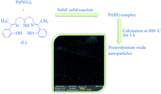
Praseodymium oxide micro/nanostructures were prepared by subjecting 0.1 g of the as-prepared Pr(III) complex powders to heat treatment at (600 °C) in the air. An average temperature increase of 30 °C is recorded every minute, before the temperature reached 600 °C, and after keeping the thermal treatment at 600 °C for 5 h, it was allowed to cool to room temperature. Schematic diagram of formation of praseodymium oxide micro/nanostructures is depicted in Scheme 1. To study the effect of Schiff base ligand (L), a blank test was performed. In the blank test, 0.1 g of praseodymium nitrate was subjected to heat treatment at (600 °C) in the air. The preparation conditions of samples were illustrated in Table 1. The as-obtained nanostructures were characterized by EDS, SEM, TEM, FT-IR, PL, XRD, UV-vis techniques.
image file: c5ra00817d-s1.tif
Scheme 1 Schematic diagram of the synthesis of Pr6O11 micro/nanostructures.
Table 1 Preparation conditions of Pr6O11 samples
Sample no. Precursor Molar ratio of Pr(NO3)3·6H2O/Schiff base ligand (L) SEM
a Blank test, in the absence of Schiff base ligand (L).
1 Pr(III) complex 1[thin space (1/6-em)]:[thin space (1/6-em)]0.5 image file: c5ra00817d-u1.tif
2 Pr(III) complex 1[thin space (1/6-em)]:[thin space (1/6-em)]1 image file: c5ra00817d-u2.tif
3 Pr(III) complex 1[thin space (1/6-em)]:[thin space (1/6-em)]2 image file: c5ra00817d-u3.tif
4 Pr(III) complex 1[thin space (1/6-em)]:[thin space (1/6-em)]3 image file: c5ra00817d-u4.tif
5 Pr(III) complex 1[thin space (1/6-em)]:[thin space (1/6-em)]4 image file: c5ra00817d-u5.tif
6a Pr(NO3)3·6H2O image file: c5ra00817d-u6.tif


Characterization

Thermogravimetric-differential thermal analysis (TG-DTA) was done by a thermal gravimetric analysis instrument (Shimadzu TGA-50H) with a flow rate of 20.0 ml min−1 and a heating rate of 10 °C min−1. Room temperature photoluminescence (PL) was investigated on a Perkin Elmer (LS 55) fluorescence spectrophotometer. A Tescan mira3 field emission scanning electron microscope (FESEM) was utilized to visualize FESEM images of samples. Fourier transform infrared spectra were recorded using a Magna-IR, spectrometer 550 Nicolet in KBr pellets in the range of 400–4000 cm−1. The energy dispersive spectrometry (EDS) analysis was studied by XL30, Philips microscope. The UV-vis diffuse reflectance spectra of the as-prepared of the products were obtained on a UV-vis spectrophotometer (Shimadzu, UV-2550, Japan). Powder X-ray diffraction (XRD) patterns were collected with a diffractometer of Philips Company with X'PertPro monochromatized Cu Kα radiation (l = 1.54 Å). Transmission electron microscope (TEM) images of Pr6O11 were obtained on a JEM-2100 with an accelerating voltage of 200 kV equipped with a high resolution CCD Camera.

Photocatalytic measurements

The photocatalytic activity of Pr6O11 obtained from sample nos. 2 and 6 was examined by monitoring the degradation of 2-naphthol solution as organic contaminant in an aqueous solution. A quartz photocatalytic reactor was utilized to carry out the degradation reaction. The photocatalytic degradation was performed by utilizing 0.0012 g of 2-naphthol solution including 0.05 g of Pr6O11 nanoparticles at room temperature. To reach adsorption equilibrium, this mixture was aerated for 30 min. Later, the mixture was placed inside the photoreactor in which the vessel was 40 cm away from the UV source of 400 W mercury lamps. To hinder UV leakage, the light source and quartz vessel were placed inside a black box that equipped with a fan. Aliquots of the mixture were taken at definite interval of times during the irradiation, and after centrifugation they were analyzed by a UV-vis spectrometer. The 2-naphthol degradation percentage was calculated as follow:
 
image file: c5ra00817d-t1.tif(1)
where A0 and At are the absorbance value of solution at 0 and t min, respectively.

Results and discussion

In this investigation, the bis-(2-hydroxy phenyl methyl ketone)-dipropylene triamin ligand was firstly synthesized, to be utilized in the preparation of Pr6O11 micro/nanostructures. In order to ensure an adequate purity of the prepared (L), 1H-NMR analysis was performed. Fig. 1 exhibits the 1H-NMR spectrum of the (L) ligand. 1H-NMR data of the Schiff base ligand (CDCl3, δ, ppm):
image file: c5ra00817d-u7.tif
As observed, 1H-NMR result confirmed the high purity of the as-synthesized Schiff base ligand.

image file: c5ra00817d-f1.tif
Fig. 1 1H-NMR of the Schiff base ligand (L).

FT-IR spectroscopy is well known as helpful and applicable device to understand the functional group of any organic molecule. The infrared spectra of Schiff base ligand, Pr(III) complex and Pr6O11 nanoparticles are illustrated in Fig. 2. Two strong peaks at 1610 and 1263 cm−1 in Fig. 2a are ascribed to the ν(C[double bond, length as m-dash]N) of the azomethines and ν(Ar–O) of the phenolic hydroxyl substituent, respectively.17 The FT-IR spectrum of Pr(III) complex illustrates the (L) ligand characteristic peaks with several different shifts because of complex formation. The FT-IR spectrum of the Pr(III) complex displays an increase in the C–O stretching frequency (12 cm−1) compared to the free (L) Schiff base ligand (Fig. 2b). This indicate that the coordination to the Pr(III) ion takes place by means of the oxygen atoms of the hydroxyl benzene of the (L) Schiff base ligand.18 This is additional proved by using the peak located at 573 cm−1 which can be attributed to ν(Pr–O) vibration (Fig. 2b). In comparison with the free (L) Schiff base ligand, the ν(C[double bond, length as m-dash]N) peak in the Pr(III) complex shifted to 1615 cm−1. This blue shift (5 cm−1) shows a stronger double bond characteristic of the iminic bonds as well as contribution of the azomethines nitrogen atoms in the coordination.17,18 This result is additional proved by using the peak located at 410 cm−1 which can be related to ν(Pr–N) vibration (Fig. 2b). The different coordination modes of the nitrate groups to the praseodymium metal are detected by the differences in the two bands |ν4ν1|. The FT-IR spectrum of the Pr(III) complex illustrates several peaks located at 1463 cm−1 (ν1), 1031 cm−1 (ν2), 851 cm−1 (ν3) and 1304 cm−1 (ν4) related to the coordinated nitrate ions (Fig. 2b). The difference between ν4 and ν1 is nearly 159 cm−1 showing that the coordinated NO3 ions in the complex may act as a bidentate ligand.19,20 The (ν0) free nitrate located at 1385 cm−1 in the spectrum of the complex. The peak located at 3442 cm−1 and a weak peak at 1635 cm−1 ascribed to the v(OH) stretching and bending vibrations, respectively, which shows the presence of physisorbed water molecules linked to Pr6O11 product.20 The characteristic peak of Pr6O11 seen at 432 cm−1 is related to Pr–O vibration21 (Fig. 2c).


image file: c5ra00817d-f2.tif
Fig. 2 FT-IR spectra of (a) Schiff base ligand (L), (b) Pr(III) complex, and (c) Pr6O11 nanoparticles obtained from Pr(III) complex in molar ratio of 1[thin space (1/6-em)]:[thin space (1/6-em)]1 by thermal treatment at 600 °C for 5 h.

The thermal observation of the as-prepared Pr(III) complex was studied with thermal gravimetric analysis (TGA). Fig. 3 reveals the TGA curve of the as-prepared Pr(III) complex. As shown in Fig. 3, there are three weight loss steps. The first weight losses happens in the temperature range 60–150 °C, and displays 6.13% weight loss, which corresponds to surface moisture evaporating. The second step happens at the temperature range 150–400 °C (exhibiting 41.51% weight loss), which corresponds to the loss of the (L) ligand. The third step happens in the temperature range 400–750 °C (exhibiting 26.05% weight loss), which corresponds to the loss of two and one nitrate species from the inner and the outer coordination sphere of the as-prepared Pr(III) complex, respectively, and the formation of praseodymium oxide.


image file: c5ra00817d-f3.tif
Fig. 3 TGA of the Pr(III) complex precursor.

Fig. 4a reveals a typical XRD pattern of the Pr(III) complex precursor prepared in a molar ratio of 1[thin space (1/6-em)]:[thin space (1/6-em)]1. Fig. 4a clearly shows that the precursor is amorphous. The crystalline structures of the as-prepared praseodymium oxide nanostructures are confirmed by XRD. The XRD patterns shown in Fig. 4(b–e) correspond to the samples obtained by thermal treatment of Pr(III) complexes, prepared in Pr(NO3)3·6H2O to (L) molar ratios of 1[thin space (1/6-em)]:[thin space (1/6-em)]1, 1[thin space (1/6-em)]:[thin space (1/6-em)]2, 1[thin space (1/6-em)]:[thin space (1/6-em)]3 and 1[thin space (1/6-em)]:[thin space (1/6-em)]4 at 600 °C for 5 h. All of the reflection peaks can be readily indexed to the cubic phase of Pr6O11 with the space group of Fm3m and cell constants a = b = c = 5.4678 Å (JCPDS: 42-1121). In these patterns, no peak of any impurity is seen, indicating the high purity of the as-prepared products. The average crystallite sizes calculated by the Scherrer formula22 of the Pr6O11 prepared from Pr(III) complex with Pr(NO3)3·6H2O to (L) molar ratios of 1[thin space (1/6-em)]:[thin space (1/6-em)]1, 1[thin space (1/6-em)]:[thin space (1/6-em)]2, 1[thin space (1/6-em)]:[thin space (1/6-em)]3 and 1[thin space (1/6-em)]:[thin space (1/6-em)]4 are 23, 21.9, 20.5 and 19.1 nm, respectively. These results indicate that the crystallite sizes of Pr6O11 decreases with decreasing Pr(NO3)3·6H2O to (L) molar ratio.


image file: c5ra00817d-f4.tif
Fig. 4 XRD patterns of the Pr(III) complex in a molar ratio 1[thin space (1/6-em)]:[thin space (1/6-em)]1 (a) and Pr6O11 nanostructures obtained by thermal treatment of Pr(III) complex in molar ratio of 1[thin space (1/6-em)]:[thin space (1/6-em)]1 (b), 1[thin space (1/6-em)]:[thin space (1/6-em)]2 (c), 1[thin space (1/6-em)]:[thin space (1/6-em)]3 (d) and 1[thin space (1/6-em)]:[thin space (1/6-em)]4 (e) at 600 °C for 5 h.

EDS measurement provide additional data for the evaluation of the purity and composition of praseodymium oxide. In the EDS spectrum of sample no. 2 as illustrated in Fig. 5, the characteristic peaks of Pr and O are detected, which confirm the preparation of pure Pr6O11.


image file: c5ra00817d-f5.tif
Fig. 5 EDS pattern of Pr6O11 nanoparticles obtained by thermal treatment of Pr(III) complex in molar ratio of 1[thin space (1/6-em)]:[thin space (1/6-em)]1 at 600 °C for 5 h.

To study the effect of the amount of (L) Schiff base ligand on the morphology of the products, experiments were performed with different molar ratios of Pr(NO3)3·6H2O to (L) (Table 1). Fig. 6 and 7 shows FESEM images of the Pr6O11 obtained from Pr(III) complexes in the Pr(NO3)3·6H2O to (L) molar ratios of 1[thin space (1/6-em)]:[thin space (1/6-em)]0.5, 1[thin space (1/6-em)]:[thin space (1/6-em)]1, 1[thin space (1/6-em)]:[thin space (1/6-em)]2, 1[thin space (1/6-em)]:[thin space (1/6-em)]3 and 1[thin space (1/6-em)]:[thin space (1/6-em)]4, prepared by using the solid state route and calcined at 600 °C for 5 h. As previously described, privilege of this study is application of a novel Schiff-base as complexing and capping agent for preparing of Pr6O11 nanoparticles. This Schiff-base with high steric hindrance is an appropriate capping agent for synthesis of nanoparticles (Scheme 2). It seems that Schiff-base group with high steric hindrance causes nucleation to be happened rather than the growth. In our experiment, different molar ratios of Pr(NO3)3·6H2O to (L) were utilized to study (L) amount effect on Pr6O11 morphology. From the FESEM images, it can be seen that by utilizing molar ratio of 1[thin space (1/6-em)]:[thin space (1/6-em)]0.5 (Pr(NO3)3·6H2O to (L)), the nanoparticles were not form well (samples no. 1). With increasing the (L) content (samples no. 2), with molar ratio of 1[thin space (1/6-em)]:[thin space (1/6-em)]1 (Pr(NO3)3·6H2O to (L)), agglomerated uniform spherical nanoparticles formed (Fig. 6a and b). By increasing the molar ratio from 1[thin space (1/6-em)]:[thin space (1/6-em)]1 to 1[thin space (1/6-em)]:[thin space (1/6-em)]4, corrugated-like and fig-leaf-like Pr6O11 nanostructures (sample nos. 3 and 4) as well as separated nanoparticles with less uniform spherical morphology (sample no. 5) were obtained, respectively (Fig. 6c and 7a and b). It seems that when (L) content increases, the aggregation between Pr6O11 nanoparticles decreases because of steric hindrance effect of (L). Moreover, images indicate that the particle size of Pr6O11 decreases with decreasing Pr(NO3)3·6H2O to (L) molar ratio. It seems that when (L) amount increases, nucleation to be happened rather than the particle growth because of steric hindrance effect of (L). So the molar ratio of Pr(NO3)3·6H2O to (L) plays an important role to control particle size and morphology of Pr6O11 nanostructures.


image file: c5ra00817d-f6.tif
Fig. 6 SEM images of the products prepared from Pr(III) complex in molar ratios of (a) 1[thin space (1/6-em)]:[thin space (1/6-em)]0.5, (b) 1[thin space (1/6-em)]:[thin space (1/6-em)]1, and (c) 1[thin space (1/6-em)]:[thin space (1/6-em)]2, by thermal treatment of precursor at 600 °C for 5 h.

image file: c5ra00817d-f7.tif
Fig. 7 SEM images of the samples prepared from Pr(III) complex in molar ratios of (a) 1[thin space (1/6-em)]:[thin space (1/6-em)]3 and (b) 1[thin space (1/6-em)]:[thin space (1/6-em)]4 by thermal treatment of precursor at 600 °C for 5 h.

image file: c5ra00817d-s2.tif
Scheme 2 Schematic diagram illustrating the formation of Pr6O11 samples at various conditions.

The structural information of the as-prepared Pr6O11 nanoparticles were assessed with transmission electron microscopy (TEM) and high resolution TEM (HRTEM). The typical TEM image of the Pr6O11 nanoparticles in a molar ratio 1[thin space (1/6-em)]:[thin space (1/6-em)]1 (Pr(NO3)3·6H2O to (L)) is illustrated in Fig. 8a. The TEM image of the Pr6O11 shows that quasi-spherical nanoparticles are sintered together. Moreover, the image indicates that as-synthesized nanoparticles have size in the range of 30–80 nm. The high-resolution TEM (HRTEM) images of a single nanoparticle in Fig. 8b and c show that the nanoparticle is highly crystalline. The marked lattice fringes correspond to (220) plane in cubic phase of Pr6O11 with a d-spacing of 0.193 nm.


image file: c5ra00817d-f8.tif
Fig. 8 TEM image (a) and HRTEM images (b and c) of Pr6O11 nanoparticles obtained by thermal treatment of Pr(III) complex in molar ratio of 1[thin space (1/6-em)]:[thin space (1/6-em)]1 at 600 °C for 5 h.

To investigate the effect of the (L) Schiff base ligand on the morphology, sample no. 6 was synthesized as blank sample without utilizing any Schiff base ligand at 600 °C. Fig. 9 shows SEM images of sample no. 6. It can be seen that in the absence of (L), bulk structures are obtained. This result indicates that utilizing (L) Schiff base ligand with high steric hindrance led to synthesize nanoparticles at 600 °C, and we find out that huge ligands may be utilized instead of common capping agents to control the morphology and particle size.


image file: c5ra00817d-f9.tif
Fig. 9 SEM images of Pr6O11 prepared in the absence of Schiff base ligand.

The optical characteristics of the Pr6O11 nanoparticles were examined by PL and UV-vis diffuse reflectance analyses. The PL spectrum of the sphere-like Pr6O11 nanoparticles excited at wavelength of 280 nm shows a emission peak at 410 nm (Fig. 10a), which can be ascribed to charge transition from the 4f band to the valence band of Pr6O11 nanostructures. This result is similar to the previous report.23


image file: c5ra00817d-f10.tif
Fig. 10 PL spectrum (a), UV-vis diffuse reflectance spectrum (b) and plot to determine the band gap (c) of the Pr6O11 obtained by thermal treatment of Pr(III) complex in molar ratio of 1[thin space (1/6-em)]:[thin space (1/6-em)]1 at 600 °C for 5 h.

Fig. 10b shows the UV-vis diffuse reflectance spectrum of the sphere-like Pr6O11 nanoparticles (sample no. 2). In the UV-vis diffuse reflectance spectrum, the absorption band is seen at 352 nm. Using Tauc's formula, the band gap can be obtained from the absorption data.24 The energy gap (Eg) of the as-prepared Pr6O11 has been estimated by extrapolating the linear portion of the plot of (αhν)2 against to the energy axis (Fig. 10c). The Eg value of the as-prepared praseodymium oxide calculated to be 3.3 eV.

The influence of morphology on photocatalytic activity was investigated by monitoring the photo-oxidation of 2-naphthol as organic contaminant in an aqueous solution over as-prepared sample nos. 2 (obtained by thermal treatment of Pr(III) complex in molar ratio of 1[thin space (1/6-em)]:[thin space (1/6-em)]1) and 6 (prepared in the absence of Schiff base ligand) with different morphology under UV light irradiation (Fig. 11, Scheme 3). No 2-naphthol was practically broken down after 12 min without using UV light irradiation or Pr6O11. This observation indicated that the contribution of self-degradation was insignificant. According to photocatalytic calculations by eqn (1), the 2-naphthol degradation was about 100 and 10.2% over sample nos. 2 and 6, respectively, after 12 min irradiation of UV light, and Pr6O11 with uniform sphere-like morphology presented high photocatalytic activity. The surface properties of the sample no. 2 are illustrated in Table 2. Fig. 12a and b depicts the nitrogen adsorption/desorption isotherm and pore size distribution of the sample no. 2, respectively. As seen in Fig. 12b, for sample no. 2 the distribution of the pores is multi-peak. It has been shown that the heterogeneous photocatalytic processes comprise various steps (diffusion, adsorption, reaction, and etc.), and suitable distribution of the pore is effective and useful to diffusion of reactants and products, which prefer the photocatalytic reaction. In this study, the enhanced photocatalytic activity of sample no. 2 with uniform sphere-like morphology can be attributed to appropriate distribution of the pore, high hydroxyl amount and high separation rate of charge carriers.25


image file: c5ra00817d-f11.tif
Fig. 11 Photocatalytic 2-naphthol degradation of sample nos. 2 and 6 under UV light.

image file: c5ra00817d-s3.tif
Scheme 3 Reaction mechanism of 2-naphthol photodegradation over Pr6O11 nanoparticles under UV light irradiation.
Table 2 The surface properties of the sample no. 2
BET area (m2 g−1) Pore volume (cm3 g−1) Pore diameter (nm)
14.68 0.126 4.2


image file: c5ra00817d-f12.tif
Fig. 12 N2 adsorption/desorption isotherm (a) and pore size distribution curve (b) of the Pr6O11 obtained by thermal treatment of Pr(III) complex in molar ratio of 1[thin space (1/6-em)]:[thin space (1/6-em)]1 at 600 °C for 5 h.

Conclusions

Pr6O11 nanostructures with different morphologies were successfully synthesized via a novel and simple route. First, solid complex molecular precursor [Pr L(NO3)2]NO3 (L = bis-(2-hydroxy phenyl methyl ketone)-dipropylene triamin Schiff base ligand) was successfully synthesized by a mechano-chemical solid–solid route, and then Pr6O11 nanostructures were obtained by thermal treatment of [Pr L(NO3)2]NO3 at 600 °C for 5 h. In this work, solid state route coupled with thermal treatment in the absence of any other template or surfactant, is considered a convenient, reproducible, facile and practical method with various advantages such as saving of time and energy, freedom from utilizing organic solvent and ease of controlling reaction temperature, all of which contribute to large-scale production. Several techniques such as XRD, FESEM, TEM, PL, FT-IR, UV-vis, and EDS were utilized to characterize the structural and optical properties of the as-synthesized nanostructures. The obtained praseodymium oxide nanoparticles can be used as an interesting candidate for photocatalytic applications under UV light such as removal of 2-naphthol, since the degradation percentage of the 2-naphthol was found to be 100 within 12 min.

Acknowledgements

Authors are grateful to the council of Council of University of Kashan on this work by Grant number (22134324/345).

Notes and references

  1. S. Zinatloo-Ajabshir and M. Salavati-Niasari, Int. J. Appl. Ceram. Technol., 2014, 11, 654 CrossRef CAS PubMed.
  2. M. Salavati-Niasari, A. Khansari and F. Davar, Inorg. Chim. Acta, 2009, 362, 4937 CrossRef CAS PubMed.
  3. S. Zinatloo-Ajabshir and M. Salavati-Niasari, J. Ind. Eng. Chem., 2014, 20, 3313 CrossRef CAS PubMed.
  4. A. Sobhani and M. Salavati-Niasari, Mater. Res. Bull., 2012, 47, 1905 CrossRef CAS PubMed.
  5. S. Zinatloo-Ajabshir and M. Salavati-Niasari, Ceram. Int., 2015, 41, 567 CrossRef CAS PubMed.
  6. M. Kawabe, H. Ono, T. Sano, M. Tsuji and Y. Tamaura, Energy, 1997, 22, 1049 CrossRef.
  7. P. Šulcová, J. Therm. Anal. Calorim., 2005, 82, 51 CrossRef.
  8. K. Asami, K. Kusakabe, N. Ashi and Y. Ohtsuka, Appl. Catal., A, 1997, 156, 43 CrossRef CAS.
  9. S. Shrestha, C. M. Y. Yeung, C. Nunnerley and S. C. Tsang, Sens. Actuators, A, 2007, 136, 191 CrossRef CAS PubMed.
  10. S. Bernal, F. J. Botana, G. Cifredo, J. J. Calvino, A. Jobacho and J. M. Rodriguez-Izquierdo, J. Alloys Compd., 1992, 180, 271 CrossRef CAS.
  11. B. M. Abu-Zied, Y. A. Mohamed and A. M. Asiri, J. Rare Earths, 2013, 31, 701 CrossRef CAS.
  12. B. A. A. Balboul, J. Anal. Appl. Pyrolysis, 2010, 88, 192 CrossRef CAS PubMed.
  13. X. Sun, T. Zhai, X. Lu, S. Xie, P. Zhang, C. Wang, W. Zhao, P. Liu and Y. Tong, Mater. Res. Bull., 2012, 47, 1783 CrossRef CAS PubMed.
  14. X. Wang, J. Zhuang and Y. Li, Eur. J. Inorg. Chem., 2004, 5, 946 CrossRef PubMed.
  15. M. Shamshi Hassan, Y.-S. Kang, B.-S. Kim, I.-S. Kim, H.-Y. Kim and M.-S. Khil, Superlattices Microstruct., 2011, 50, 139 CrossRef PubMed.
  16. Y. Zhang, K. Han, X. Yin, Z. Fang, Z. Xu and W. Zhu, J. Cryst. Growth, 2009, 311, 3883 CrossRef CAS PubMed.
  17. T. A. Youssef, J. Coord. Chem., 2008, 61, 816 CrossRef CAS.
  18. W. B. Sun, P. F. Yan, G. M. Li, H. Xu and J. W. Zhang, J. Solid State Chem., 2009, 182, 381 CrossRef CAS PubMed.
  19. W. Wang, Y. Huang and N. Tang, Spectrochim. Acta, Part A, 2007, 66, 1058 CrossRef PubMed.
  20. Y. L. Zhang, W. W. Qin, W. S. Liu, M. Y. Tan and N. Tang, Spectrochim. Acta, Part A, 2002, 58, 2153 CrossRef.
  21. M. Popa and M. Kakihana, Solid State Ionics, 2001, 141–142, 265 CrossRef CAS.
  22. R. L. Snyder Jenkins, Chemical Analysis: Introduction to X-ray Powder Diffractometry, John Wiley & Sons, Inc., New York, 1996 Search PubMed.
  23. N. Krishna Chandar and R. Jayavel, Mater. Res. Bull., 2014, 50, 417 CrossRef CAS PubMed.
  24. M. Salavati-Niasari, D. Ghanbari and M. R. Loghman-Estarki, Polyhedron, 2012, 35, 149 CrossRef CAS PubMed.
  25. J. Zhong, J. Li, F. Feng, Y. Lu, J. Zeng, W. Hu and Z. Tang, J. Mol. Catal. A: Chem., 2012, 357, 101 CrossRef CAS PubMed.

This journal is © The Royal Society of Chemistry 2015