DOI:
10.1039/C5RA00701A
(Paper)
RSC Adv., 2015,
5, 22365-22372
DNA-capped Fe3O4/SiO2 magnetic mesoporous silica nanoparticles for potential controlled drug release and hyperthermia
Received
13th January 2015
, Accepted 16th February 2015
First published on 18th February 2015
Abstract
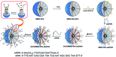
We proposed a strategy to construct DNA-capped Fe3O4/SiO2 magnetic mesoporous silica (MMS) nanoparticles for potential temperature controlled drug release and magnetic hyperthermia. Drug release behavior, magnetic heating capacity, in vitro cytotoxicity, and cell uptake of the MMS-based nanocarriers were evaluated. The results showed that the DOX/MMS–NH2–dsDNA complexes could release DOX fast at 50 °C, but very slow at 37 °C. Also, MMS-based nanocarriers could efficiently generate heat upon exposure to an alternating magnetic field due to the superparamagnetic behavior. Furthermore, the MMS–NH2–dsDNA complexes could be effectively taken up by murine breast cancer 4T1 cells, and negligible cytotoxicity of the MMS–NH2–dsDNA complexes has been observed. Therefore, DNA-capped MMS nanoparticles had potential for cancer therapy with temperature controlled drug release and magnetic hyperthermia.
1. Introduction
Controlled drug release systems are of great importance in drug delivery. To date, various organic and inorganic carriers have been investigated for controlled drug release systems.1 Among inorganic carriers, biocompatible and biodegradable hydroxyapatite (HA) nanoparticles have been widely studied for drug delivery, and HA nanoparticles showed great potential as drug carriers with high drug loading and pH-responsive features for future intracellular drug delivery system.2–6 Mesoporous silica nanoparticles (MSN) are another promising candidate carriers for drug delivery due to their high surface area, large pore volume, good biocompatibility and ease of surface functionalization.7–9 In particular, the readily functionalized surface allows MSN to construct smart caps on the mesopore outlets to control their opening or closing state, resulting in the controlled drug release. Many studies have reported the design of smart caps, such as nanoparticles, cyclodextrin, gelatin, supramolecular assembles and biomolecules, to control drug release from MSN in response to different stimuli including light, pH, temperature, redox activation, competitive binding and enzymes.10–18
Temperature is an external stimulus and provides a great opportunity to release a drug at a desired time. For example, Fu et al., Zhou et al. and You et al. reported the temperature-responsive controlled release systems based on poly(N-isopropylacrylamide) (PNIPAM)-modified MCM-41 and mesostructure cellular foam materials (MCFs).19–21 Jiao et al. prepared the copolymer of 2-(2-methoxyethoxy)ethyl methacrylate (MEO2MA) and oligo(ethylene glycol) methacrylate (OEGMA) cross-linked by disulfide bonds (P(MEO2MA–s–s–OEGMA)) to coat hollow MSN, and found that the loaded drugs could be released rapidly at a temperature higher than 37 °C.22 However, these temperature-controlled drug release systems showed the complicated synthesis and coating process of the temperature-responsive polymers on MSN. Also, Aznar et al. and Liu et al. designed temperature-controlled drug release systems based on MSN with phase-change molecules as caps,23,24 but the limited phase-change molecules could be used to construct the controlled drug release systems due to the requirements of biocompatibility, biosafety and phase-change temperature in the range of body temperature.
Recently, DNA-capped MSN as the controlled release nanocarriers have attracted more attention due to the easy synthesis and programmability, good biocompatibility and high cellular uptake.25–34 Most important, DNA molecules as caps could response to many stimuli, such as temperature, pH, enzyme and competitive binding.25–34 For example, Chen et al. used i-motif quadruplex DNA to cap the mesopores and triggered pH-controlled release based on the morphology change of DNA chains.26 Zhu et al. reported an enzyme-triggered drug release system that is based on CpG oligodeoxynucleotide-capped hollow mesoporous silica particles.34 Zhang et al. developed a nanocarrier by capping MSN with a programmable DNA hybrid, and the drug release was controlled by microRNA owning to the competitive binding.18 From the viewpoint of practical application, these methods to trigger drug release with the stimuli of pH, enzyme and competitive binding are challenging because of the complexity of the in vivo environment.
However, the temperature-responsive controlled drug release from DNA-caped MSN is promising for clinical applications. On one hand, the DNA duplex endows the feature with temperature tunability in the range of body temperature through changes in chain length, variations in G/C content, or even the size and surface density of oligonucleotides attached to the nanoparticles.31,35 On the other hand, many possible stimuli, such as near-infrared (NIR) light and magnetic field can trigger the DNA-capped MSN-based nanocarriers to generate heat for the uncapping of mesopores, and thereby release drugs.31–33 For example, Chang et al. and Li et al. developed NIR light-triggered nanocarriers for controlled drug release, in which the drugs can be loaded into mesoporous silica coated Au nanorods and then capped with DNA,32,33 because Au nanorods with strong surface plasmon resonance are highly absorbent of light in the NIR region and can act as a nanoheater by absorbing the NIR laser. However, the practical application of light-triggered drug delivery systems could be limited due to the low tissue penetration depth of light (several mm for NIR light).36
In comparison, human tissues are transparent to the magnetic field, thus the use of a magnetic field can serve as an alternative approach to overcome the limitation of low tissue penetration depth of light.37 On one hand, magnetic mesoporous nanoparticles can generate heat under an alternating magnetic field due to the Néel and Brownian relaxation or hysteresis loss of magnetic nanoparticles,38 which can induce the DNA-capped magnetic mesoporous nanoparticles to denaturize the DNA capping. For example, Ruiz–Hernandez et al. conjugated double-stranded DNA (dsDNA) together with spherical γ-Fe2O3 magnetic nanoparticles to cap the mesopores of MSN, and utilized a magnetic field to induce heat for triggering the uncapping of mesopores for drug release.31 On the other hand, magnetic hyperthermia, a type of cancer therapy in which body tissue is exposed to magnetic field for increasing the temperature to be higher than normal body temperature (≈41–46 °C), has been developed as an efficacious treatment modality for localized or deeply exist cancer.39–43 Moreover, magnetic hyperthermia could combine chemotherapy to improve cancer therapeutic efficiency, because hyperthermia can render tumor cells temporarily more sensitive to anticancer drugs.44 Therefore, it can be imagined that DNA-capped magnetic mesoporous silica (MMS) nanoparticles could be a promising multifunctional platform for potential cancer therapy with temperature controlled drug release and hyperthermia under an alternating magnetic field. That is to say, after the DNA-capped MMS nanoparticles-based drug delivery system arrive the tumor sites, the heat will be generated from MMS nanoparticles to raise the temperature of the surrounding tumor organ/cells for hyperthermia therapy when applied an alternating magnetic field. At the same time, the generated heat increases the temperature of the DNA-capped MMS nanocarrier, resulting in the opening of the mesopores to release drugs for chemotherapy.
Herein we proposed a concept to develop DNA-capped Fe3O4/SiO2 MMS nanoparticles as nanocarriers for potential temperature controlled drug release and hyperthermia. As shown in Fig. 1, Fe3O4 nanoparticles were prepared and incorporated into mesoporous silica matrix to form Fe3O4/SiO2 MMS nanoparticles. MMS nanoparticles are functionalized with single-stranded DNA (ssDNA) through the modification of amino groups on MMS nanoparticles and the grafting of 6-maleimidocaproic acid sulfo-N-succinimidyl ester (sulfo-EMCS) linkers with amino groups. After loading of doxorubicin (an anticancer drug), ssDNA functionalized MMS nanoparticles are capped with complementary ssDNA (cDNA) through the formation of double-stranded DNA (dsDNA) for capping the mesopores. When applied an alternating magnetic field, the DNA-capped MMS nanoparticles generate heat for hyperthermia, and the loaded drugs could be simultaneously released by the heat-triggered denaturation of the dsDNA capping chains.
 |
| Fig. 1 Schematic illustration of preparation of temperature controlled drug release system based on DNA-capped Fe3O4/SiO2 MMS nanoparticles. | |
2. Experimental
2.1. Materials
Tetraethyl orthosilicate (TEOS), triethanolamine (TEA), ethanol, hydrochloric acid (HCl, 37%), potassium dihydrogen phosphate (KH2PO4), sodium hydroxide (NaOH), ferric chloride (FeCl3·6H2O), ferrous chloride (FeCl2·4H2O) were obtained from Sinopharm Chemical Reagent Co. Ltd., PBS (pH 7.4), HEPES buffer, doxorubicin hydrochloride (DOX), and EDTA disodium salt dihydrate were obtained from Sangon Biotech (Shanghai) Co. Ltd., Hexadecyltrimethylammonium p-toluenesulfonate (CTAT) and 3-aminopropyltriethoxysilane (APTES) were obtained from Sigma-Aldrich. 6-Maleimidocaproic acid sulfo-N-succinimidyl ester (sulfo-EMCS) was purchased from Toronto Research chemicals Inc. Ultrapure water was obtained from Millipore pure water system. All chemicals were of analytical-reagent grade and used without further purification. Oligonucleotides were purchased from Sangon Biotech (Shanghai) Co. Ltd., and the sequences are 5′-SH(CH2)6TTATCGCTGATTCAA-3′ (ssDNA) and 5′-TTGAATCAGCGATAATCGAATAGCGACTAAGTT-3′ (cDNA).
2.2. Synthesis of aminated magnetic mesoporous silica nanoparticles (MMS–NH2)
MMS nanoparticles were synthesized according to the previously reported method.45 Amino groups were grafted onto MMS nanoparticles to form MMS–NH2 nanoparticles as follows: 0.5 g of MMS nanoparticles was homogeneously dispersed in 100 ml ethanol and sonicated for 30 min. Subsequently, 1.5 ml of APTES was added to the suspension with mild stirring for 24 h at the room temperature. The suspension was filtered off and washed extensively with ethanol to remove the residual APTES. Finally, the MMS–NH2 nanoparticles were dried in vacuum at 50 °C for 12 h.
2.3. Characterization
The wide angle X-ray diffraction (WAXRD) patterns were obtained on a D8 ADVANCE powder diffractometer using Cu Kα1 radiation (1.5405 Å). Transmission electron microscopy (TEM) images were obtained using a JEM-2100F transmission electron microscope at an acceleration voltage of 200 kV. N2 adsorption–desorption isotherms were obtained on a Micromeritics Tristar 3020 automated surface area and pore size analyzer at −196 °C under continuous adsorption conditions. Brunauer–Emmett–Teller (BET) and Barrett–Joyner–Halenda (BJH) methods were used to determine the surface area and mesopore size distribution. Zeta potential measurements were performed on a Malvern zeta sizer Nano-ZS90. Fourier transform infrared (FTIR) spectra were recorded on a LAM750(s) spectrometer in transmission mode. UV-vis analysis was measured on a Nanodrop 2000C spectrophotometer. Magnetization curve was carried out using a TF-WI-ZDYP V 3.0.4 vibrating sample magnetometer (VSM) at 298 K.
2.4. Functionalization of MMS–NH2 with ssDNA (MMS–ssDNA)
5′-SH(CH2)6TTATCGCTGATTCAA-3′ (ssDNA) coupling to MMS–NH2 nanoparticles was performed using sulfo-EMCS as a cross-linker according to the reported method.33 Typically, 10 mg of MMS–NH2 nanoparticles was first dispersed to 950 μl of HEPES buffer (20 mM, pH 7.0) by ultrasonication for 10 min. Subsequently, the suspension was reacted with 50 μl of sulfo-EMCS for 1 h, and the sulfo-EMCS linked nanoparticles were isolated by centrifugation and washed with hybridization buffer. To functionalize nanoparticles with ssDNA, the sulfo-EMCS linked nanoparticles were dispersed into another 950 ul of HEPES buffer, following by the addition of 30 μl of ssDNA (1 mmol l−1). The suspension was shaken at room temperature for 3 h. Finally, the ssDNA-functionalized MMS–NH2 (MMS–ssDNA) nanoparticles were filtered off and washed with hybridization buffer to remove the residual ssDNA.
2.5. Drug loading and complementary ssDNA (cDNA) capping (DOX/MMS–dsDNA)
DOX, an anticancer drug, was loaded into mesopores by soaking the obtained MMS–ssDNA nanoparticles into 1 ml of DOX solution (0.5 mol l−1 in HEPES buffer) under dark conditions at the room temperature for 24 h. Subsequently, 30 μl of complementary ssDNA (5′-TTGAATCAGCGATAATCGAATAGCGACTAAGTT-3′, 1 mmol l−1) was added in the mixture with continuous stirring for another 3 h to allow the hybridization. The DOX/MMS–dsDNA complexes were collected by centrifugation at 12
000 rpm for 10 min and washed extensively with hybridization buffer to remove the residual DOX and cDNA. The supernatant was collected for UV-vis analysis at the wavelength of 488 nm to estimate the DOX loading capacity.
2.6. DOX release from the DOX/MMS–dsDNA complexes
In vitro release of DOX from the DOX/MMS–dsDNA complexes was carried out in the release solution of pH 7.4 or pH 5.0 with shaking at 100 rpm. The temperature of the release solution was kept at 37 °C or 50 °C. DOX release from the DOX/MMS–dsDNA complexes in the release solution of pH 7.4 at 37 °C as an example: the obtained DOX/MMS–dsDNA complexes were dispersed into 1 ml of PBS solution with pH 7.4 at 37 °C. At predetermined time intervals, the release system was centrifuged and 20 μl of the supernatant solution was taken out for UV-vis analysis to determine the released DOX amount, and replaced with the same amount of fresh medium each time. Before determination, a calibration curve was recorded by measuring the absorbance values at the absorbance of DOX at 488 nm.
2.7. Magnetic heating capacity of MMS–NH2 nanoparticles
The magnetic heating capacity of MMS–NH2 nanoparticles were evaluated using a DM100 System (NanoScale Biomagnetics, Spain) and the temperature was measured with an optical fiber temperature sensor. MMS–NH2 nanoparticles were dispersed in water to a concentration of 50 mg ml−1. The suspension was heated under an alternating magnetic field with the strength of 90–180 Gauss and the frequency of 409 kHz for 20 min, respectively. The upper limit of temperature was set to be 80 °C. The capacity of MMS–NH2 nanoparticles to absorb energy from an alternating magnetic field is quantified by the specific absorption rate (SAR).
2.8. Cell culture
Murine breast cancer 4T1 cells were maintained in RPMI-1640 medium containing 10% fetal bovine serum, 100 units/ml penicillin, and 100 mg ml−1 streptomycin. Cells were cultured with the complete medium in 5% CO2 at 37 °C. For all experiments, cells were harvested from sub-confluent cultures by the use of trypsin and were resuspended in fresh complete medium before plating.
2.9. In vitro cytotoxicity assay
An in vitro cytotoxicity assay for MMS nanoparticles was using a 3-[4,5-dimethylthiazol-2-yl]-2,5-diphenyltetrazolium bromide (MTT) assay. 4T1 cells were seeded into a 96-well plate at a density of 1 × 10⁴ cells per well. After seeding the cells, the MMS–dsDNA complexes solution (1 mg ml−1 in RPMI-1640 medium) was immediately added into a 96-well plate. The final concentrations of the MMS–dsDNA complexes were 0, 25, 50, 75, 100 and 200 μg ml−1, and the final medium volume in each well was 100 μl. After incubation of cells for 24 h, the medium containing the MMS–dsDNA complexes was removed, and 10 μl of MTT solution was added to each well. After incubation at 37 °C for another 4 h, 100 μl of dimethyl sulfoxide (DMSO) was added to each well to dissolve the pure formazan crystal. Then, the plates were read at 570 nm using a microplate reader (MK3, Thermo). The cytotoxicity was expressed as the percentage of cell viability compared to the untreated control cells.
2.10. Cell uptake assay
For investigation on cell uptake of the MMS–dsDNA complexes, the cDNA was labeled with FITC to hybridize with ssDNA to form the MMS–dsDNA–FITC complexes. In a typical procedure, coverslips with a diameter of 14 mm were pretreated with 5% HCl, 30% HNO3, and 75% alcohol and then fixed in a 12-well tissue culture plate. 5 × 10⁴ 4T1 cells were seeded into each well and cultured for about 48 h to allow the 4T1 cells to attach onto the coverslip. After the cells were washed twice with PBS, the MMS–dsDNA–FITC complexes were added to each well in a concentration of 100 μg ml−1. After incubation for 6 h, the 4T1 cells were fixed with glutaraldehyde (4%) for 15 min at 4 °C and stained with DAPI (1 μg ml−1) for 15 min at 4 °C using a standard procedure. Finally, the samples were imaged using a 63 × oil-immersion objective lens on a confocal laser scanning microscopy (Carl Zeiss LSM 700, Jena, Germany).
3. Results and discussion
3.1. Characterization of MMS–NH2 nanoparticles
Preparation of MMS–NH2 nanoparticles involved the incorporation of Fe3O4 nanoparticles into mesoporous silica matrix with hexadecyltrimethylammonium p-toluenesulfonate as structure-directing agent46 and the functionalization of amino groups onto the surface of MMS nanoparticles through the grafting method using 3-aminopropyltriethoxysilane. As shown in Fig. 2A, a broad reflection at 2θ = 20–25° is attributed to the amorphous mesoporous silica matrix, and other well-resolved diffraction peaks can be indexed to Fe3O4 phase according to the reflection peak positions and relative intensities. The magnetization curve of the MMS–NH2 nanoparticles shows a very small hysteresis loop with the coercivity of 3.5 Oe and the remanence of 0.2 emu g−1, suggesting the superparamagnetic behavior of MMS–NH2 nanoparticles (Fig. 2B). The saturation magnetization value is estimated to be 4.2 emu g−1. Therefore, MMS–NH2 nanoparticles have potential to generate heat under alternating magnetic field due to their superparamagntic behavior. TEM image shows that MMS–NH2 nanoparticles are spherical and highly monodisperse, and the particle sizes are estimated to be about 100–200 nm (Fig. 2C). Moreover, Fe3O4 nanoparticles are embedded in each MMS–NH2 nanoparticle, and the mesopores can be clearly observed on MMS–NH2 nanoparticles. N2 adsorption–desorption measurement shows a typical type IV isotherm, indicative of a mesoporous structure. The BET surface area and the pore volume are found to be about 372 m2 g−1 and 1.44 cm3 g−1, respectively. The BJH pore sizes are mainly distributed in 2–4 nm. It suggested that the mesoporous channels and high surface area allow drug molecules to load in MMS–NH2 nanoparticles.
 |
| Fig. 2 (A) wide-angle XRD pattern, (B) magnetization curve measured at room temperature, (C) TEM image and (D) N2 adsorption–desorption isotherm and the corresponding pore size distribution of MMS–NH2 nanoparticles. | |
On the other hand, FTIR spectra and zeta potential were used to verify the grafting of amino groups onto the surface of MMS nanoparticles. As shown in Fig. 3, after MMS were modified with amino groups, vibration peaks can be observed at 1340 and 1385 cm−1 assigned to the stretching bands of C–N groups, and the peaks at 1495 and 1560 cm−1 can be attributed to the bending vibration of N–H groups. At the same time, the Si–OH band at 960 cm−1 in the MMS spectrum became significantly weaker after modifying with amino groups. These results indicated that amino groups have been modified onto MMS nanoparticles. Zeta potential measurements further confirmed the amino modification of MSN. As shown in Fig. 4, zeta potential value of the nanoparticles was reversed from a negative value of −14.88 ± 0.19 mV to a positive one of 6.09 ± 0.25 mV due to the grafting of 3-aminopropyltriethoxysilane, suggesting the grafting of positively charged amino groups onto the surface of MMS nanoparticles to form MMS–NH2 nanoparticles.
 |
| Fig. 3 FTIR spectra of MMS, MMS–NH2, MMS–NH2–ssDNA, DOX/MMS–NH2–ssDNA and DOX/MMS–NH2–dsDNA nanoparticles. | |
 |
| Fig. 4 Zeta potentials of MMS, MMS–NH2, MMS–NH2–ssDNA, DOX/MMS–NH2–ssDNA and DOX/MMS–NH2–dsDNA nanoparticles in H2O. | |
3.2. Preparation of the DOX/MMS–NH2–dsDNA complexes and the temperature controlled DOX release behavior
For capping the mesopore outlets of MMS–NH2 nanoparticles with dsDNA, 15-mer ssDNA was firstly conjugated onto the surface of MMS–NH2 nanoparticles to form MMS–NH2–ssDNA complexes by using sulfo-EMCS linkers. As shown in Fig. 4, zeta potential value of the MMS–NH2–ssDNA complexes decreased from 6.09 ± 0.25 mV to −6.32 ± 0.03 mV due to the conjugation of negative charged ssDNA on MMS–NH2 nanoparticles. On the other hand, the appearance of a minor peak at 2510 cm−1 in the FTIR spectrum of the MMS–NH2–ssDNA complexes also suggests the successful conjugation of the SH-labeled ssDNA on MMS–NH2 nanoparticles.
To investigate temperature controlled drug release, we used anti-cancer drug, doxorubicin (DOX), as a model drug to introduce into MMS–NH2–ssDNA complexes, and then cap the mesoporous outlets through the hybridization of 33-mer complementary ssDNA (cDNA) with the 15-mer ssDNA chain on the MMS surface. As shown in Fig. 4, zeta potential value further decreased to −16.27 ± 0.22 mV owning to the hybridization of 33-mer cDNA with 15-mer ssDNA. The loaded amount of DOX in the DOX/MMS–NH2–dsDNA complexes was estimated to be ca. 40 μg mg−1 MMS–ssDNA nanoparticles, which was determined from the difference in the DOX concentration between the initial solution and the residual supernatant using UV-vis analysis at the characteristic absorbance peak of DOX (488 nm) according to the Beer–Lambert law.
With the aim to verify the performance of dsDNA capping in response to changes in temperature, the DOX/MMS–NH2–dsDNA complexes were dispersed into the release solution (pH 7.4 or pH 5.0) at 37 or 50 °C, respectively, in order to investigate the drug release behavior. Fig. 5 shows the DOX release profiles of the DOX/MMS–NH2–dsDNA complexes under different pH and temperature conditions. At the temperature of 37 °C, less than 7% DOX was released from the DOX/MMS–NH2–dsDNA complexes in the release solution of pH 7.4, and only 17% DOX was released in the release solution of pH 5.0, which indicated that the dsDNA could cap the mesoporous outlets to prevent the DOX molecules from leaking effectively. While at the temperature of 50 °C, much more DOX were released from the DOX/MMS–NH2–dsDNA complexes at both pH conditions. About 50% DOX was released from the DOX/MMS–NH2–dsDNA complexes within 9 h in the release solution of pH 5.0, and about 27% DOX could be released from the complexes within 9 h in the release solution of pH 7.4. The results indicated that the DOX/MMS–NH2–dsDNA complexes exhibited temperature controlled drug release behavior due to the denaturation of the dsDNA capping chains at 50 °C, higher than the melting temperature of the dsDNA (40.3 °C).29 On the other hand, at the same temperature, the DOX release rate in the release solution of pH 5.0 was faster than that in the release solution of pH 7.4. It might be that DOX molecules were adsorbed on the mesopore surfaces of MMS–NH2 nanoparticles via electrostatic interaction. When the pH value of the release solution decreased, the interaction forces between DOX and MMS–NH2 nanoparticles could decrease due to the protonation of the aminated surfaces, resulting in easier detachment of DOX molecules and thereby increase the DOX release rate.
 |
| Fig. 5 The DOX release profiles of the DOX/MMS–NH2–dsDNA complexes under different pH and temperature conditions. | |
It has demonstrated that the pH environment of cancer cells was nearly pH 7.4, while that of the endosome/lysosome and cytosol in cells was estimated to be pH 5.0–5.5. Therefore, such a drug release behavior is very useful for an efficient drug delivery system, because the very slow DOX release at pH 7.4 and 37 °C could decrease the side effect of DOX release during the transport of the drug delivery system in bloodstream. While much faster DOX release occurs at acid pH and hyperthermia temperature after drug delivery system arrives at the targeted cancer cells, which could enhance the therapeutic efficiency. Therefore, dsDNA-capped MMS nanoparticles as carriers for DOX delivery had temperature controlled drug release behavior and would be beneficial for chemotherapy.
3.3. Magnetic heating capacity of MMS–NH2 nanoparticles
The presence of magnetic Fe3O4 nanoparticles in mesoporous silica matrix ensures the potential of the MMS–NH2 nanoparticles to reach hyperthermia temperature range under an alternating magnetic field. Fig. 6 shows the magnetic heating capacity of the MMS–NH2 nanoparticles with a concentration of 50 mg ml−1 in H2O evaluated under the magnetic field with a frequency of 409 kHz and magnetic field strength of 90–180 Gauss. From the temperature kinetic curves (Fig. 6A), the MMS–NH2 nanoparticles can generate heat to increase the surrounding temperature. With the increase of magnetic field strength, the temperature increase became faster. The absolute temperature increases in 20 min was estimated to be 13.1, 21.2, 31.6 and 46.2 °C when the magnetic field strength was 90, 120, 150 and 180 Gauss, respectively (insert in Fig. 6A). Furthermore, the magnetic heating capacity of the MMS–NH2 nanoparticles was quantified through the specific absorption rate (SAR). It can be found that the magnetic heating capacity of the MMS–NH2 nanoparticles was dependent on the magnetic field strength. As shown in Fig. 6B, the SAR values increased with an increase of magnetic field strength. When magnetic field strength was increased from 90 to 180 Gauss, SAR value increased from 1.44 to 8.74 W g−1. Martín-Saavedra et al. investigated the ability of magnetic mesoporous silica spheres to conduct magnetic hyperthermia upon exposure to an alternating magnetic field using cancer cells, and the results showed the ability to control the temperature rise in the cell culture environment after the treatment of magnetic mesoporous silica spheres under alternating magnetic field, and thereby induced the significant decrease in cell viability.47 Therefore, the MMS–NH2–dsDNA complexes had potential to apply for magnetic hyperthermia therapy, although the value of SAR should be as high as possible to minimize the amount of magnetic material applied for hyperthermia.
 |
| Fig. 6 The magnetic heating capacity of the MMS–NH2 nanoparticles with a concentration of 50 mg ml−1 in H2O evaluated under the magnetic field with a frequency of 409 kHz and magnetic field strength of 90–180 Gauss: (A) temperature kinetic curves; (B) the specific absorption rate (SAR) of the MMS–NH2 nanoparticles under different magnetic field strength. | |
3.4. In vitro cytotoxicity and cell uptake of the MMS–NH2–dsDNA complexes
Evaluation of the cytotoxicity of drug delivery carriers is important for drug delivery systems. In this study, in vitro cytotoxicity of the MMS–NH2–dsDNA complexes to murine breast cancer 4T1 cells was evaluated using MTT assay. As shown in Fig. 7, after incubation of cells with the MMS–NH2–dsDNA complexes for 24 h, cell viabilities did not show a significant difference even up to a concentration of 200 μg ml−1 compared to the control group, which indicated that the MMS–NH2–dsDNA complexes had low cytotoxicity and could be used as carriers for DOX delivery.
 |
| Fig. 7 In vitro cytotoxicity evaluation of the MMS–NH2–dsDNA complexes to murine breast cancer 4T1 cells, as measured by MTT assay. | |
It is desirable for drug carriers to be taken up by cancer cells. In this study, cell uptake of the MMS–NH2–dsDNA complexes could facilitate local magnetic hyperthermia owning to magnetic heating in cancer cells. On the other hand, cell uptake of the MMS–NH2–dsDNA complexes could also be beneficial for chemotherapy due to the enhancement of the intracellular delivery of DOX, and thereby enhancing the anticancer activity of DOX. To verify the cell uptake of the MMS–NH2–dsDNA complexes, the MMS–NH2–ssDNA nanoparticles hybridized with FITC-labeled 33-mer cDNA to form the MMS–NH2–dsDNA–FITC complexes, and the MMS–NH2–dsDNA–FITC complexes were incubated with 4T1 cells for 6 h. As shown in Fig. 8, green fluorescence from the MMS–NH2–dsDNA–FITC complexes were distributed in the cells and primarily located between cell membrane and nucleus, which suggest that the MMS–NH2–dsDNA–FITC complexes were taken up into 4T1 cells after endocytosis. Therefore, loading of anticancer drugs in the MMS–NH2–dsDNA complexes to deliver in cancer cells could significantly enhance the efficiency of drug delivery and magnetic hyperthermia, and thereby promote cancer therapy.
 |
| Fig. 8 Confocal laser scanning microscope (CLSM) images of 4T1 cells after 6 h of incubation with the MMS–NH2–dsDNA–FITC complexes: (A) bright field; (B) DAPI channel; (C) FITC channel and (D) merged from bright, DAPI and FITC channels. | |
4. Conclusions
In this study, a temperature controlled release nanocarrier based on DNA-capped Fe3O4/SiO2 magnetic mesoporous silica (MMS) nanoparticles has been developed. The results indicated that DOX, a model anticancer drug, was loaded in MMS nanoparticles and could be blocked by dsDNA through the hybridization of 15-mer ssDNA and 33-mer cDNA. DOX release could be controlled by the temperature-triggered denaturation of dsDNA capping agent. An in vitro study showed effective cell uptake of the MMS–NH2–dsDNA complexes in murine breast cancer 4T1 cells, and negligible cytotoxicity of the MMS–NH2–dsDNA complexes has been observed. Furthermore, MMS–NH2 nanoparticles could efficiently generate heat upon exposure to an alternating magnetic field due to the superparamagnetic behavior. Therefore, DNA-capped MMS nanoparticles could be a promising multifunctional platform for potential cancer therapy with temperature controlled drug release and magnetic hyperthermia.
Acknowledgements
The authors gratefully acknowledge the support by the Program for Professor of Special Appointment (Eastern Scholar) at Shanghai Institutions of Higher Learning, National Natural Science Foundation of China (no. 51102166), Program for New Century Excellent Talent in University (no. NCET-12-1053), Shanghai Shuguang Project (no. 12SG39), Key Project of Chinese Ministry of Education (no. 212055) and the Hujiang Foundation of China (no. B14006).
References
- K. Cho, X. Wang and S. Nie, Clin. Cancer. Res., 2008, 14, 1310–1316 CrossRef CAS PubMed.
- K. C.-W. Wu, Y.-H. Yang, Y.-H. Liang, H.-Y. Chen, E. Sung, Y. Yamauchi and F.-H. Lin, Current NanoSci., 2011, 7, 926–931 CrossRef CAS.
- M. Okada and T. Furuzono, Sci. Technol. Adv. Mater., 2012, 13, 064103 CrossRef (14pp).
- B. P. Bastakoti, Y.-C. Hsu, S.-H. Liao, K. C.-W. Wu, M. Inoue, S.-I. Yusa, K. Nakashima and Y. Yamauchi, Chem. - Asian J., 2013, 8, 1301–1305 CrossRef CAS PubMed.
- Y.-H. Yang, C.-H. Liu, Y.-H. Liang, F.-H. Lin and K. C.-W. Wu, J. Mater. Chem. B, 2013, 1, 2447–2450 RSC.
- Z. Li, T. Wen, Y. Su, X. Wei, C. He and D. Wang, CrystEngComm, 2014, 16, 4202–4209 RSC.
- I. I. Slowing, J. L. Vivero-Escoto, C. W. Wu and V. S. Y. Lin, Adv. Drug Delivery Rev., 2008, 60, 1278–1288 CrossRef CAS PubMed.
- Q. He and J. Shi, J. Mater. Chem., 2011, 21, 5845–5855 RSC.
- P. Yang, S. Gai and J. Lin, Chem. Soc. Rev., 2012, 41, 3679–3698 RSC.
- C. Y. Lai, B. G. Trewyn, D. M. Jeftinija, K. Jeftinija, S. Xu, S. Jeftinija and V. S. Y. Lin, J. Am. Chem. Soc., 2003, 125, 4451–4459 CrossRef CAS PubMed.
- T. D. Nguyen, K. C. F. Leung, M. Liong, Y. Liu, J. F. Stoddart and J. I. Zink, Adv. Funct. Mater., 2007, 17, 2101–2110 CrossRef CAS.
- Y. Zhu, S. Kaskel, T. Ikoma and N. Hanagata, Microporous Mesoporous Mater., 2009, 123, 107–112 CrossRef CAS PubMed.
- C. Park, K. Lee and C. Kim, Angew. Chem., Int. Ed., 2009, 48, 1275–1278 CrossRef CAS PubMed.
- Z. Luo, K. Y. Cai, Y. Hu, L. Zhao, P. Liu, L. Duan and W. H. Yang, Angew. Chem., Int. Ed., 2011, 50, 640–643 CrossRef CAS PubMed.
- Y. Zhu, W. Meng, H. Gao and N. Hanagata, J. Phys. Chem. C, 2011, 115, 13630–13636 CAS.
- Z. Zou, D. G. He, X. X. He, K. M. Wang, X. Yang, Z. H. Qiang and Q. Zhou, Langmuir, 2013, 29, 12804–12840 CrossRef CAS PubMed.
- Z. Zhang, D. Balogh, F. Wang, S. Y. Sung, R. Nechushtai and I. Willner, ACS Nano, 2013, 7, 8455–8468 CrossRef CAS PubMed.
- P. Zhang, F. Cheng, R. Zhou, J. Cao, J. Li, C. Burda, Q. Min and J.-J. Zhu, Angew. Chem., Int. Ed., 2014, 53, 2371–2375 CrossRef CAS PubMed.
- Q. Fu, G. V. R. Rao, L. K. Ista, Y. Wu, B. P. Andrzejewski, L. A. Sklar, T. L. Ward and G. P. López, Adv. Mater., 2003, 15, 1262 CrossRef CAS.
- Z. Zhou, S. Zhu and D. Zhang, J. Mater. Chem., 2007, 17, 2428 RSC.
- Y. You, K. K. Kalebaila, S. L. Brock and D. Oupický, Chem. Mater., 2008, 20, 3354 CrossRef CAS.
- Y. F. Jiao, Y. F. Sun, B. S. Chang, D. R. Lu and W. L. Yang, Chem.–Eur. J., 2013, 19, 15410–15420 CrossRef CAS PubMed.
- E. Aznar, L. Mondragon, J. V. Ros-Lis, F. Sancenon, M. D. Marcos, R. Martinez-Manez, J. Soto, E. Perez-Paya and P. Amoros, Angew. Chem., Int. Ed., 2011, 50, 11172–11175 CrossRef CAS PubMed.
- J. Liu, C. Detrembleur, M.-C. De Pauw-Gillet, S. Mornet, L. V. Elst, S. Laurent, C. Jérôme and E. Duguet, J. Mater. Chem. B, 2014, 2, 59–70 RSC.
- Z. Yu, N. Li, P. Zheng, W. Pan and B. Tang, Chem. Commun., 2014, 50, 3494–3497 RSC.
- C. Chen, F. Pu, Z. Huang, Z. Liu, J. Ren and X. Qu, Nucleic Acids Res., 2011, 39, 1638–1644 CrossRef CAS PubMed.
- A. Schlossbauer, S. Warncke, P. M. E. Gramlich, J. Kecht, A. Manetto, T. Carell and T. Bein, Angew. Chem., Int. Ed., 2010, 49, 4734–4737 CrossRef CAS PubMed.
- E. Climent, R. Martinez-Máñez, F. Sancenón, M. D. Marcos, J. Soto, A. Maquieira and P. Amorós, Angew. Chem., Int. Ed., 2010, 49, 7281–7283 CrossRef CAS PubMed.
- X. Ma, O. S. Ong and Y. Zhao, Biomater. Sci., 2013, 1, 912–917 RSC.
- J. Yang, X. Liu, Z. Liu, F. Pu, J. S. Ren and X. G. Qu, Adv. Mater., 2012, 24, 2890–2895 CrossRef PubMed.
- E. Ruiz-Hernandez, A. Baeza and M. Vallet-RegÍ, ACS Nano, 2011, 5, 1259–1266 CrossRef CAS PubMed.
- N. Li, Z. Yu, W. Pan, Y. Han, T. Zhang and B. Tang, Adv. Funct. Mater., 2013, 23, 2255–2262 CrossRef CAS.
- Y.-T. Chang, P.-Y. Liao, H.-S. Sheu, Y.-J. Tseng, F.-Y. Cheng and C.-S. Yeh, Adv. Mater., 2014, 24, 3309–3314 CrossRef PubMed.
- Y. Zhu, W. Meng and N. Hanagata, Dalton Trans., 2011, 10203–10208 RSC.
- R. Jin, G. Wu, Z. Li, C. A. Mirkin and G. C. Schatz, J. Am. Chem. Soc., 2003, 125, 1643–1654 CrossRef CAS PubMed.
- J. Lammertyn, A. Peirs, J. De Baerdemaeker and B. Nicolai, Postharvest Biol. Technol., 2000, 18, 121–132 CrossRef.
- M. H. Cho, E. J. Lee, M. Son, J.-H. Lee, D. Yoo, J. Kim, S. W. Park, J.-S. Shin and J. Cheon, Nat. Mater., 2012, 11, 1038–1043 CrossRef CAS PubMed.
- C. S. S. R. Kumar and F. Mohammad, Adv. Drug Delivery Rev., 2011, 63, 789–808 CrossRef CAS PubMed.
- A. P. Khandhar, R. M. Ferguson, J. A. Simon and K. M. Krishnan, J. Appl. Phys., 2012, 111, 07B306 CrossRef PubMed.
- T.-C. Lin, F.-H. Lin and J.-C. Lin, Acta Biomater, 2012, 8, 2704–2711 CrossRef CAS PubMed.
- D. Yoo, H. Jeong, C. Preihs, J.-S. Choi, T.-H. Shin, J.-L. Sessler and J. Cheon, Angew. Chem., Int. Ed., 2012, 51, 12482–12485 CrossRef CAS PubMed.
- F. Lu, A. Popa, S. Zhou, J.-J. Zhu and A. C. S. Samia, Chem. Commun., 2013, 49, 11436–11438 RSC.
- Y.-J. Kim, M. Ebara and T. Aoyagi, Adv. Funct. Mater., 2013, 23, 5753–5761 CrossRef CAS.
- C. D. Kowal and J. R. Bertino, Cancer Res., 1979, 39, 2285 CAS.
- C. Tao and Y. Zhu, Dalton Trans., 2014, 15482–15490 RSC.
- K. Zhang, L.-L. Xu, J.-G. Jiang, N. Calin, K.-F. Lam, S.-J. Zhang, H.-H. Wu, G.-D. Wu, B. Albela, L. Bonneviot and P. Wu, J. Am. Chem. Soc., 2013, 135, 2427–2430 CrossRef CAS PubMed.
- F. M. Martín-Saavedra, E. Ruíz-Hernández, A. Boré, D. Arcos, M. Vallet-Regí and N. Vilaboa, Acta Biomater, 2010, 6, 4522–4531 CrossRef PubMed.
|
This journal is © The Royal Society of Chemistry 2015 |
Click here to see how this site uses Cookies. View our privacy policy here.