Design, synthesis and biological evaluation of novel C3-functionalized oxindoles as potential Pim-1 kinase inhibitors

Hong-bao Sun a, Xiao-yan Wangb, Guo-bo Lia, Li-dan Zhangc, Jie Liu*a and Li-feng Zhao*a
aState Key Laboratory of Biotherapy and Cancer Center, West China Hospital, Sichuan University, and Collaborative Innovation Center for Biotherapy, Chengdu 610041, China. E-mail: liujie2011@scu.edu.cn; lifengzhao@scu.edu.cn; Fax: +86-28-8550-3817; Tel: +86-28-8550-3817
bAnalytical & Testing Center, Sichuan University, Chengdu 610064, China
cCollege of Chemical Engineering, Sichuan University, Chengdu 610064, China

Received 5th January 2015 , Accepted 12th March 2015

First published on 18th March 2015


Abstract

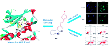
A novel series of C3-functionalized oxindoles, 3-(2-oxo-4-phenylbut-3-en-1-ylidene)indolin-2-ones, were designed, synthesized and investigated for inhibition of cell proliferation against different types of human cancer cell lines, including SW620, HeLa and A549. This biological study showed that these compounds containing the scaffolds of indole and aromatic α,β-unsaturated ketone had moderate to significant antitumor activities. Further study suggested that compound 4b, as one of this kind of structure derivative, showed broad-spectrum antitumor activities against MCF-7, PC-3, SKOV-3, U87, SMMC-7721, SY5Y and A875 cancer cell lines. Besides, the results of the inhibition of Pim kinases indicated that compound 4b showed selective and efficient anti-Pim-1 kinase activity (IC50 = 5 μM). Docking simulation, flow cytometry (FCM), and Hoechst 33342 staining assay suggested that the most active compound 4b induced cell death through apoptosis via binding to the active ATP pocket of Pim-1. Moreover, it showed that compound 4b had strong inhibition of tubulin polymerization which may be caused by inhibiting Pim-1.


Introduction

Pim-1, a member of the Pim kinases family (Pim-1, Pim-2 and Pim-3), is a serine/threonine protein kinase identified by cloning the retroviral integration sites in Moloney murine leukemia virus induced lymphomas. Pim-1 has been implicated to function in several normal biological process including cell cycle regulation, cell survival, proliferation and differentiation. It can phosphorylate Cdc25A and Cdc25C to enhance G1/S transition and G2/M transition, respectively.1,2 The phosphorylation of the pro-apoptotic protein Bad at Ser112 induced by Pim-1 blocks apoptotic cell death.3 Pim-1 also inactivates ASK1 through phosphorylation at Ser83 leading to less caspase3 activation and reducing cell death.4 Moreover, Pim-1 associates with tubulin and plays a vital role during mitosis.5 However, when these processes become disrupted or hyper-activated, Pim-1 expresses several hallmarks of cancer. Numerous evidences confirmed Pim-1 kinase as a proto-oncogene. Pim-1 was found over-expression in many of human tumor diseases, such as hematologic malignancies,6,7 bladder cancer,8 prostate cancer,9 gastric cancer.10 Thus, inhibition of Pim-1 is considered as a valuable target for therapy of cancer.11,12 It has attracted great interesting in developing novel inhibitors of Pim-1, and a large amount structurally different small molecule inhibitors have been obtained, such as SGI-1776,13 SMI-4a,14 DHPCC-9,15 K00486,16 CX-6258,17 Cpd 14 (ref. 18) (Fig. 1). Despite these successes, only a few of them have been tested in cell-based assays or the related mechanisms of Pim kinases function to demonstrate anti-cancer activity. Only understanding the processes that can be targeted by Pim inhibition is of great importance for defining the potential activity of these compounds. Therefore, designing novel Pim-1 inhibitors that targeted Pim and inhibited Pim kinases related function will still be a challenge for current anti-cancer drug discovery.
image file: c5ra00177c-f1.tif
Fig. 1 Small molecule inhibitors targeting Pim family kinases.

C3-functionalized oxindole is being continuously discovered to be at the core of several natural products, and becomes an emerging new scaffold for drug discovery with potential anti-cancer and other biological activities.19–22 Among the representative examples are spiro-tetrahydrofuryl oxindole as anti-cancer agent,23 spiro-isoxazolidinyl oxindole with anti-cancer and anti-tubercular activities,24–26 pyrrolidinooxindole-type alkaloid CPC-1,27 convolutamydine A with potent activity in the differentiation of HL-60 cells,28 celogentin K as potential inhibitor of tubulin polymerization,29 CX-6258 as Pim-1, 2, 3 inhibitor,17 maremycins with anti-cancer activity30 (Fig. 2). Another privileged skeletons, known chemically as chalcones, are biogenetic precursors of flavonoids in higher plants. These structures contain a ketone functionality and an unsaturated group (double bond), which are in conjugated arrangement among the naturally occurring or synthetic anti-cancer agents. The much flexible structure made it easier to combine with different protein receptors and exhibited good anti-canter activity (Fig. 2). Chalcone inhibited the proliferation of human breast cancer cell (MCF-7) by blocking cell cycle progression and inducing apoptosis.31 Licochalcone A had excellent activities against MCF-7 and HL-60.32 Isoliquiritigenin (ISL), isolated from Licorice, suppressed pulmonary metastasis of mouse renal cell carcinoma,33 induced apoptosis and cell cycle arrest through p53 dependent pathway in HepG2.34 The plant polyphenol butein, another nature product of derivative of chalcone, inhibited testosterone-induced proliferation in breast cancer cells expressing aromatase.35 These privileged structures provide us new idea for designing novel drugs.


image file: c5ra00177c-f2.tif
Fig. 2 Biologically important molecules containing C3-functionalized oxindoles or α,β-unsaturated ketones, and the design strategy of combinatorial chemistry for new compound.

In the past decades, plentiful efforts have been started for treating a wide range and types of cancer, and chemotherapy had become a common treatment option for cancer patients.36 One of well-known methods for primary anti-cancer drug discovery is combinatorial chemistry.37 As it's a part of our own interest to develop some C3-functionalized oxindoles,38 we conceived that combine C3-functionlized oxindoles with α,β-unsaturated ketones to obtain a novel series of compounds as potential Pim-1 inhibitors with great biological activities, as shown in Fig. 2. Based on our previous synthesis efforts,39 herein we report the efficient synthesis of 3-(2-oxo-4-phenylbut-3-en-1-ylidene)indolin-2-ones with efficient and broad-spectrum anti-tumor activities in vitro and described the structure–activity relationship (SAR). The most effective compound 4b was selected for further study. It showed selective and good inhibition against Pim-1 (IC50 = 5 μM). Docking simulation also suggested that Pim-1 is the potential target for compound 4b. Further study, including flow cytometry (FCM), Hoechst 33342 staining assay and immunohistochemistry of tubulin indicated that compound 4b caused cell arrest in G2/M phase, induced cell apoptosis and had significant inhibition on tubulin polymerization, which may be all associated with the inhibition of Pim-1.

Result and discussion

The synthetic strategy to prepare the target compounds is depicted in Scheme 1. The key intermediates 3a–y were prepared from benzylidene acetones and isatins as previously reported.39 Then, targeted compounds 4a–y were obtained, respectively, by the reaction with corresponding 3a–y in the presence of CCl4 and PPh3 refluxing in THF, in moderate to high (up to 96%) yields. All the products were characterized by 1H NMR, 13C NMR, 19F NMR and ESI-MS analysis.
image file: c5ra00177c-s1.tif
Scheme 1 Reagents and conditions: (a) CH3COCH3, H2O, 0.5% NaOH, r. t, overnight; (b) 10% L-Arg, MeOH, r. t, overnight; (c) CCl4, PPh3, THF, reflux, overnight.

All the newly synthesized compounds 4a–y were investigated for their anti-proliferative activity on three human cancer cell lines including SW620, HeLa, and A549 in vitro by MTT assay method. The half maximal inhibitory concentrations (IC50) of the tested compounds were listed in Table 1. These compounds exhibited good anti-tumor activities against SW620, and most of them had moderate to remarkable activities against A549. Their inhibitory effect on HeLa changed obviously with the different groups in the positions of R1, R2 and R3. The structure–activity relationships (SAR) could be observed mainly related to the influence of various substituents in R1, R2 and R3. The electronic properties and steric hindrance of the substituents at the aromatic ring had great influence on anti-tumor ability. The comparison of the activities of the pair of compounds 4b/4j and 4c/4h, the first of each pair component with a substituent at para-position (4b and 4c) and the second with the corresponding substituent at meta-position (4j) or ortho-position (4h), showed that the compounds with substituents at para-position had higher activities (4b/4j IC50 = 1.53 and 6.72 μM, 4c/4h IC50 = 2.20 and 2.96 μM, respectively). The activities of 4b, 4c and 4d were 1.53 μM, 2.20 μM, and >20 μM, suggesting that the size of substituent at para-position had a huge influence on the activity, the bigger the substituent was, the lower the activity would be, and even lost activity. The IC50 of compound 4g was 4.15 μM and 4d was >20 μM, meaning that the activity changed little with the size of substituent at meta-position changing. Compounds 4q–t and 4y lost activities but compound 4p had a good activity of 3.45 μM, indicating that R1 group must be aromatic ring and there should be no substituent at the nitrogen.

Table 1 In vitro activities of compounds 4a–y

image file: c5ra00177c-u1.tif

Compound R1 R2 R3 IC50a (μM)
SW620 HeLa A549
a The anti-proliferation activities of compounds 4a–y in vitro were determined by the MTT assay on Sw620, HeLa, A549 cell lines, IC50 was average of three determinations and deviation from the average was <5% of the average value.
4a Ph H H 0.19 3.18 5.03
4b 4-F–C6H4 H H 0.13 1.53 2.02
4c 4-Cl–C6H4 H H 1.68 2.20 3.86
4d 4-Br–C6H4 H H 1.97 >20 15.41
4e 4-CH(CH3)2–C6H4 H H 1.56 2.64 3.97
4f 4-MeO–C6H4 H H 1.98 8.46 17.32
4g 3-Br–C6H4 H H 1.34 3.15 7.31
4h 3-Cl–C6H4 H H 1.21 2.96 3.01
4i 3-CF3–C6H4 H H 0.57 6.13 3.15
4j 2-F–C6H4 H H 0.46 6.72 5.58
4k 3,4-DiCl–C6H4 H H 0.49 >20 8.86
4l 3,4-DiMe–C6H4 H H 0.76 2.43 10.21
4m 2,4-DiCl–C6H4 H H 0.54 >20 4.34
4n 2-F–4-Br–C6H4 H H 2.31 >20 >20
4o 2,3,4-TriMeO–C6H4 H H 0.91 >20 4.95
4p 2-Naphthyl H H 0.28 3.45 2.57
4q 2-Furyl H H 2.03 >20 3.85
4r 2-Thienyl H H 2.54 >20 4.92
4s Cyclohexyl H H 3.21 >20 10.22
4t CH(CH3)2 H H 3.87 >20 >20
4u Ph H 4-Cl 0.67 7.96 7.81
4v Ph H 6-Cl 0.35 3.92 4.56
4w Ph H 5-Br 3.91 7.52 >20
4x Ph H 5-Me 0.03 >20 3.14
4y Ph Me H 1.10 >20 5.50


To further study the anti-proliferative activity of this series of compounds, 4b, 4c and 4p were selected for evaluation of their inhibitory activities against a panel of different types of human cancer cell lines: A549, HeLa, MCF-7, PC-3, SKOV-3, U87, SMMC-7721, SY5Y, A875. As shown in Table 2, generally speaking, all these compounds had much stronger inhibition activities than the positive control cisplatin. Compound 4b (IC50 = 1.53 μM, 0.47 μM, 4.67 μM, 3.29 μM and 1.13 μM for HeLa, PC-3, U87, SMMC-7721 and SY5Y, respectively) showed the best anti-proliferative activities against the cancer cell lines of HeLa, PC-3, U87, SMMC-7721 and SY5Y. Meanwhile, compound 4p (IC50 = 3.21 μM, 4.02 μM, 2.2 μM and 0.22 μM against A549, MCF-7, skov-3 and A875, respectively) had most effective activities on A549, MCF-7, skov-3 and A875 cell lines. It indicated that these compounds had broad-spectrum anti-proliferative activity against human cancers.

Table 2 The anti-proliferation activities of compounds 4b,c, 4p and cisplatin against various cancer cell lines
Compound IC50a (μM)
A549 HeLa MCF-7 PC-3 SKOV-3 U87 SMMC-7721 SY5Y A875
a Values were average of three determinations and deviation from the average is <5% of the average value.b Positive control.
4b 3.53 1.53 4.86 0.47 2.39 4.67 3.29 1.13 1.03
4c 4.74 2.2 10.93 3.28 3.22 9.94 6.84 1.74 1.78
4p 3.21 3.45 4.02 2.47 2.2 9.08 4.32 1.16 0.22
Cisplatinb 10.05 5.02 >20 10 5.25 11.08 6.35 1.71 4.15


At the beginning of design these compounds, our project was to develop new Pim-1 inhibitors as anti-tumor agents. Therefore we performed kinase assay to examine whether the compounds (4b, 4c, 4p) interact with Pim kinases and inhibit Pim kinases, in vitro. Compound 4b (IC50 = 5 μM) showed more effective than compound 4c (IC50 = 7 μM) and 4p (IC50 = 10 μM) against Pim-1. In addition, the IC50 of compound 4b against Pim-1, -2, and -3 were 5 μM, >20 μM and 15 μM, respectively. It suggested that compound 4b had selective and good inhibition against Pim-1.

Based on these results, compound 4b was selected as a lead compound for further study because of its best potency against most of the cancer cells and selective inhibition of Pim-1, in vitro. For better understanding of the potency of compound 4b and guiding further SAR studies, we evaluated the interaction between compound 4b and Pim kinases (Pim-1, Pim-2 and Pim-3). The compound 4b bound to the active site of ATP and the binding modes of compound 4b to Pim kinases were exhibited in Fig. 3. Firstly, we used molecular docking to analyze the binding mode of compound 4b with Pim-1. A total of 20 docking poses were generated by GOLD program. According to the RMSD values, two docking pose clusters (cluster I and cluster II) were obtained. Cluster I contained 16 docking poses, while cluster II only had 4 docking poses. Therefore, we considered that the docking poses in cluster I might be the true binding modes of compound 4b with Pim-1. Fig. 3a depicted the binding pose of compound 4b with the highest Goldscore value of 48.231. The amide group of compound 4b was observed to form strong hydrogen-bonding interactions with Lys67 and Glu89. The fluorine atom of compound 4b formed halogen-bonding interaction with Leu44. In addition, compound 4b formed intensive hydrophobic interactions with residues Leu93, Leu120, Leu44, Val52, Lys67 and Ile185. The interaction mode between compound 4b and Pim-1 was corresponded to the SAR. Then, we analyzed the binding modes of compound 4b with Pim-2 and Pim-3. The results showed that the compound 4b did not perfectly match with Pim-2 and Pim-3, comparing with Pim-1 (see Fig. 3b and c). Goldscore for the compound 4b binding with Pim-2 and Pim-3 were 45.241 and 41.398, respectively, which slightly lower than that with Pim-1. These results may be used for explaining why the inhibitory activity of compound 4b against Pim-1 was better than Pim-2 and Pim-3. Finally, we also further predicted the other possible kinase targets for compound 4b using a reverse molecule docking method.40 The top 10 kinases ranked by the reverse molecule docking method were AKT-1, BCR-ABL, CLK3, KIT, CK2, MAPK14, PEPCK, ALK5, CaMKK2, TGFR1, respectively. These kinases may be the targets of compound 4b.


image file: c5ra00177c-f3.tif
Fig. 3 Molecular docking compound 4b with Pim kinases (a) binding mode of compound 4b (represented with stick) to the ATP binding pocket of Pim-1 (PDB ID: 1XWS). (b) Binding mode of compound 4b (represented with stick) to the ATP binding pocket of Pim-2. (c) Binding mode of compound 4b (represented with stick) to the ATP binding pocket of Pim-3.

Next, compound 4b was used to evaluate other biological functions associated with Pim-1. Flow cytometric analysis was used to quantitatively assess and the cell cycle after PI-stained. As shown in Fig. 4a significant accumulation of cells in the S phase, accompanied by a decrease in G0/G1 phase was observed after treatment with compound 4b in HeLa cancer cells at 1 μM. However a large amount of tetraploid and polyploidies were appeared in contrast with a sharply decreasing cells in G0/G1 phase and S phase from 2 μM to 3 μM. The results suggested that compound 4b arrested the cell divided during the DNA replicating. In addition, fluorescence microscopic examination of Hoechst 33342 stained cell confirmed the inducing apoptosis effect of compound 4b. The apoptotic cells are often characterized by its morphological, including cell shrinkage, membrane blabbing, chromatin condensation and DNA fragmentation. After treated with compound 4b for 48 hours, HeLa cells shrank and rounded up. With the increasing of concentration, it became more significant. In contrast, the untreated HeLa cells retained their normal size and shape, kept proliferating with time and became over-crowded by 36 hours (Fig. 5A). Morphological changes were also characteristic of apoptosis after Hoechst 33342 staining. Apoptosis cells, bright blue fluorescent and condensed nuclei, were observed in a large number of treated cells in HeLa, and the change was concentration-dependent, whereas there was no significant apoptosis in untreated HeLa cells that showed blue, diffusely stained intact nuclei (Fig. 5B). These results suggested that compound 4b induced apoptosis in HeLa cells.


image file: c5ra00177c-f4.tif
Fig. 4 Effect of compound 4b on cell cycle distribution: DNA fluorescence histograms of PI-stained HeLa cells. Cells were treated with 1 μM, 2 μM, 3 μM for 48 h.

image file: c5ra00177c-f5.tif
Fig. 5 Effect of compound 4b on cell morphology: (A) bright-field microscopy images and (B) fluorescence microscopic appearance of Hoechst 33342 staining nuclei of HeLa cells, after incubation with compound 4b for 48 h at varying concentrations: 2 μM, 4 μM, 8 μM. Apoptosis cells were observed in treated cells containing condensed and fragmented fluorescent nuclei.

To further test the apoptosis inducing ability of 4b, an Annexin-V/PI fluorescence staining was analyzed by flow cytometry in HeLa cells. As shown in Fig. 6, HeLa cells were treated with 4b at the concentrations of 0, 2, 4 and 8 μM for 48 h. We found an obvious concentration-dependent reduction in the percentage of surviving cells. As the concentration of compound 4b increased, the early and the late stage apoptotic cells increased from 9.04% at 2 μM to 62.31% at 8 μM. This result indicated that compound 4b induced apoptosis of HeLa cells in a dose-dependent manner.


image file: c5ra00177c-f6.tif
Fig. 6 Compound 4b induces apoptosis in HeLa cells. Flow cytometric analysis of cells stained with Annexin V-FITC/PI after treatment with various concentrations (0 μM, 2 μM, 4 μM and 8 μM) of compound 4b for 48 hours.

We also validated the effect of compound 4b on cellular tubulin immunohistochemistry. Studies were carried out to examine the in situ effects of compound 4b on cellular microtubules in HeLa cells. HeLa cells seeded on sterile cover slips were treated with compound 4b at 2 μM for 48 h. As shown in Fig. 7, untreated HeLa cells displayed the normal distribution of microtubules. However, cells treated with compound 4b showed disrupted microtubule organization, thus demonstrated the inhibition of tubulin polymerization, influenced the mitosis and affected the cell division in an orderly and programmed manner. This process may be all associated with the inhibition of Pim-1. In dividing cells, Pim-1 is located in the mitotic spindle and specifically the spindle poles, which plays the key role of influencing the function of mitotic spindle.5 When Pim-1 was inhibited by compound 4b, the function of spindle was disturbed and then the microtubule constituted spindle fibers was disorder just as observed in Fig. 7. Finally the cells were arrested in G2/M phase and apoptosis. The result also explained the appearance of polyploidy in flow cytometric analysis.


image file: c5ra00177c-f7.tif
Fig. 7 HeLa cells were treated with compounds 4b with 8 μM for 24 h followed by staining with α-tubulin antibody. Chromosomal DNA (blue) was stained with DAPI. Microtubule organization was clearly observed by green tubulin network-like structures in control cells, and was found to be disrupted in cells treated with compound 4b.

Experimental

Materials and measurements

Melting points were recorded at SGW X-4 Melting point instrument (Shanghai precision & scientific instrument Co., Ltd, Shanghai, China). 1H NMR spectra were recorded at 400 MHz. The chemical shifts were recorded in ppm relative to tetramethylsilane and with the solvent resonance as the internal standard. Data were reported as follows: chemical shift, multiplicity (s = singlet, d = doublet, t = triplet, q = quartet, m = multiplet), coupling constants (Hz), integration. 13C NMR data were collected at 100 MHz with complete proton decoupling. Chemical shifts were reported in ppm from the tetramethylsilane with the solvent resonance as internal standard. 19F NMR data were collected at 376 MHz with complete proton decoupling. ESI-HRMS spectra were recorded on a commercial apparatus and methanol was used to dissolve the sample. All chemicals were obtained from commercial sources and used without further purification. Column chromatography was carried out on silica gel (300–400 mesh, Qingdao Marine Chemical Ltd, Qingdao, China). Thin layer chromatography (TLC) was performed on TLC silica gel 60 F254 plates.

General procedure for the synthesis of benzylidene acetones (2a–t)

To a stirred solution of 1a–t (10 mmol) in 2 mL acetone and 1 mL H2O was added 0.25 mL 10% NaOH aqua and stirred at room temperature for 12 hours. Then the solvent was removed under reduced pressure and the residue was extracted with acetic ether. The combined organic layers were dried (MgSO4), filtered and the filtrate was concentrated under reduced pressure. The crude product was used for next step.

General procedure for the synthesis of 3-hydroxy-3-(2-oxo-4-phenylbut-3-en-1-yl)indolin-2-ones (3a–y)

A mixture of isatin (0.1 mmol), (E)-4-phenylbut-3-en-2-one (2a, 0.1 mmol), arginine (0.01 mmol) in MeOH (1.0 mL) was stirred for 48 hours at 25 °C. After completion of the reaction (TLC), the solvent was removed under vacuum. The crude product was subjected to column chromatography on silica gel using petroleum ether/ethyl acetate = 1[thin space (1/6-em)]:[thin space (1/6-em)]1 as the eluent to give 3a.

Compounds 3b–y were synthesized by a similar procedure as described for compound 3a. For the separation of these compounds, the eluent of silica gel column chromatography consisted of appropriate mixtures of petroleum ether and ethyl acetate.

General procedure for the synthesis of 3-(2-oxo-4-phenylbut-3-en-1-ylidene)indolin-2-ones (4a–y)

Compound 3a–y (0.2 mmol) was dissolved in anhydrous THF (4 mL). Ph3P (78.7 mg, 0.3 mmol) and CCl4 (0.1 mL) were then added. The solution was heated to reflux for 12 hours. After completion of the reaction (TLC), the solvent was removed under vacuum. The crude product was subjected to column chromatography on silica gel using petroleum ether/ethyl acetate = 2[thin space (1/6-em)]:[thin space (1/6-em)]1 as the eluent to give 4a–y.

(Z)-3-((E)-2-Oxo-4-phenylbut-3-en-1-ylidene)indolin-2-one (4a)

Red solid, yield 89%, m. p. 176–177 °C; 1H-NMR (400 MHz, DMSO) δ 10.78 (s, 1H), 8.29 (d, J = 8.0 Hz, 1H), 7.74 (dd, J = 10.0, 6.0 Hz, 3H), 7.44–7.38 (m, 4H), 7.32 (dd, J = 21.2, 12.0 Hz, 2H), 6.97 (t, J = 7.6 Hz, 1H), 6.88 (d, J = 7.6 Hz, 1H); 13C-NMR (100 MHz, DMSO) δ 190.1, 168.6, 144.8, 144.6, 136.0, 134.1, 133.1, 131.1, 129.1, 128.8, 127.80, 127.5, 127.2, 121.8, 120.1, 110.3; ESI-MS: m/z 274.1 [M − H].

(Z)-3-((E)-4-(4-Fluorophenyl)-2-oxobut-3-en-1-ylidene)indolin-2-one (4b)

Red solid, yield 96%, m. p. 204–205 °C; 1H-NMR (400 MHz, DMSO) δ 10.77 (s, 1H), 8.36 (d, J = 8.0 Hz, 1H), 7.90 (dd, J = 8.4, 6.0 Hz, 2H), 7.79 (d, J = 16.0 Hz, 1H), 7.42–7.26 (m, 5H), 6.99 (t, J = 8.0 Hz, 1H), 6.88 (d, J = 8.0 Hz, 1H); 13C-NMR (100 MHz, DMSO) δ 189.9, 168.5, 163.6 (d, J = 248 Hz), 145.0, 143.2, 136.2, 133.1, 131.3 (d, J = 8.6 Hz), 131.0 (d, J = 2.9 Hz), 127.8, 127.7 (d, J = 2.3 Hz), 127.4, 121.7, 120.3, 116.1 (d, J = 21.8 Hz), 110.3; 19F-NMR (376 MHz, DMSO) δ −108.9; ESI-MS: m/z 292.1 [M − H].

(Z)-3-((E)-4-(4-Chlorophenyl)-2-oxobut-3-en-1-ylidene)indolin-2-one (4c)

Red solid, yield 93%, m. p. 215–216 °C; 1H-NMR (400 MHz, DMSO) δ 10.78 (s, 1H), 8.38 (d, J = 7.6 Hz, 1H), 7.86 (d, J = 8.4 Hz, 2H), 7.78 (d, J = 16.0 Hz, 1H), 7.53 (d, J = 8.4 Hz, 2H), 7.45–7.35 (m, 3H), 7.00 (t, J = 7.6 Hz, 1H), 6.88 (d, J = 7.6 Hz, 1H); 13C-NMR (100 MHz, DMSO) δ 189.8, 168.4, 145.1, 142.9, 136.3, 135.5, 133.3, 133.2, 130.6, 129.1, 128.4, 127.6, 127.4, 121.7, 120.2, 110.3; ESI-MS: m/z 308.2 [M − H].

(Z)-3-((E)-4-(4-Bromophenyl)-2-oxobut-3-en-1-ylidene)indolin-2-one (4d)

Red solid, yield 92%, m. p. 222–223 °C; 1H-NMR (400 MHz, DMSO) δ 10.78 (s, 1H), 8.38 (d, J = 7.6 Hz, 1H), 7.78 (dd, J = 12.4, 8.0 Hz, 3H), 7.67 (d, J = 8.4 Hz, 2H), 7.48–7.33 (m, 3H), 7.00 (t, J = 7.6 Hz, 1H), 6.88 (d, J = 7.6 Hz, 1H); 13C-NMR (100 MHz, DMSO) δ 189.81, 168.4, 145.1, 143.0, 136.3, 133.6, 133.2, 132.0, 130.8, 128.4, 127.6, 127.4, 124.4, 121.7, 120.2, 110.3; ESI-MS: m/z 352.2 [M − H].

(Z)-3-((E)-4-(4-Isopropylphenyl)-2-oxobut-3-en-1-ylidene)indolin-2-one (4e)

Red solid, yield 91%, m. p. 184–185 °C; 1H-NMR (400 MHz, DMSO) δ 10.77 (s, 1H), 8.37 (d, J = 7.6 Hz, 1H), 7.76 (dd, J = 12.0, 10.4 Hz, 3H), 7.42 (s, 1H), 7.39–7.27 (m, 4H), 6.99 (t, J = 7.6 Hz, 1H), 6.89 (d, J = 7.6 Hz, 1H), 2.92 (m, 1H), 1.22 (d, J = 6.8 Hz, 6H); 13C-NMR (100 MHz, DMSO) δ 189.9, 168.5, 151.9, 145.0, 144.5, 136.0, 133.0, 132.0, 129.1, 127.9, 127.3, 127.0, 126.9, 121.7, 120.3, 110.2, 33.4, 23.5; ESI-MS: m/z 316.1 [M − H].

(Z)-3-((E)-4-(4-Methoxyphenyl)-2-oxobut-3-en-1-ylidene)indolin-2-one (4f)

Red solid, yield 65%, m. p. 180–181 °C; 1H-NMR (400 MHz, DMSO) δ 10.76 (s, 1H), 8.36 (d, J = 7.6 Hz, 1H), 7.84–7.72 (m, 3H), 7.40–7.33 (m, 2H), 7.27 (d, J = 16.0 Hz, 1H), 7.06–6.95 (m, 3H), 6.88 (d, J = 7.6 Hz, 1H), 3.83 (s, 3H); 13C-NMR (100 MHz, DMSO) δ 189.7, 168.5, 161.7, 144.9, 144.6, 135.7, 132.9, 130.9, 128.2, 127.3, 126.8, 125.4, 121.7, 120.3, 114.6, 110.2, 55.4; ESI-MS: m/z 304.0 [M − H].

(Z)-3-((E)-4-(3-Bromophenyl)-2-oxobut-3-en-1-ylidene)indolin-2-one (4g)

Red solid, yield 84%, m. p. 192–193 °C; 1H-NMR (400 MHz, DMSO) δ 10.78 (s, 1H), 8.39 (d, J = 7.6 Hz, 1H), 8.10 (s, 1H), 7.83 (d, J = 7.6 Hz, 1H), 7.76 (d, J = 16.0 Hz, 1H), 7.65 (dd, J = 8.0, 0.8 Hz, 1H), 7.50 (d, J = 16.0 Hz, 1H), 7.46–7.34 (m, 3H), 7.00 (t, J = 7.6 Hz, 1H), 6.88 (d, J = 7.6 Hz, 1H); 13C-NMR (100 MHz, DMSO) δ 189.8, 168.4, 145.1, 142.5, 136.9, 136.4, 133.33, 133.2, 131.2, 131.0, 129.1, 127.9, 127.6, 127.4, 122.4, 121.7, 120.2, 110.3. ESI-MS: m/z 352.0 [M − H].

(Z)-3-((E)-4-(3-Chlorophenyl)-2-oxobut-3-en-1-ylidene)indolin-2-one (4h)

Red solid, yield 83%, m. p. 192–193 °C; 1H-NMR (400 MHz, DMSO) δ 10.77 (s, 1H), 8.39 (d, J = 7.6 Hz, 1H), 7.95 (s, 1H), 7.78 (dd, J = 11.6, 8.4 Hz, 2H), 7.58–7.44 (m, 3H), 7.44–7.30 (m, 2H), 7.00 (t, J = 7.6 Hz, 1H), 6.89 (d, J = 7.6 Hz, 1H); 13C-NMR (100 MHz, DMSO) δ 189.8, 168.4, 145.1, 142.5, 136.6, 136.4, 133.8, 133.2, 130.8, 130.4, 129.1, 128.3, 127.5, 127.5, 127.4, 121.7, 120.2, 110.3; ESI-MS: m/z 308.1 [M − H].

(Z)-3-((E)-2-Oxo-4-(3-(trifluoromethyl)phenyl)but-3-en-1-ylidene)indolin-2-one (4i)

Red solid, yield 79%, m. p. 178–179 °C; 1H-NMR (400 MHz, DMSO) δ 10.79 (s, 1H), 8.32 (d, J = 7.6 Hz, 1H), 8.15 (s, 1H), 8.06 (d, J = 7.6 Hz, 1H), 7.84–7.76 (m, 2H), 7.66 (t, J = 7.6 Hz, 1H), 7.48 (d, J = 16.0 Hz, 1H), 7.41–7.29 (m, 2H), 6.97 (dd, J = 11.2, 4.4 Hz, 1H), 6.88 (d, J = 7.6 Hz, 1H); 13C-NMR (100 MHz, DMSO) δ 190.0, 168.6, 144.9, 142.5, 136.4, 135.3, 133.3, 132.4, 130.1, 130.0, 129.3, 127.5, 127.3, 127.1 (q, J = 6.3 Hz), 125.3 (q, J = 7.4 Hz), 121.9, 120.1, 110.4, 48.6; 19F-NMR (376 MHz, DMSO) δ −61.2; ESI-MS: m/z 342.1 [M − H].

(Z)-3-((E)-4-(2-Fluorophenyl)-2-oxobut-3-en-1-ylidene)indolin-2-one (4j)

Red solid, yield 94%, m. p. 186–187 °C; 1H-NMR (400 MHz, DMSO) δ 10.79 (s, 1H), 8.32 (d, J = 7.6 Hz, 1H), 7.88 (t, J = 7.6 Hz, 1H), 7.76 (d, J = 16.0 Hz, 1H), 7.59–7.17 (m, 6H), 6.97 (t, J = 7.6 Hz, 1H), 6.87 (d, J = 7.6 Hz, 1H); 13C-NMR (100 MHz, DMSO) δ 189.7, 168.6, 160.9 (d, J = 250.6 Hz), 144.9, 136.4, 135.8 (d, J = 3.3 Hz), 133.3, 133.1 (d, J = 9.0 Hz), 129.4 (d, J = 4.8 Hz), 129.2 (d, J = 1.7 Hz), 127.5, 127.4, 125.1 (d, J = 3.1 Hz), 121.9, 121.8 (d, J = 11.2 Hz), 120.0, 116.1 (d, J = 21.4 Hz), 110.4; 19F-NMR (376 MHz, DMSO) δ −115.4; ESI-MS: m/z 292.1 [M − H].

(Z)-3-((E)-4-(3,4-Dichlorophenyl)-2-oxobut-3-en-1-ylidene)indolin-2-one (4k)

Red solid, yield 77%, m. p. 221–222 °C; 1H-NMR (400 MHz, DMSO) δ 10.79 (s, 1H), 8.39 (d, J = 7.6 Hz, 1H), 8.17 (d, J = 2.0 Hz, 1H), 7.83 (dd, J = 8.4, 2.0 Hz, 1H), 7.75 (t, J = 12.4 Hz, 2H), 7.52 (d, J = 16.0 Hz, 1H), 7.43–7.34 (m, 2H), 7.00 (t, J = 7.6 Hz, 1H), 6.89 (d, J = 7.6 Hz, 1H); 13C-NMR (100 MHz, DMSO) δ 189.7, 168.4, 145.1, 141.4, 136.5, 135.3, 133.3, 133.1, 131.8, 131.1, 130.5, 129.6, 128.8, 127.4, 127.4, 121.7, 120.2, 110.3; ESI-MS: m/z 342.1 [M − H].

(Z)-3-((E)-4-(3,4-Dimethylphenyl)-2-oxobut-3-en-1-ylidene)indolin-2-one (4l)

Red solid, yield 74%, m. p. 198–199 °C; 1H-NMR (400 MHz, DMSO) δ 10.76 (s, 1H), 8.36 (d, J = 7.6 Hz, 1H), 7.72 (d, J = 16.0 Hz, 1H), 7.61 (s, 1H), 7.53 (d, J = 7.6 Hz, 1H), 7.44–7.28 (m, 3H), 7.22 (d, J = 7.6 Hz, 1H), 6.99 (t, J = 7.6 Hz, 1H), 6.88 (d, J = 7.6 Hz, 1H), 2.26 (s, 6H); 13C-NMR (100 MHz, DMSO) δ 189.7, 168.4, 144.9, 144.7, 140.1, 136.9, 135.9, 132.9, 131.9, 130.1, 129.8, 127.9, 127.3, 126.7, 126.6, 121.7, 120.3, 110.2, 19.5, 19.2; ESI-MS: m/z 302.1 [M − H].

(Z)-3-((E)-4-(2,4-Dichlorophenyl)-2-oxobut-3-en-1-ylidene)indolin-2-one (4m)

Red solid, yield 78%, m. p. 214–215 °C; 1H-NMR (400 MHz, DMSO) δ 10.80 (s, 1H), 8.45 (d, J = 7.6 Hz, 1H), 8.09 (d, J = 8.4 Hz, 1H), 7.93 (d, J = 16.0 Hz, 1H), 7.79 (d, J = 2.0 Hz, 1H), 7.66–7.51 (m, 2H), 7.43–7.33 (m, 2H), 7.01 (t, J = 7.6 Hz, 1H), 6.89 (d, J = 7.6 Hz, 1H); 13C-NMR (100 MHz, DMSO) δ 189.4, 168.4, 145.2, 136.7, 134.4, 133.4, 130.6, 130.6, 130.2, 130.1, 128.4, 128.3, 127.5, 127.4, 121.8, 120.2, 119.8, 110.3; ESI-MS: m/z 342.0 [M − H].

(Z)-3-((E)-4-(4-Bromo-2-fluorophenyl)-2-oxobut-3-en-1-ylidene)indolin-2-one (4n)

Red solid, yield 75%, m. p. 201–202 °C; 1H-NMR (400 MHz, DMSO) δ 10.79 (s, 1H), 8.41 (d, J = 7.6 Hz, 1H), 7.93 (t, J = 8.0 Hz, 1H), 7.72 (dd, J = 12.4, 7.6 Hz, 2H), 7.53 (t, J = 12.8 Hz, 2H), 7.44–7.31 (m, 2H), 7.00 (t, J = 7.6 Hz, 1H), 6.88 (d, J = 7.6 Hz, 1H); 13C-NMR (100 MHz, DMSO) δ 189.4, 168.4, 160.6 (d, J = 254.8 Hz), 145.2, 136.7, 134.4 (d, J = 2.5 Hz), 133.4, 130.6 (d, J = 2.6 Hz), 130.2 (d, J = 4.7 Hz), 128.4 (d, J = 3.0 Hz), 127.5, 127.4, 124.7 (d, J = 10.0 Hz) 121.8, 121.5 (d, J = 11.4 Hz), 120.2, 119.7 (d, J = 25.5 Hz), 110.3; 19F-NMR (376 MHz, DMSO) δ −112.4; ESI-MS: m/z 370.1 [M − H].

(Z)-3-((E)-2-Oxo-4-(2,3,4-trimethoxyphenyl)but-3-en-1-ylidene)indolin-2-one (4o)

Red solid, yield 75%, m. p. 178–179 °C; 1H-NMR (400 MHz, DMSO) δ 10.78 (s, 1H), 8.31 (d, J = 7.6 Hz, 1H), 7.84 (d, J = 16.0 Hz, 1H), 7.60 (d, J = 8.8 Hz, 1H), 7.40–7.29 (m, 2H), 7.25 (d, J = 16.0 Hz, 1H), 6.98 (t, J = 7.6 Hz, 1H), 6.93–6.82 (m, 2H), 3.84 (s, 3H), 3.82 (s, 3H), 3.80 (s, 3H); 13C-NMR (100 MHz, DMSO) δ 189.9, 168.7, 156.2, 153.2, 144.6, 141.7, 139.1, 135.7, 133.0, 128.4, 127.3, 125.9, 123.7, 121.9, 120.4, 120.1, 110.3, 108.6, 61.5, 60.5, 56.0; ESI-MS: m/z 364.1 [M − H].

(Z)-3-((E)-4-(Naphthalen-2-yl)-2-oxobut-3-en-1-ylidene)indolin-2-one (4p)

Red solid, yield 93%, m. p. 199–200 °C; 1H-NMR (400 MHz, DMSO) δ 10.80 (s, 1H), 8.33 (d, J = 7.6 Hz, 1H), 8.27 (s, 1H), 7.97–7.89 (m, 5H), 7.64–7.51 (m, 2H), 7.45 (d, J = 13.6 Hz, 2H), 7.35 (t, J = 7.6 Hz, 1H), 6.99 (t, J = 7.6 Hz, 1H), 6.89 (d, J = 7.6 Hz, 1H); 13C-NMR (100 MHz, DMSO) δ 190.0, 168.6, 144.8, 144.7, 136.0, 134.0, 133.1, 132.8, 131.8, 131.2, 128.7, 128.6, 127.9, 127.8, 127.7, 127.4, 127.3, 126.9, 123.9, 121.9, 120.1, 110.4; ESI-MS: m/z 324.1 [M − H].

(Z)-3-((E)-4-(Furan-2-yl)-2-oxobut-3-en-1-ylidene)indolin-2-one (4q)

Red solid, yield 88%, m. p. 184–185 °C; 1H-NMR (400 MHz, DMSO) δ 10.76 (s, 1H), 8.29 (d, J = 6.0 Hz, 1H), 7.87 (s, 1H), 7.57 (d, J = 15.6 Hz, 1H), 7.32 (d, J = 6.8 Hz, 2H), 7.12–6.82 (m, 4H), 6.66 (s, 1H); 13C-NMR (100 MHz, DMSO) δ 189.4, 168.6, 150.5, 146.8, 144.7, 135.9, 133.1, 130.9, 127.7, 127.3, 124.2, 121.8, 120.1, 118.3, 113.4, 110.3; ESI-MS: m/z 264.1[M − H].

(Z)-3-((E)-2-Oxo-4-(thiophen-2-yl)but-3-en-1-ylidene)indolin-2-one (4r)

Red solid, yield 87%, m. p. 177–178 °C; 1H-NMR (400 MHz, DMSO) δ 10.75 (s, 1H), 8.36 (d, J = 7.6 Hz, 1H), 7.98 (d, J = 15.6 Hz, 1H), 7.83 (d, J = 5.2 Hz, 1H), 7.68 (d, J = 3.6 Hz, 1H), 7.41–7.31 (m, 2H), 7.21 (dd, J = 4.8, 3.6 Hz, 1H), 7.04 (d, J = 15.8 Hz, 1H), 6.99 (t, J = 7.6 Hz, 1H), 6.88 (d, J = 7.6 Hz, 1H); 13C-NMR (100 MHz, DMSO) δ 189.2, 168.5, 144.9, 139.4, 137.2, 135.9, 133.6, 133.0, 131.3, 128.9, 127.8, 127.4, 126.2, 121.7, 120.3, 110.2; ESI-MS: m/z 280.0 [M − H].

(Z)-3-((E)-4-Cyclohexyl-2-oxobut-3-en-1-ylidene)indolin-2-one (4s)

Red solid, yield 73%, m. p. 196–197 °C; 1H-NMR (400 MHz, DMSO) δ 11.21 (s, 1H), 7.55 (d, J = 7.6 Hz, 1H), 7.40 (s, 1H), 7.20 (t, J = 7.6 Hz, 1H), 7.03 (t, J = 7.6 Hz, 1H), 6.89 (d, J = 7.6 Hz, 1H), 6.27 (d, J = 12.0 Hz, 1H), 6.20 (d, J = 12.0 Hz, 1H), 2.31–2.21 (m, 1H), 1.65–1.47 (m, 4H), 1.36–1.07 (m, 6H); 13C-NMR (100 MHz, DMSO) δ 189.9, 170.2, 150.7, 147.6, 140.0, 135.4, 128.3, 124.3, 122.5, 119.4, 118.1, 110.2, 37.4, 29.2, 28.38, 27.6, 26.8, 26.1; ESI-MS: m/z 280.1 [M − H].

(Z)-3-((E)-5-Methyl-2-oxohex-3-en-1-ylidene)indolin-2-one (4t)

Red solid, yield 69%, m. p. 180–181 °C; 1H-NMR (400 MHz, DMSO) δ 11.20 (s, 1H), 7.57 (d, J = 7.6 Hz, 1H), 7.40 (s, 1H), 7.19 (t, J = 7.6 Hz, 1H), 7.03 (t, J = 7.6 Hz, 1H), 6.89 (d, J = 7.6 Hz, 1H), 6.32 (d, J = 10.8 Hz, 1H), 6.14 (d, J = 12.0 Hz, 1H), 2.50 (m, 1H), 1.89 (s, 3H), 1.84 (s, 3H); 13C-NMR (100 MHz, DMSO) δ 170.2, 150.3, 140.0, 139.5, 135.4, 128.3, 124.3, 122.63, 121.1, 119.5, 118.8, 110.2, 59.7, 26.6, 18.8; ESI-MS: m/z 240.2 [M − H].

(Z)-4-Chloro-3-((E)-2-oxo-4-phenylbut-3-en-1-ylidene)indolin-2-one (4u)

Yellow solid, yield 95%, m. p. 217–218 °C; 1H-NMR (400 MHz, DMSO) δ 10.90 (s, 1H), 7.80–7.69 (m, 3H), 7.57 (d, J = 16.4 Hz, 1H), 7.43 (dd, J = 5.2, 1.6 Hz, 3H), 7.32 (t, J = 8.0 Hz, 1H), 7.07 (t, J = 12.4 Hz, 2H), 6.86 (d, J = 7.6 Hz, 1H); 13C-NMR (100 MHz, DMSO) δ 194.9, 165.7, 144.7, 144.2, 136.2, 134.3, 131.7, 130.7, 130.2, 129.2, 128.9, 128.6, 126.8, 122.7, 117.9, 109.0; ESI-MS: m/z 308.2 [M − H].

(Z)-6-Chloro-3-((E)-2-oxo-4-phenylbut-3-en-1-ylidene)indolin-2-one (4v)

Red solid, yield 94%, m. p. 229–230 °C; 1H-NMR (400 MHz, DMSO) δ 11.01 (s, 1H), 8.41 (d, J = 8.4 Hz, 1H), 7.83–7.78 (m, 3H), 7.51–7.36 (m, 5H), 7.07 (dd, J = 8.4, 2.0 Hz, 1H), 6.91 (t, J = 5.6 Hz, 1H); 13C-NMR (100 MHz, DMSO) δ 189.7, 168.5, 146.4, 144.6, 137.1, 135.1, 134.2, 131.1, 129.0, 128.9, 128.8, 128.2, 127.7, 121.5, 119.1, 110.3; ESI-MS: m/z 308.1 [M − H].

(Z)-5-Bromo-3-((E)-2-oxo-4-phenylbut-3-en-1-ylidene)indolin-2-one (4w)

Red solid, yield 91%, m. p. 193–194 °C; 1H-NMR (400 MHz, DMSO) δ 10.93 (s, 1H), 8.61 (d, J = 1.6 Hz, 1H), 7.94–7.74 (m, 3H), 7.55 (dd, J = 8.4, 2.0 Hz, 1H), 7.49–7.43 (m, 5H), 6.86 (d, J = 8.4 Hz, 1H); 13C-NMR (100 MHz, DMSO) δ 189.7, 168.0, 144.9, 144.2, 135.3, 135.3, 134.3, 131.1, 129.7, 129.2, 129.0, 128.9, 127.6, 122.1, 113.2, 112.2; ESI-MS: m/z 352.1 [M − H].

(Z)-5-Methyl-3-((E)-2-oxo-4-phenylbut-3-en-1-ylidene)indolin-2-one (4x)

Red solid, yield 81%, m. p. 169–170 °C; 1H-NMR (400 MHz, DMSO) δ 10.67 (s, 1H), 8.24 (s, 1H), 7.86–7.77 (m, 3H), 7.50–7.45 (m, 3H), 7.41 (d, J = 14.8 Hz, 2H), 7.18 (dd, J = 8.0, 0.8 Hz, 1H), 6.78 (d, J = 8.0 Hz, 1H), 2.27 (s, 3H); 13C-NMR (100 MHz, DMSO) δ 189.8, 168.5, 144.3, 142.8, 136.5, 134.3, 133.5, 130.9, 130.4, 129.0, 128.9, 127.8, 127.7, 127.6, 120.3, 109.9, 20.7; ESI-MS: m/z 288.1 [M − H].

(Z)-1-Methyl-3-((E)-2-oxo-4-phenylbut-3-en-1-ylidene)indolin-2-one (4y)

Red solid, yield 90%, m. p. 114–115 °C; 1H-NMR (400 MHz, DMSO) δ 8.30 (d, J = 7.6 Hz, 1H), 7.81–7.72 (m, 3H), 7.49–7.40 (m, 5H), 7.32 (d, J = 16.0 Hz, 1H), 7.03 (dd, J = 14.8, 7.6 Hz, 2H), 3.16 (s, 4H); 13C-NMR (100 MHz, DMSO) δ 190.1, 167.2, 145.8, 144.9, 135.0, 134.1, 133.0, 131.2, 129.1, 128.9, 128.4, 127.4, 126.8, 122.4, 119.4, 109.1, 26.1; ESI-MS: m/z 290.3 [M + H]+.

Anti-tumor assay

Briefly, targeted cancer cells lines (3 × 10³ per well) were seeded in 96-well plates and cultured for 24 h, followed by compounds treatment for 48 h. A volume of 20 μL of 5 mg mL−1 MTT was added per well and incubated for another 2 h at 37 °C, then the supernatant fluid was removed and DMSO was added 150 μL per well for 15–20 min. The absorptions (OD) were measured at 570 nm with Spectra MAX M5 microplate spectrophotometer (Molecular Devices). The effect of compounds on tumor cells viability was expressed by IC50 of each cell line. Each assay was carried out at least three times. The results are summarized in Tables 1 and 2.

Docking simulation

The molecular docking method was used for the analysis of compound 4b binding with Pim-1, Pim-2 and Pim-3. The GOLD docking program (version 5.0) was adopted here. The X-ray crystal structure of Pim-1 (PDB ID: 1XWS) was taken from PDB database, and the 3D structure of Pim-2 and Pim-3 were built using the homology modelling module in Discovery Studio 3.1. The preparation of protein structure, including adding hydrogen atoms, removing water molecules, and assigning Charmm force field, were carried out by using Discovery Studio 3.1 software package. A sphere containing the residues in ATP binding pocket of protein structure were defined as the binding site. Goldscore was selected as the score function, and the other parameters were set as default. For each docking study, a total of 20 docking poses were retained. The root-mean square deviation (RMSD) between docking poses were calculated.

A reverse docking method was used for predicting the other possible kinase targets for compound 4b. Detailed description of the reverse docking method please see ref. 40 (Journal of Molecular Graphics and Modelling, 2013, 44, 278–285). Here the target database only includes top 10 kinases.

Hoechst staining

Briefly, cells (1 × 10⁵ per well) were seeded in 6-well plates and cultured for 24 h, followed by 4b treatment for another 48 h. After rinsing with PBS, the cells were fixed using 75% of ethanol. The morphological change of cell was examined by inverted microscope, then the cells were stained with Hoechst 33342 (2 lg mL−1, in PBS) and analyzed under fluorescence microscope (Zeiss, Axiovert 200, Germany) to identify the apoptotic cells.

Cell-cycle distribution by flow cytometry

Briefly, cells were seeded in 6-well plates at the density of (1 × 10⁵ per well) and cultured for 24 h, followed by compound 4b treatment for another 48 h. At the indicated intervals, both attached cells and floating cells were harvested, sediments were resuspended in 1 mL hypotonic fluorochrome solution containing 50 μg mL−1 PI in 0.1% sodium citrate plus 0.1% Triton X-100, and then analyzed by flow cytometer (ESP Elite, Beckman-Coulter, Miami, FL). Cell cycle distribution was estimated with Listmode software.

Flow cytometric analysis of apoptosis

For Annexin V/PI assays, HeLa cells were stained with Annexin V-FITC and PI and then monitored for apoptosis by flow cytometry. Briefly, 5 × 10³ cells were seeded in 6-well plates for 24 hours and then were treated with compound 4b (0–8 μM) for 48 hours. Then cells were collected and washed twice with PBS and stained with 5 μL of Annexin V-FITC and 2.5 μL of PI (5 μg mL−1) in 1 × binding buffer (10 mM HEPES, pH 7.4, 140 mM NaOH, 2.5 mM CaCl2) for 30 minutes at room temperature in the dark. Apoptotic cells were quantified using a FACS can cytofluorometer. Statistical analysis was done using Flowjo 7.6.1 software. Both early apoptotic (Annexin V-positive, PI-negative) and late apoptotic (double positive of Annexin V and PI) cells were detected.

Immunohistochemistry of tubulin

HeLa cells were seeded on glass cover slips, incubated for 48 h in the presence or absence of test compound 4b. Following the termination of incubation, cells were fixed with 3% formaldehyde, 0.02% glutaraldehyde in PBS and permeabilized by dipping the cells in 100% methanol (−20 °C). Later, cover slips were blocked with 1% BSA in phosphate buffered saline for 1 h followed by incubation with a primary anti-tubulin (mouse monoclonal) antibody and FITC conjugated secondary mouse anti IgG antibody. Photographs were taken using the confocal microscope, equipped with FITC settings and the pictures were analyzed for the integrity of microtubule network.

Conclusions

In conclusion, we have designed and synthesized a new series C3-functionalized oxindoles, 3-(2-oxo-4-phenylbut-3-en-1-ylidene)indolin-2-ones, and evaluated their anti-cancer activities against three cancer cell lines. Most of these compounds exhibited pronounced anti-tumor with IC50 values ranging from 0.03 to 17.32 μM. The most potent compound 4b displayed remarkable inhibitory activities against a panel of different types of human cancer cell lines with IC50 values in the low micromolar range and showed much stronger effect than Cisplatin, as a positive control, on each cell line. The inhibition of Pim-1 and the molecular docking to Pim-1, suggested that compound 4b was a potential Pim-1 inhibitor. Furthermore, flow cytometry (FCM), Hoechst 33342 staining assay and immunohistochemistry of tubulin indicated that compound 4b induces cell death by apoptosis and compound 4b leads to apoptotic cell death by inhibition of tubulin polymerization and influencing cell division, which were accordant to the biological phenomena resulted from the inhibition of Pim-1. Based on these results, it's evident that the novel series of 3-(2-oxo-4-phenylbut-3-en-1-ylidene)indolin-2-ones, particularly compound 4b, have the potential to be developed as a new class of inhibitors of Pim-1 with efficient anti-cancer activity by further structural modifications. Further study on the anti-tumor activities in vivo and exact biological mechanism is underway.

Acknowledgements

We appreciate the financial support of National Natural Science Foundation of China (no. 81202403). We also thank Sichuan University Analytical & Testing Center for the NMR analysis.

Notes and references

  1. T. Mochizuki, C. Kitanaka, K. Noguchi, T. Muramatsu, A. Asai and Y. Kuchino, J. Biol. Chem., 1999, 274, 18659–18666 CrossRef CAS PubMed.
  2. M. Bachmann, C. Kosan, P. X. Xing, M. Montenarh, I. Hoffmann and T. Möröy, Int. J. Biochem. Cell Biol., 2006, 38, 430–443 CrossRef CAS PubMed.
  3. T. L. Aho, J. Sandholm, K. J. Peltola, H. P. Mankonen, M. Lilly and P. J. Koskinen, FEBS Lett., 2004, 571, 43–49 CrossRef CAS PubMed.
  4. J. J. Gu, Z. Wang, R. Reeves and N. S. Magnuson, Oncogene, 2009, 28, 4261–4271 CrossRef CAS PubMed.
  5. B. Nandini, Z. P. Wang, C. Davitt, I.F. C. Mckenzie, P. X. Xing and N. S. Magnuson, Chromosoma, 2002, 111, 80–95 CrossRef PubMed.
  6. R. Amson, F. Sigaux, S. Przedborski, G. Flandrin, D. Givol and A. Telerman, Proc. Natl. Acad. Sci. U. S. A., 1989, 86, 8857–8861 CrossRef CAS.
  7. E. D. Hsi, S. H. Jung, R. Lai, J. L. Johnson, J. R. Cook, D. Jones, S. Devos, B. D. Cheson, L. E. Damon and J. Said, Leuk. Lymphoma, 2008, 49, 2081–2090 CrossRef CAS PubMed.
  8. S. J. Guo, X. P. Mao, J. X. Chen, B. Huang, C. Jin, Z. B. Xu and S. P. Qiu, J. Exp. Clin. Cancer Res., 2010, 29, 161 CrossRef CAS PubMed.
  9. S. M. Dhanasekaran, T. R. Barrette, D. Ghosh, R. Shah, S. Varambally, K. Kurachi, K. J. Pienta, M. A. Rubin and A. M. Chinnaiyan, Nature, 2001, 412, 822–826 CrossRef CAS PubMed.
  10. B. Yan, E. X. Yau, S. Samanta, C. W. Ong, K. J. Yong, L. K. Ng, B. Bhattacharya, K. H. Lim, R. Soong, K. G. Yeoh, N. Deng, P. Tan, Y. L. Lam and M. Salto-Tellez, Gastric Cancer, 2012, 15, 188–197 CrossRef CAS PubMed.
  11. C. Blanco-Aparicio and A. Carnero, Biochem. Pharmacol., 2013, 85, 629–643 CrossRef CAS PubMed.
  12. M. Bachmann, H. Hennemann, P. X. Xing, I. Hoffmann and T. Möröy, J. Biol. Chem., 2004, 279, 48319–48328 CrossRef CAS PubMed.
  13. L. S. Chen, S. Redkar, P. Taverna, J. E. Cortes and V. Gandhi, Blood, 2011, 118, 693–702 CrossRef CAS PubMed.
  14. Z. Xia, C. Knaak, J. Ma, Z. M. Beharry, C. McInnes, W. X. Wang, A. S. Kraft and C. D. Smith, J. Med. Chem., 2009, 52, 74–86 CrossRef CAS PubMed.
  15. N. M. Santio, R. L. Vahakoski, E. M. Rainio, J. A. Sandholm, S. S. Virtanen, M. Prudhomme, F. Anizon, P. Moreau and P. J. Koskinen, Mol. Cancer, 2010, 9, 279–291 CrossRef PubMed.
  16. V. Pogacic, A. N. Bullock, O. Fedorov, P. Filippakopoulos, C. Gasser, A. Biondi, S. Meyer-Monard, S. Knapp and J. Schwaller, Cancer Res., 2007, 67, 6916–6924 CrossRef CAS PubMed.
  17. M. Haddach, J. Michaux, M. K. Schwaebe, F. Pierre, S. E. O'Brien, C. Borsan, J. Tran, N. Raffaele, S. Ravula, D. Drygin, A. Siddiqui-Jain, L. Darjania, R. Stansfield, C. Proffitt, D. Macalino, N. Streiner, J. Bliesath, M. Omori, J. P. Whitten, K. Anderes, W. G. Rice and D. M. Ryckman, ACS Med. Chem. Lett., 2012, 3, 135–139 CrossRef CAS PubMed.
  18. H. Nakano, N. Saito, L. Parker, Y. Tada, M. Abe, K. Tsuganezawa, S. Yokoyama, A. Tanaka, H. Kojima, T. Okabe and T. Nagano, J. Med. Chem., 2012, 55, 5151–5164 CrossRef CAS PubMed.
  19. S. Peddibhotla, Curr. Bioact. Compd., 2009, 5, 20–38 CrossRef CAS.
  20. D. J. Faulkner, Nat. Prod. Rep., 2001, 18, 1–49 RSC.
  21. I. Ninomiya, J. Nat. Prod., 1992, 55, 541–564 CrossRef CAS.
  22. F. Zhou, Y. L. Liu and J. Zhou, Adv. Synth. Catal., 2010, 352, 1381–1407 CrossRef CAS.
  23. A. K. Franz, P. D. Dreyfuss and S. L. Schreiber, J. Am. Chem. Soc., 2007, 129, 1020–1021 CrossRef CAS PubMed.
  24. S. R. Yong, A. T. Ung, S. G. Pyne, B. W. Skeltonb and A. H. White, Tetrahedron, 2007, 63, 5579–5586 CrossRef CAS PubMed.
  25. Raunak, V. Kumar, S. Mukherjee, Poonam, A. K. Prasad, C. E. Olsen, S. J. C. Schäffer, S. K. Sharma, A. C. Watterson, W. Erringtone and V. S. Parmar, Tetrahedron, 2005, 61, 5687–5697 CrossRef CAS PubMed.
  26. V. Sharma, S. Peddibhotla and J. J. Tepe, J. Am. Chem. Soc., 2006, 128, 9137–9143 CrossRef CAS PubMed.
  27. M. Kitajima, I. Mori, K. Arai, N. Kogure and H. Takayama, Tetrahedron Lett., 2006, 47, 3199–3202 CrossRef CAS PubMed.
  28. Y. Kamano, H. P. Zhang, Y. Ichihara, H. Kizu, K. Komiyama and G. R. Pettit, Tetrahedron Lett., 1995, 36, 2783–2784 CrossRef CAS.
  29. H. Suzuki, H. Morita, M. Shiro and J. Kobayashi, Tetrahedron, 2004, 60, 2489–2495 CrossRef CAS PubMed.
  30. Y. Q. Tang, I. Sattler, R. Thiericke, S. Grabley and X. Z. Feng, Eur. J. Org. Chem., 2001, 2001, 261–267 CrossRef.
  31. Y. L. Hsu, P. L. Kuo, W. S. Tzeng and C. C. Lin, Food Chem. Toxicol., 2006, 44, 704–713 CrossRef CAS PubMed.
  32. M. M. Rafi, R. T. Rosen, A. Vassil, C. T. Ho, H. Zhang, G. Ghai, G. Lambert and R. S. Dipaola, Anticancer Res., 2000, 20, 2653–2658 CAS.
  33. S. Yamazaki, T. Morita, H. Endo, T. Hamamoto, M. Baba, Y. Joichi, S. Kaneko, Y. Okada, T. Okuyama, H. Nishino and A. Tokue, Cancer Lett., 2002, 183, 23–30 CrossRef CAS.
  34. Y. L. Hsu, P. L. Kuo and C. C. Lin, Life Sci., 2005, 77, 279–292 CrossRef CAS PubMed.
  35. Y. Wang, F. L. Chan, S. Chen and L. K. Leung, Life Sci., 2005, 77, 39–51 CrossRef CAS PubMed.
  36. A. Jemal, R. Siegel, J. Xu and E. Ward, Ca-Cancer J. Clin., 2010, 60, 277–300 CrossRef PubMed.
  37. J. K. Buolamwini, Curr. Opin. Chem. Biol., 1999, 77, 500–509 CrossRef.
  38. J. Liu, H. B. Sun, X. J. Liu, L. Ouyang, T. R. Kang, Y. M. Xie and X. Y. Wang, Tetrahedron Lett., 2012, 53, 2336–2340 CrossRef CAS PubMed.
  39. T. T. Yan, X. Y. Wang, H. B. Sun, J. Liu and Y. M. Xie, Molecules, 2013, 18, 14505–14518 CrossRef PubMed.
  40. G. B. Li, L. L. Yang, Y. Xu, W. J. Wang, L. L. Li and S. Y. Yang, J. Mol. Graph. Model., 2013, 44, 278–285 CrossRef CAS PubMed.

Footnote

These authors made equal contributions to this work.

This journal is © The Royal Society of Chemistry 2015
Click here to see how this site uses Cookies. View our privacy policy here.