
In situ optical emission spectroscopy diagnostics of glow discharges in SiH4/GeH4/H2
Guanghong Wang*a,
Chengying Shib,
Ruidan Hua,
Lei Zhaoa,
Ge Wanga,
Hongwei Diaoa and
Wenjing Wanga
aKey Laboratory of Solar Thermal Energy and Photovoltaic System of Chinese Academy of Sciences, Institute of Electrical Engineering, The Chinese Academy of Sciences, Beijing 100190, P. R. China. E-mail: wangguanghong@mail.iee.ac.cn; Fax: +86 10 82547041; Tel: +86 10 82547041
bInformation and Post & Telecommunications Industry Products Quality Surveillance & Inspection Center, China Telecommunication Technology Labs, China Academy of Research of MIIT, Beijing 100015, P. R. China
First published on 3rd February 2015
Abstract
An optical emission spectroscopic study identifies transient and steady-state behavior of the excited H*α/H*β/SiH*/GeH* emission in parallel plate SiH4/GeH4/H2 plasma. The effect of deposition parameters on the radical density in plasma is determined. To clarify the radical production mechanism further, the optoelectronic properties of the hydrogenated amorphous silicon–germanium thin films deposited at different hydrogen flow rates in 3 Torr pressure are investigated. The results show a low hydrogen flow rate improves the optoelectronic properties of the thin films whereas a high flow rate leads to a much higher defect density in the thin films. The ratio of GeH* to SiH* emission intensity is related to the germanium content in the plasma. The germanium content is changed for the different hydrogen flow rates, thereby adjusting the optical bandgap, with 1.32 ± 0.20 eV at a high 200 sccm hydrogen flow rate.
1. Introduction
Amorphous silicon–germanium (a-SiGe) alloys have widely been used as narrow bandgap i-layers in multi-junction silicon based solar cells because their optical bandgap (Eg) shifts to lower energies with increasing germanium (Ge) content.1–4 A lot of work has been done to obtain high quality films and high-efficiency solar cells, which has mainly focused on the effect of deposition parameters.5–7 However, it is necessary to clarify the radical production mechanism behind the growth of thin films by changing parameters.For many years, optical emission spectroscopy (OES) has been employed to investigate plasma during the deposition process.8–12 The OES study reveals radical density in plasma and allows for species identification in the gas phase. By observing the species in plasma, it is possible to determinate some reactions occurring in the process. In previous work, an OES study was carried out to enhance the deposition rate of microcrystalline silicon.8,9 This transient depletion of the SiH4 source gas in the initial processing stage induces the formation of an additional incubation layer. The initial processing stage is defined as the period after processing has started but before a steady-state gas composition is reached. The kinetics of the process is monitored by recorded SiH* emission so as to have a good control of the interface properties.10–12 Takai et al.13 measured electron temperature by OES in amorphous silicon thin films deposition process. Zhang et al.14 analyzed the growth mechanism from the effect of helium flow rate on the radical emission intensity for the deposition of microcrystalline silicon germanium thin films assisted by helium. Moon et al.15 monitored optical emission properties of plasmas with germane (GeH4) flow rate and detected the bandgap variation by the emission intensity Ge*/Si*.
One promising technique to improve the quality of a-SiGe:H thin film is a hydrogen dilution method. Many groups have reported on this technique from the effect of hydrogen dilution on thin film properties.16,17 Different optical emission characteristic peaks originated from a dissociation–excitation process of SiH4, GeH4 and H2 are influenced by the hydrogen flow rate. The effect on plasma of a-SiGe:H deposition process is worth investigating.
In this work, the transient behavior of source gases is used to study gas transient depletion. The effects of different hydrogen flow rate in 2 Torr/3 Torr pressure on radical density in plasma are investigated. To clarify the radical production mechanism further, the a-SiGe:H films are deposited at different hydrogen flow rates in 3 Torr pressure and their properties are measured. According to the analysis, the small Eg a-SiGe:H thin film is applied to the deposition of a single junction solar cell.
2. Experiments and measurements
2.1 Experimental details
The a-SiGe:H thin films are fabricated in a capacitively coupled radio frequency plasma enhanced chemical vapor deposition (RF-PECVD) system with a base vacuum of 10−5 Pa. The schematic drawing of PECVD reactor is shown in Fig. 1. The substrate temperature is fixed at 270 °C. The gap between the cathode and the substrate is 17 mm. The reactive gases for the deposition of thin films includes SiH4, GeH4 and H2. The pressure and flow rates are independently controlled by a downstream throttle valve controller and upstream mass flow controllers respectively. | ||
| Fig. 1 The schematic drawing of PECVD reactor for a-SiGe:H thin films deposition. | ||
The a-SiGe:H thin films are deposited on the glass substrate. Their electrical properties of a-SiGe:H thin films are measured with a co-planar Al electrode configuration. The length of Al electrode is about 1.60 cm and the distance between the two Al electrodes is about 0.05 cm. The thickness of the films is measured by Veeco Dektak 150 surface profiler. The optical reflection and transmission of thin films is measured by Varian Excalibur HE 3100 UV-Vis-NIR Spectrophotometer and the bandgap of the thin films has been estimated from these data. The Raman or Fourier Transform Infrared spectroscopy (FTIR) spectroscopy is utilized for the layers deposited on quartz or c-Si substrates to reveal their microstructure. FTIR measurement is taken on a Varian 3100 Excalibur system. Raman measurement is carried out on a LabRAM HR system from HORIBA Scientific, operating at a wavelength of 532 nm.
In situ optical emission spectroscopy diagnostics of the plasma during a-SiGe:H thin films deposition process are performed by AvaSpec-2048-USB2-RM Fiber Optic Spectrometer.
2.2 Emission spectrum in H2/SiH4/GeH4 plasmas
In this work, several characteristic optical emission peaks are recorded at ∼656 nm, ∼486 nm, ∼387 nm and ∼412 nm corresponding to H*α, H*β, GeH* and SiH*, as shown in Fig. 2. The integral intensity is recorded in our work. The broadenings are reported as a function of temperature and electron density for the emission lines, in which indirectly reflect radical density in plasma.18 | ||
| Fig. 2 Emission spectrum obtained from H2/SiH4/GeH4 plasmas. | ||
3. Results and discussion
3.1 Transient behavior of source gases
The kinetics after plasma ignition for a-SiGe:H thin films deposition is monitored by OES to obtain emission intensity in steady-state gas composition. Fig. 3 shows recorded H*α, H*β, SiH*, and GeH* emission intensity with germane gas flow of 8 sccm and silane gas flow of 10 sccm as function of time in (a) 2 Torr and (b) 3 Torr. The initial emission intensity is higher than the stable one in 2 Torr. The effect occurs on a time scale of ∼30 s for H*α, H*β and SiH*, but ∼10 s for GeH*, however, ∼36 s for SiH* in 3 Torr. There is about ∼5 s response time for H*α, H*β and GeH* because of the delay between switching on the power and supplying the power to the deposition system. Initial diffusion flux of SiH4 and GeH4 from the reactor's dead volume back into the plasma zone because of mixture gas transient depletion is responsible for the initial enhancement. The reactor volume is defined as a ‘dead’ region in which no processing takes place.10–12 The behavior can not happen except SiH4 gas because of much more reactant gas at 3 Torr. The bonding energy of the Si–H bond is stronger than that of the Ge–H one. So the Si–H is favored if there is competition to form hydrogen bonded state during the deposition. In addition, SiH* emission intensity is higher than GeH* one because of a preferential attachment of H to Si over Ge.19 | ||
| Fig. 3 Recorded H*α, H*β, SiH*, and GeH* emission intensity as function of time in (a) 2 Torr and (b) 3 Torr. | ||
3.2 Effect of hydrogen flow rate on radical density
Fig. 4 shows relative emission intensity of SiH*, GeH*, H*α, and H*β with germane gas flow of 8 sccm and silane gas flow of 10 sccm at different hydrogen flow rate in (a) 2 Torr/80 W and (b) 3 Torr/80 W. On increasing hydrogen flow rate, the emission intensity decreases in 2 Torr pressure. However, the one increases in 3 Torr pressure. The more hydrogen radicals in plasma are supplied to the growing surface with the addition of hydrogen flow rate in 3 Torr. In addition, the SiH* emission intensity is higher than the H*β one when hydrogen flow rate is below 140 sccm in 3 Torr. But the Hβ* emission intensity is higher above 140 sccm. | ||
| Fig. 4 Emission intensity of H*α, H*β, SiH*, and GeH* at different hydrogen flow rate in (a) 2 Torr/80 W and (b) 3 Torr/80 W. Lines are drawn as guides for the eyes. | ||
The steady-state depletion of the source gases is monitored by optical emission intensity changes with power. Fig. 5 shows relative emission intensity of H*α, H*β, SiH*, and GeH* at different power in (a) 2 Torr/200 sccm H2 and (b) 3 Torr/200 sccm H2. The emission intensity changes hardly except for H atom in 2 Torr. The emission intensity all linearly increase as a function of the power in 3 Torr, which shows the SiH4 and GeH4 gases are depleted from plasma ignition to steady-state in 2 Torr/80 W and do not fully decompose in 3 Torr/80 W.
 | ||
| Fig. 5 Emission intensity of H*α, H*β, SiH*, and GeH* at different power in (a) 2 Torr/200 sccm H2 and (b) 3 Torr/200 sccm H2. Lines are drawn as guides for the eyes. | ||
From the deposition parameters the residence time, tres, of gas particles in the space can be estimated using the following formula20
 | (1) |
 | ||
| Fig. 6 The tres at different hydrogen flow rate in 2 Torr and 3 Torr. | ||
The tres reduction can explain the decreased emission intensity when the reaction source gases are depleted in 2 Torr pressure, since it lessens the probability that the significant chemical reaction takes place in SiH4/GeH4 plasma dominated by electron collisional dissociation. However, the emission intensity increases when the tres reduces in 3 Torr. In 1990, Tochikubo et al.21 reported that three excitation or dissociation processes are excitation by accelerated electrons. In 1992, Tochikubo et al.22 accounted for the emission peak observed in front of the instantaneous anode by the faster electron and ion drift flows in H2. In 1995, Leroy et al.23 interpreted the mechanisms by simplified numerical modes based on both a fluid model and a particle-in-cell-Monte Carlo one. The enhanced H positive ions density with high drift velocity induces much more losses in charge particles in front of the anode as function of hydrogen flow rate, so the ionization of source gases increases because electrons drift faster toward anode, which leads to the increased SiH* and GeH* radical emission intensity in plasma. The H*α and H*β emission intensity increase can be interpreted in terms of an increase in the contribution of H2, SiH4 and GeH4 dissociation to generate atomic hydrogen.24
3.3 Thin films properties
To clarify the radical production mechanism further, the properties of the a-SiGe:H thin films deposited at different hydrogen flow rate in 3 Torr pressure are investigated. Fig. 7 shows the deposition rate (Rd) at different hydrogen flow rate. The Rd changes lightly at low hydrogen flow rate, then dramatically decreases above 140 sccm. The more hydrogen radicals in plasma are supplied to the growing surface with hydrogen flow rate (in Fig. 4(b)), however, atomic hydrogen etching led to low Rd. | ||
| Fig. 7 Deposition rate (Å s−1) at different hydrogen flow rate. Lines are drawn as guides for the eyes. | ||
The ratio of GeH* to SiH* emission intensity at different hydrogen flow rate is calculated from Fig. 4(b), as shown in Fig. 8. The ratio increases with hydrogen flow rate in 3 Torr pressure. One possible explanation is that the application of the high hydrogen dilution method causes an increase of Ge–Ge bonds in the amorphous network, which will make Eg shift to lower energies.1–4,25
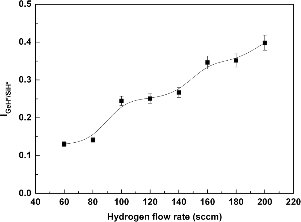 | ||
| Fig. 8 IGeH*/SiH* obtained from Fig. 4(b) at different hydrogen flow rate. Lines are drawn as guides for the eyes. | ||
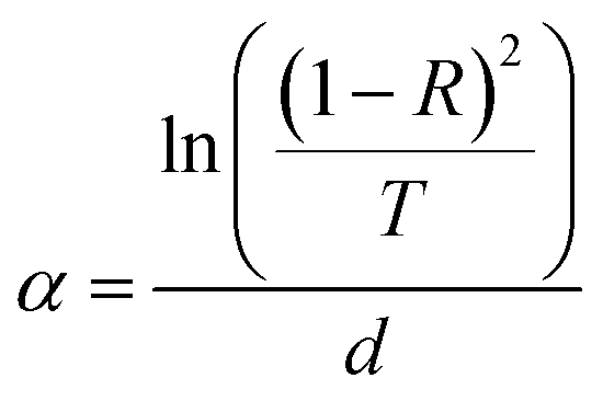
The Eg is deduced from a linear fit of the absorption data according to the well-known relation proposed by Tauc26
| (αhν)1/2 = B(hν − Eg) | (2) |
 | (3) |
Eg is obtained by extrapolation of the linear fit to α = 0. Fig. 9 shows the Eg of a-SiGe:H thin films deposited at the hydrogen flow rate. The Eg decreases with the hydrogen flow rate, which further shows an increase of Ge–Ge in the amorphous network. The Eg of thin film is 1.32 ± 0.20 eV at 200 sccm high hydrogen flow rate.
 | ||
| Fig. 9 The Eg of a-SiGe:H thin films at different hydrogen flow rate. Lines are drawn as guides for the eyes. | ||
Fig. 10 shows the Raman spectra of thin films. The broad and smooth band nature of all the spectra indicates that the thin films are amorphous. Three broad peaks near 480, 390, and 280 cm−1 are assigned to Si–Si, Si–Ge, and Ge–Ge transverse optical vibrations respectively.27 The emission intensity of the Si–Ge and Ge–Ge modes intensifies at 180 and 200 sccm hydrogen flow rate, which also corresponds to the increase of Ge content in the a-SiGe:H thin films. This is consistent with the Eg change of thin films (in Fig. 9).
 | ||
| Fig. 10 Raman spectra of thin films deposited at different hydrogen flow rate. The curves have been shifted vertically for clarity. | ||
The infrared absorption spectra of thin films deposited at different hydrogen flow rate are further utilized to check their microstructure, as shown in Fig. 11. Each thin film has been fitted with a pair of Gaussian peaks centered at 2090 cm−1 and 2000 cm−1, corresponding to Si–H2 and Si–H stretching vibrations respectively. The peak at 1880 cm−1 corresponds to Ge–H stretching vibrations. At high hydrogen flow rate, 180 or 200 sccm, the Si–H2 absorption significantly increases and the Si–H absorption in turn decreases, which means porous amorphous material with more voids.28 The deep defect density increases with the Ge fraction, which is identical to those found by Cohen.29 The a-SiGe:H thin films deposited at the high hydrogen flow rate shows a corresponding reduction in their performance.
 | ||
| Fig. 11 Infrared spectra of thin films deposited at different hydrogen flow rate. The curves have been shifted vertically for clarity. | ||
Fig. 12 shows the dark and photo conductivity at different hydrogen flow rate. The photo conductivity decreases one order of magnitude, and the ratio of photo to dark conductivity decreases by a factor of 17 with hydrogen flow rate, which means low performance thin films deposited at high hydrogen flow rate. This is consistent with the results obtained by infrared spectra.
 | ||
| Fig. 12 Dark and photo conductivity at different hydrogen flow rate. Lines are drawn as guides for the eyes. | ||
4. Summary
The back diffusion of GeH4/SiH4 source gas is suppressed in high pressure. The steady-state depletion of the gases in 2 Torr pressure is monitored by optical emission intensity changes with power. The reduction of gas residence time with hydrogen flow rate leads to decreased H*α/H*β/SiH*/GeH* emission intensity. Although the residence time reduces, their emission intensity increases when the source gases do not fully decompose in 3 Torr. SiH* emission intensity is higher than GeH* one because of a preferential attachment of H to Si over Ge. Based on optoelectronic properties analysis of thin films, we conclude that the ratio of GeH* to SiH* obtained by OES will be a good monitoring parameter to control Ge content during process without any plasma disturbances. The ratio increase shows the increase of Ge content in the a-SiGe:H thin films, thereby the Eg decreases. The appropriate hydrogen flow rate improves the electrical and structural properties of the a-SiGe:H thin films. The deep defect density increases with the Ge content at high hydrogen flow rate. The ratio of photo to dark conductivity decreases.Acknowledgements
This work is supported by the 973 National Basic Research Program of China (2011CBA00705) and the 863 High Technology Research Program of China (2011AA050502).References
- P. Agarwal, H. Povolny, S. Han and X. Deng, J. Non-Cryst. Solids, 2002, 299–302(2), 1213–1218 CrossRef CAS.
- S. Okamoto, E. Maruyama, A. Terakawa, W. Shinohara, S. Nakano, Y. Hishikawa, K. Wakisaka and S. Kiyama, Sol. Energy Mater. Sol. Cells, 2001, 66(1–4), 85–94 CrossRef CAS.
- X. Deng, X. Liao, S. Han, H. Povolny and P. Agarwal, Sol. Energy Mater. Sol. Cells, 2000, 62, 89–95 CrossRef CAS.
- X. Liao, W. Du, X. Yang, H. Povolny, X. Xiang and X. Deng, IEEE, 2008, 1444–1447 Search PubMed.
- G. H. Wang, C. Y. Shi, L. Zhao, B. J. Yan, G. Wang, J. W. Chen, Z. C. Li, H. W. Diao and W. J. Wang, Thin Solid Films, 2013, 534, 591–593 CrossRef CAS PubMed.
- D. Lundszien, F. Finger and H. Wagner, Sol. Energy Mater. Sol. Cells, 2002, 74, 365–372 CrossRef CAS.
- N. Maley and J. S. Lannin, Phys. Rev. B: Condens. Matter Mater. Phys., 1987, 36(2), 1146–1152 CrossRef CAS.
- Y. Fukuda, Y. Sakuma, C. Fukai, Y. Fujimura, K. Azuma and H. Shirai, Thin Solid Films, 2001, 386, 256–260 CrossRef CAS.
- T. Kilper, M. N. Van den Donker, R. Carius, B. Rech, G. Brauer and T. Repmann, Thin Solid Films, 2008, 516, 4633–4638 CrossRef CAS PubMed.
- M. N. Van den Donker, B. Rech, F. Finger, W. M. M. Kessels and M. C. M. Van de Sanden, Appl. Phys. Lett., 2005, 87, 263503 CrossRef PubMed.
- M. N. Van den Donker, B. Rech, W. M. M. Kessels and M. C. M. Van de Sanden, New J. Phys., 2007, 9, 280 CrossRef.
- M. N. Van den Donker, B. Rech, F. Finger, L. Houben, W. M. M. Kessels and M. C. M. Van de Sanden, Prog. Photovolt: Res. Appl., 2007, 15, 291–301 CrossRef CAS.
- M. Takai, T. NIshimoto, M. Kondo and A. Mstsuda, Appl. Phys. Lett., 2000, 77(18), 2828–2830 CrossRef CAS PubMed.
- L. P. Zhang, J. j. Zhang, X. Zhang, Y. Cao and Y. Zhao, Thin Solid Films, 2012, 520, 5940–5945 CrossRef CAS PubMed.
- S. Youn Moon, D. J. You, S. E. Lee and H. Lee, Curr. Appl. Phys., 2013, 13, 1502–1505 CrossRef PubMed.
- S. Oda, S. Ishihara, N. Shibata, S. Takagi, H. Shirai, A. Miyauchi and I. Shimizu, J. Non-Cryst. Solids, 1985, 77–78, 877–880 CrossRef CAS.
- M. Shima, A. Terakawa, M. Isomura, H. Haku, M. Tanaka, K. Wakisaka, S. Kiyama and S. Tsuda, J. Non-Cryst. Solids, 1998, 227–230, 442–446 CrossRef.
- L. Pardini, S. Legnaioli, G. Lorenzetti, V. Palleschi, R. Gaudiuso, A. De Giacomo, D. M. Diaz Pace, F. Anabitarte Garcia, G. De Holanda Cavalcanti and C. Parigger, Spectrochim. Acta, Part B, 2013, 88, 98–103 CrossRef CAS PubMed.
- J. Tauc, Optical Propertiesof Solid, Plenum Press, New York, 1974, pp. 11 Search PubMed.
- T. Roschek, B. Rech, J. Muller, R. Schmitz and H. Wagner, Thin Solid Films, 2004, 451–52, 466–469 CrossRef PubMed.
- F. Tochikubo, A. Suzuki, S. Kakuta, Y. Terazono and T. Makabe, J. Appl. Phys., 1990, 68(11), 5532–5539 CrossRef CAS PubMed.
- F. Tochikubo, T. Makabe, S. Kakuta and A. Sukuzi, J. Appl. Phys., 1992, 71(5), 2143–2150 CrossRef CAS PubMed.
- O. Leroy, P. Stratil, J. Perrin, J. Jolly and P. Belengue, J. Phys. D: Appl. Phys., 1995, 28, 500–507 CrossRef CAS.
- J. R. Doyle, D. A. Doughty and A. Gallagher, J. Appl. Phys., 1992, 71(10), 4727–4738 CrossRef CAS PubMed.
- A. M. Pérez, C. Zuñiga, F. J. Renero and A. Torres, Opt. Eng., 2005, 44(4), 043801 CrossRef PubMed.
- J. Tauc and A. Menth, J. Non-Cryst. Solids, 1972, 8–10, 569–585 CrossRef CAS.
- Y. P. Chou and S. C. Lee, J. Appl. Phys., 1998, 83(8), 4111–4123 CrossRef CAS PubMed.
- H. Wagner and W. Beyer, Solid State Commun., 1983, 48, 585–587 CrossRef CAS.
- J. David Cohen, Sol. Energy Mater. Sol. Cells, 2003, 78, 399–424 CrossRef.
| This journal is © The Royal Society of Chemistry 2015 |