New insights into fluorinated TiO2 (brookite, anatase and rutile) nanoparticles as efficient photocatalytic redox catalysts

Yafang Wangab, Liping Lia, Xinsong Huanga, Qi Lia and Guangshe Li*a
aKey Laboratory of Design and Assembly of Functional Nanostructures, Fujian Institute of Research on the Structure of Matter, Chinese Academy of Sciences, Fuzhou 350002, P. R. China.. E-mail: guangshe@fjirsm.ac.cn; Fax: 86-591-63179426
bSchool of Chemistry and Chemical Engineering, Inner Mongolia University, Hohhot 010021, P. R. China

Received 26th December 2014 , Accepted 7th April 2015

First published on 7th April 2015


Abstract

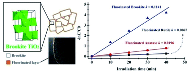
The synthesis of anionic-modified nanostructures with specific properties is often hindered by difficulty in tuning the material compositions without sacrificing phase purity and sample uniformity. Here, we present a novel methodology using NH4F as fluorine source and a high-temperature ionic diffusion that facilitate surface fluorination of TiO2 without changes of phase structure. Using this method, we prepared fluorinated TiO2 nanostructures of different phases (brookite, anatase and rutile) and investigated their structural and photocatalytic redox properties. Photocatalytic selective oxidation of MO and selective reduction of water in producing hydrogen under UV-light irradiation are employed as probe reactions to test the redox properties of the fluorinated TiO2 of different phases. The results show that the photocatalytic redox properties of TiO2 are highly dependent on phase structure and surface fluorination. Among all the phases, the oxidation activity of fluorinated TiO2 can be ranked as brookite > anatase > rutile. Strikingly, the photocatalytic reduction activity for these fluorinated TiO2 followed the same sequence. The variations of photocatalytic redox activity are most likely due to the synergism of phase composition control and surface fluorination. This could effectively delay electron–hole recombination, and generate more free ˙OH radicals but less free electrons. The methodology reported here is simple and general, which might be used to design and control the structures and properties of anionic-modified nanostructures for a variety of applications in environmental cleaning and energy conversion.


1. Introduction

Among all advanced oxidation and reduction processes, heterogeneous photocatalysis is popularly recognized as an effective method for dye degradation,1 and photocatalytic water splitting, which is based on semiconductor oxides (e.g., TiO2, ZnO, WO3, etc.).2–4 Presently, the full exploitation of photocatalytic processes is largely hampered by the lack of efficient photocatalysts.5–7 Surface modification is promising method in preparing high-performance catalysts, since it could alter the adsorption properties of inorganic8 and organic species9 on the semiconductors' surface, and thus influence the rate of photo-induced reactions and their pathways. One may expect that the optimized surface charge distributions and tuned substrate–surface interactions can promote the interfacial electron transfer rates for effective organic degradation.7 Recently, modification of TiO2 surface by F species has attracted remarkable attention.10–12 It is important to figure out how and why the presence of F anions can affect the photocatalytic redox processes, so as to lay a foundation for further development in photocatalysis. However, despite of extensive investigation, for TiO2 of different phases, the understanding on the operative mechanism of fluorination effect are still unsystematic and ambiguous.13 Diverse interpretations have been proposed, but no consensus is reached so far. For instance, adsorption of F anions on the TiO2 surface decreases the amount of surface hydroxyl groups, leading to the formation of ≡Ti–F groups which are able to serve as effective trapping sites by tightly holding the electrons due to the strong electronegativity of fluorine, and consequently depressed the recombination of electrons and holes.14 Xu et al. suggested that the enhanced production of free ˙OH radicals is due to desorption of surface-bound ˙OH radicals by F anions in the solution through the formation of ˙OH-F hydrogen bond.15 Minero et al. reported the fluorination effect and suggested that the displacement of surface hydroxyl groups by fluoride suppressed the formation of surface-trapped radicals (TiO˙) but enhanced the formation of free ˙OH radicals.16

Among the three main crystallographic forms of TiO2, naturally metastable brookite is the least investigated in comparison with the comprehensively studied anatase and rutile phases because of the difficulties encountered in acquiring its pure form.17 In the recent years, in spite of the study of brookite TiO2 is increasing,18,19 photocatalytic redox performance of fluorinated brookite is rare. One may wonder if fluorinated brookite TiO2 can show a photocatalytic activity superior to other counterparts. Then, one may find new routes of improving photocatalytic redox performance for obtain efficient energy and environmental cleaning.

In this work, we conducted a comprehensive study on a series of fluorinated TiO2 of different phases (brookite, anatase and rutile), in order to provide better understanding into photocatalytic processes. These fluorinated TiO2 were prepared with a novel methodology which enables us to realize surface fluorination while maintain their pristine phase structure unchanged at the same time. Photocatalytic selective oxidation methyl orange degradation and selective reduction hydrogen production from water are employed as probe reaction to test the redox properties of the fluorinated TiO2 (brookite, anatase and rutile). These two reactions can serve as the criterion of oxidation and reduction abilities, respectively. Among all phases, fluorinated brookite TiO2 photocatalyst is highly stable, and both the photocatalytic oxidation and reduction activity of fluorinated TiO2 can be ranked as brookite > anatase > rutile. The reasons underlying these experimental observations were discussed in terms of the synergism of phase composition control and surface fluorination.

2. Experimental section

2.1 Materials

All chemical reagents used were analytical grade, purchased from Sinopharm Chemical Reagent Corp, P. R. China, and were employed without further purification. Water purified by a Milli-Q water system (Millipore) was used throughout.

2.2 Preparation of fluorinated brookite TiO2

The sample synthesis involves two-processes. The first process is the synthesis of pure-phase brookite using a solvothermal method. In a typical synthesis, 5 mL of tetra-butyl titanate (TBOT) was added in 25 mL absolute ethyl alcohol. After sufficient stirring, this mixed solution was added dropwise into 30 mL deionized water under vigorous stirring. Subsequently, 5 g of urea were mixed and dissolved in this solution with agitation. Finally, 5 mL of sodium lactate liquor (60%) were dropped in the mixed solution while stirring for about 30 min. All these mixed solutions were removed to autoclaves, which were sealed and heated in an oven at 200 °C for 20 h. After reactions, a white product of brookite phase was separated, washed respectively with ethanol and deionized water for 3 times, and dried in an oven at 60 °C.

The second process is the surface fluorination. 0.6 g the as-prepared brookite TiO2 was mixed with 0.10932 g NH4F. After sufficient grinding in an agate mortar, the mixture was calcined in N2 atmosphere at given temperatures of 400, 500, 600, and 700 °C, respectively, for 4 h. These samples were named B-FT400-700, respectively.

To study the effect of surface fluorination, a reference sample was also prepared using the above procedure (calcination in N2 atmosphere at given temperatures), while NH4F was not involved. Named as B-T400.

2.3 Preparation of fluorinated anatase TiO2

The sample was also prepared using the similar two-step processes, but with some modifications. Firstly, anatase TiO2 nanoparticles were prepared, based on an approach reported by Xing et al.20 A mixture of 35 ml absolute ethyl alcohol (EtOH), 2.0 mL H2O, and 0.5 mL HNO3 was named as solution A, and a mixture of 8.0 mL TBOT and 32.0 mL EtOH was named as solution B. Solution B was added dropwise to solution A under vigorous stirring, and this mixture was stirred continuously for 30 min and immediately transferred to a 100 mL Teflon-lined stainless steel autoclave, which was allowed to react at 180 °C, for 12 h. After solvothermal reactions, the precipitate was washed with water and EtOH, dried, and ground to obtain the final anatase nanoparticles.

During the second surface fluorination process, 0.6 g of the as-prepared anatase TiO2 was mixed with 0.10932 g NH4F, and sufficiently ground in agate mortar. After that, the mixture was calcined in N2 atmosphere at a given temperature of 400 °C for 4 h. The sample was named A-FT400.

For comparison, anatase nanoparticles without surface fluorination were also prepared after calcination in N2 atmosphere at given temperatures but without NH4F involved. Named as A-T400.

2.4 Preparation of fluorinated rutile TiO2

The sample was also prepared using the similar two-step processes, but the titanium source was replaced by potassium titanyl oxalate dihydrate for the first step of hydrothermal reactions. 1 mmol potassium titanyl oxalate dihydrate was added to 60 mL HCl solution (1 M) under stirring. Sometime later, 6 mmol oxalic acid were added to this mixed solution, and transferred to a 100 mL Teflon-lined stainless steel autoclave. After reaction at 180 °C for 12 h, the precipitate was washed with water, dried, and ground to obtain nanoparticles.

Similar to the procedures for fluorinated brookite and anatase, 0.6 g of rutile TiO2 was mixed with 0.10932 g NH4F. After sufficient grinding in agate mortar and subsequent calcinations in N2 atmosphere at 400 °C for 4 h, fluorinated rutile TiO2 was obtained. The sample was named R-FT400.

For comparison, rutile nanoparticles without surface fluorination were also prepared, according to the procedures mentioned above for brookite and anatase. Named as R-T400.

2.5 Sample characterization

XRD analysis was conducted on a Rigaku MiniFlex II diffractometer at a voltage of 30 kV and a current of 15 mA with Cu-Kα radiation (λ = 1.5406 Å), employing a scanning step width of 0.02° and 8 s measurement times per step in the range of 10–80°. 10–15 wt% Ni powder was added in the samples to act as an internal standard for peak positions calibration. The lattice parameters for all samples were further calculated by Rietveld refinement method using GSAS program. The Raman spectra were recorded at room temperature with a HORIBA LabRAM HR confocal microscope spectrograph with a spectral resolution of 0.6 cm−1 and excitation lines at 532 nm. TEM graphs of the samples were obtained on a Tecnai G2 F20 S-TWIN field emission transmission electron microscope with a maximum acceleration voltage of 200 kV. Prior to TEM measurements, samples were dispersed in absolute ethanol and deposited on a carbon film coated copper grid. UV-visible diffuse reflectance spectra of the samples were collected using a Varian Cary 500 UV-vis-NIR spectrometer, and were converted from reflection to absorption according to the Kubelka–Munk method. An X-ray photoelectron spectroscopy (XPS) measurement was carried out on an ESCA-LAB MK II apparatus using a monochromatic Al Kα X-ray source operating at 150 W. During the XPS measurements, emission lines were calibrated using the C 1s signal at 284.6 eV. Photoluminescence (PL) spectra of the samples were carried out at room temperature by a Varian Cary Eclipse Fluorescence Spectrometer. Temperature programmed reduction (TPR) tests were carried out in a conventional low apparatus with a TCD detector at a heating rate of 5 °C min−1 using 10 vol% H2 in Ar to examine the redox behaviour of the samples. The flowing rate was set at 30 mL min−1. Before TPR tests, 50 mg catalyst was pretreated in Ar at 200 °C for 2 h.

2.6 Photocatalytic redox activity tests of the samples

Methyl orange degradation. Photocatalytic oxidation activities of the samples were assessed by methyl orange (MO) degradation as a probe reaction under UV-light irradiation (>300 nm). UV-light was obtained by employing a 300 W Xe lamp equipped with a 300 nm cut-off filter to remove the undesired radiations. The quantity ratio of catalyst to MO solution (10 mg L−1) was set at 0.1 g[thin space (1/6-em)]:[thin space (1/6-em)]100 mL, and MO solution containing the catalyst was magnetically stirred for 3 h to establish an adsorption and desorption equilibrium before illumination. The lamp was located at a distance of 15 cm away from the surface of the suspension placed in a beaker open to air. Sampling was proceeded from the illuminated suspension at a certain time interval, and the extracted suspension was centrifugated at a rate of 8000 rpm to remove the solid powders. The resulting supernatant was measured by a Perkin-Elmer UV lambda 35 spectrophotometer to acquire its UV-vis adsorption spectra.
Hydrogen generation. The photocatalytic reduction activities of hydrogen production for the photocatalyst samples were examined. In a typical photocatalytic experiment, 50 mg samples were added into the reactor with 60 mL de-ionized water in it, then 20 mL methanol were added to the solution which acted as sacrificial agent to capture photo-generated holes. In order to improve the photocatalytic activity of the samples, a certain amount of H2PtCl6 solution as co-catalyst was added to the reactor. Thermostatic water flowed through a jacket of the Pyrex reactor remove extra heat produced by the lamp and keep the temperature at 5 °C The light source used was a 300 W xenon lamp equipped with a UV-cutoff filter (λ > 300 nm). Before visible light irradiation, we must ensure that the photocatalytic reactor was sealed, and the residual air was deprived by vacuum pump. The experiment time was also set at 3 h. The photo-generated gas was detected in every 1 h by a gas chromatograph (Fuli 9790II) with TCD detector, which purchased from Fuli Analytical Instrument Corp, Zhejiang, PR China.

2.7 Determination of reactive species (hydroxyl radical, ˙OH)

In order to measure the relative concentration of hydroxyl radicals (˙OH), the terephthalic acid (TA) fluorescence method was employed in this study since TA can react with ˙OH to form highly fluorescent 2-hydroxyterephthalic acid (TA-OH). The mixture of TA solution was prepared from 5 × 10⁻⁴ molar of TA and 2 × 10⁻³ molar of NaOH in deionized water. Then, 0.01 g of the fluorinated brookite TiO2 at 400 °C was dispersed in 50 mL of the TA aqueous solution. The solution was collected at every 5 min during the irradiation procedure in order to estimate the generated TA-OH, which can be detected by fluorescence spectroscopy at 425 nm.

3. Results and discussion

3.1 Crystallinity, specific surface area, and structure properties for fluorinated TiO2 of different phases

Fig. 1 shows the XRD patterns of the fluorinated samples obtained after calcinations under N2 flow. All diffraction lines are relatively strong, indicating a high crystallinity for all samples. Further, the peak positions and relative intensities of the diffraction lines match well with standard diffraction data for different TiO2 phases, i.e. brookite, anatase and rutile. For instance, the peaks observed at 2θ values of 25.33, 25.68, 30.80, 36.25, 40.15, 48.01 and 55.23 for the top diffraction pattern of Fig. 1 could be indexed to the (120), (111), (121), (012), (022), (231) and (241) planes of an orthorhombic phase brookite, respectively, consistent with the standard XRD data for brookite TiO2 (JCPDS, no. 39-1360). The other two XRD patterns are consistent with the standard XRD data for tetragonal phase anatase TiO2 (JCPDS, no. 21-1272) and tetragonal rutile TiO2 (JCPDS, no. 21-1276), respectively.
image file: c4ra17076h-f1.tif
Fig. 1 XRD patterns of the fluorinated (at 400 °C) brookite, anatase and rutile TiO2.

It should be noted that the fluorinated TiO2 (brookite, anatase and rutile) samples are all present in pure phases, exactly inherited from their respective pristine structure (see XRD data of un-fluorinated TiO2 in Fig. S1), without any detectable secondary phases. It is thus clear that surface fluorination does not have significant influence on the phase structure of TiO2.

To study the impacts of calcinations and surface fluorination on the microstructures of the TiO2 with different phases, XRD patterns for all samples were carefully analyzed. The average crystallite sizes of the samples were estimated from the full width at half maxima of the respective peaks of XRD data at 2θ values of 29–60°, using Scherrer's equation in eqn (1),21

 
D = /β[thin space (1/6-em)]cos[thin space (1/6-em)]θ (1)
where k is a constant equal to 0.89, λ is the X-ray wavelength equal to 0.154 nm, β is the full width at half maximum, and θ is the half diffraction angle. The calculated crystallite size for TiO2 (brookite, anatase and rutile) were listed in Table 1. B-T, A-T and R-T are meaning of brookite, anatase or rutile TiO2 that prepared with solvothermal method; the B-T400, A-T400 and R-T400 are meaning of calcinated brookite, anatase or rutile TiO2 at 400 °C; B-FT400, A-FT400 and R-FT400 are meaning of fluorinated brookite, anatase or rutile TiO2 at 400 °C, respectively. From Table 1, one can see that the crystallite sizes for TiO2 (brookite, anatase and rutile) increased, while specific surface area decreased with calcination and fluorination.

Table 1 Lattice parameters, unit cell volume, crystalline size and specific surface area of the samples: as-prepared, after 400 °C calcinations or surface fluorination
Samples Lattice parameters Unit cell volume (Å3) Crystalline size (abc)a (nm) BET (m2 g−1)
a (Å) b (Å) c (Å)
a (abc) is (120), (101) and (110) for brookite, anatase and rutile, respectively.
B-T 9.1844(5) 5.4542(3) 5.1458(3) 257.77(3) 21.0 31
B-T400 9.1888(4) 5.4591(2) 5.1466(2) 258.17(2) 21.0 28
B-FT400 9.1982(2) 5.4649(1) 5.1536(1) 259.1(1) 27.5 17
A-T 3.7870(2) 3.7870(2) 9.5168(8) 136.49(2) 9.4 121
A-T400 3.7943(2) 3.7943(2) 9.5296(7) 137.19(2) 9.6 93
A-FT400 3.8100(2) 3.8100(2) 9.5634(7) 138.83(2) 12.4 25
R-T 4.6036(6) 4.6036(6) 2.9586(4) 62.70(3) 24.1 23
R-T400 4.6062(6) 4.6062(6) 2.9650(4) 62.91(3) 28.2 13
R-FT400 4.6062(2) 4.6062(2) 2.9640(1) 62.89(1) 26.3 13


Besides the impacts on the crystallite sizes and surface area, calcinations and surface fluorination also alter the lattice parameters of TiO2 with different phases. The lattice parameters for all samples were analyzed using GSAS structural refinement. For the as-prepared brookite TiO2, the refined lattice parameters were a = 9.1844(5), b = 5.4542(3) and c = 5.1458(3) Å, respectively, which are all compatible with those of the standard values (i.e., a = 9.18, b = 5.45, and c = 5.16 Å). After 400 °C calcinations, the lattice parameters slightly increased to a = 9.1888(4), b = 5.4591(2) and c = 5.1466(2) Å. This observation is surprising, since high temperature calcinations usually lead to lattice shrinkage due to the larger grain sizes. Surface fluorination at 400 °C led to a further increase in lattice parameters at a = 9.1982(2), b = 5.4649(1) and c = 5.1536(1) Å. Interestingly, the axial ratio, c/a, representing the lattice symmetry, remains almost constant at 0.560. The lattice expansion of brookite TiO2 observed upon high-temperature calcinations and surface fluorination should be closely related to the presence of defects.22

Similarly, for anatase and rutile TiO2, calcinations and surface fluorination led to a gradual increase in lattice parameters and unit cell volume.

Raman spectroscopy is sensitive enough to distinguish brookite, anatase and rutile TiO2. The solid was then characterized with Raman spectroscopy, and the results are shown in Fig. 2. For fluorinated brookite TiO2 at 400 °C, there were 14 Raman bands in the range from 100 to 700 cm−1, which can be assigned to A1g (133, 156, 195, 251, 414, 550, and 639 cm−1), B1g (217 and 289 cm−1), B2g (369, 465 and 586 cm−1), and B3g (326 and 505 cm−1) of brookite, respectively.23 It is worth noting that characteristic Raman vibrations for anatase at 518 cm−1 and rutile at 442 cm−1 are absent in the spectrum. This indicates that the titanium oxide presents in the form of pure brookite phase. Other TiO2 polymorphs thus obtained are also phase pure TiO2, as evidenced by their respective Raman spectrum: for example, the major Raman bands at 147, 199, 399, 518 and 641 cm−1 are associated with five Raman-active modes of anatase phase TiO2 with the symmetries of Eg, Eg, B1g, B1g and Eg, respectively;23,24 for “rutile”, the Raman peaks at 143 cm−1 is for the mode of B1g symmetry, 249 and 442 cm−1 are for Eg mode while 611 cm−1 is for A1g mode.25 Except for these distinctive vibrations, no additional peaks can be found in each spectrum. These observations confirm that fluorinated brookite, anatase and rutile synthesized in this work are in pure phase.


image file: c4ra17076h-f2.tif
Fig. 2 Raman patterns of the fluorinated (at 400 °C) brookite, anatase and rutile TiO2.

Effective strains of the samples are calculated by the broadening effects of the diffraction peaks using the Williams and Hall theorem in eqn (2),26,27

 
β[thin space (1/6-em)]cos[thin space (1/6-em)]θ/λ = 1/D + η[thin space (1/6-em)]sin[thin space (1/6-em)]θ/λ (2)
where β is the full width at half maximum (FWHM), θ is the diffraction angle, λ is the X-ray wavelength, D is the effective particle size, and η is the effective strain. The strain is calculated from the slope, and the particle size (D) is calculated from the intercept of a plot of β[thin space (1/6-em)]cos[thin space (1/6-em)]θ/λ vs. sin[thin space (1/6-em)]θ/λ.

For the as-prepared brookite TiO2, the strain was determined to be compressive at −0.07%, which became −0.01% when the brookite TiO2 was calcinated at 400 °C. Strikingly, the nature of the strain changed from the compressive to tensile at +0.15% upon surface fluorination at 400 °C (Fig. 3a1–3 and d).


image file: c4ra17076h-f3.tif
Fig. 3 Comparisons between Williamson–Hall plots of as-prepared (black), calcinated (red) and fluorinated (blue) samples for (a1–a3) brookite, (b1–b3) anatase and (c1–c3) rutile TiO2, and (d) calculated lattice strain.

Similar variation trend of strain was also observed for anatase with the exception of the magnitude. For instance, the strain was compressive at −0.36% for the as-prepared anatase TiO2, which increased to −0.09% after calcinations at 400 °C. Upon surface fluorination at 400 °C, compressive strain changes to tensile again at +0.78%. These results indicate that the lattice strain could be tuned from the compressive to tensile by fluorination for both brookite and anatase.

Different from the cases for brookite and anatase, the strain was tensile at 0.78% for the as-prepared rutile TiO2, which was increased to +0.87% after calcinations at 400 °C or +0.83% upon surface fluorination at 400 °C. No changes in the strain nature were observed. Furthermore, by comparing the lattice parameters listed in Table 1 and the strain variations in Fig. 3, one can clearly see that the increasing trend of lattice parameter is coincident with that of the strain. It is most likely that the strains varied with calcinations or surface fluorination are responsible for the increases in lattice parameters.

The surface fluorination of TiO2 nanoparticles was further investigated by TEM and HR-TEM. For fluorinated brookite TiO2 at 400 °C, the as-prepared particles are uniform, showing a relatively narrow particle size distribution (Fig. S2 and S3). The particle shape is rod-like, as indicated from TEM image in Fig. 4a. Onto the surfaces of brookite nanorods, there deposited a thin layer with a thickness of about 0.5 nm, which might be the fluorinated layers (see HRTEM image for a single brookite particle in Fig. 4b). The existence of surface fluorinated layers were also proved by a designed experiment that compared the photocatalytic oxidation performance between NaOH-treated and untreated samples (Fig. S10b). The lattice fringes with an interplanar spacing of 0.3528 nm correspond to the (120) crystal plane of orthorhombic phase brookite TiO2. The SAED pattern (inset of Fig. 3a) shows a set of rings consisted of spots, which confirms the random orientations of nanoparticles.


image file: c4ra17076h-f4.tif
Fig. 4 (a) TEM and (b) HRTEM images of brookite TiO2 after surface fluorinations at 400 °C. Insets show the corresponding SAED patterns and a schematic illustration.

The formation of the uniform fluorinated TiO2 is beneficial from the two-step reaction processes adopted in this work: the first step of pure-phase TiO2 synthesis, and the second step of uniform surface fluorination using NH4F as fluorine source and a high-temperature ionic diffusion. Take brookite TiO2 for example, the schematic reaction path of reactants during the synthesis is illustrated in Scheme 1, which is based on literature data analyses and our XRD, TEM and Raman observations. It is well documented that brookite TiO2 nanoparticles can be prepared through a low-basicity hydrothermal process utilizing urea as an in situ OH source, and sodium lactate as the complexant and surfactant.2,18 Surface fluorination increases the rate of photocatalytic oxidation process through a hydroxyl radical-mediated path.7 With these information in mind, a novel concept of establishing the fluorinated brookite TiO2 in the monomer level is proposed in the present study: tetra-butyl titanate as a titanium source, urea as an in situ OH source, sodium lactate as the complexant, and surfactant and NH4F as fluorine source are suitable starting species to facilitate the formation of surface fluorination of brookite TiO2. With these starting species, brookite TiO2 was initially formed in terms of the procedure in Scheme 1 through the reaction among various chemical ingredients when treated under autogenous pressure conditions inside the autoclave at 180 °C. The material obtained from the autoclave is subjected to calcinations at 400 °C in the presence of NH4F for 4 h in order to realize surface fluorination for brookite TiO2. In order to prove NH4F can fully participate in the reaction, keep the brookite structure and appropriate specific surface area, we select 400 °C as the optimum synthesis temperature. The present method is very attractive in surface modification of TiO2 because it won't cause phase evolution even when fluorinated at 400 °C. We have conducted a series of comparative investigations to optimize the fluorination method and eventually obtained the above preparation route, changes in any step may lead to a mixed phase of brookite and rutile TiO2 (Fig. S4 and S6).


image file: c4ra17076h-s1.tif
Scheme 1 Schematic illustration for the formation of surface fluorinated TiO2 nanoparticles after calcination at 400 °C, taking brookite as an example.

3.2 Optimum photocatalytic redox performance of fluorinated TiO2 with different phases

Photocatalytic oxidation activities of TiO2 of different phases with and without surface fluorination were comparatively studied by taking a model reaction of MO photodegradation under UV-visible light irradiation (λ > 300 nm) at room temperature. Depending on the phases of TiO2, surface fluorination may show different impacts on the photocatalytic oxidation activities. For brookite, surface fluorination led to an apparent enhancement in photocatalytic activity, as indicated by the time-dependent degradation spectra of MO aqueous solutions during UV-vis light irradiation (Fig. S5). This activity is significantly improved, when comparing to those un-fluorinated samples (either 400 °C calcinations or as-prepared brookite). Different from the case of brookite, surface fluorination of anatase or rutile led to a decrease in photocatalytic activity (Fig. S6), therefore, the photocatalytic oxidation activity is affected by the synergistic effect of phase structure control and surface fluorination. Namely, the increased photocatalytic oxidation of surface fluorination was effected by phase structure. Interestingly, when comparing all fluorinated samples, fluorinated brookite TiO2 nanoparticles showed an optimum photocatalytic oxidation activity towards MO removal: almost 100% of MO molecules were degraded in 30 min (Fig. 5a). For the calcinated TiO2, whether brookite or anatase, rutile, only partly MO was removed in the same time (Fig. 5c).
image file: c4ra17076h-f5.tif
Fig. 5 (a) and (c) Photocatalytic degradation towards MO; (b) and (d) the corresponding kinetics plots for pseudo first order reaction of decolourisation in the presence of fluorinated TiO2 (brookite, anatase and rutile) under UV-visible light irradiation.

The kinetics of MO decolourisation of fluorinated or calcinated TiO2 in different phases, brookite, anatase and rutile are presented in Fig. 5b and d. It is seen that the photocatalytic degradation process exactly follows a pseudo-first order reaction in eqn (3),28

 
−ln(C/C0) = kt (3)
where k is the apparent rate constant (min−1), C0 is the initial concentration of dye and C is the concentration of dye at time (t). As the dye concentration in these experiments remains in the regime where the Beer–Lambert law holds, the concentration can be substituted by the dye absorbance at a given wavelength (typically the peak absorption).

From Fig. 5b, for brookite, surface fluorination led to an apparent rate constant of 0.1141 min−1, which is about 6 times larger than that of 0.0196 min−1 for fluorinated anatase, and even 17 times larger than that of 0.0067 min−1 for fluorinated rutile. Such a superior photocatalytic oxidation activity towards the degradation of MO has not been ever reported before for fluorinated TiO2. One cannot explain this superior activity in terms of the surface area, since the fluorinated brookite TiO2 didn't possess the highest specific surface area (Table 1). Therefore, it is necessary to examine other factors, which will be discussed below.

In Fig. 5d, the apparent rate constants of calcinated brookite, anatase and rutile TiO2 are 0.0127, 0.0154 and 0.0067 min−1, respectively. Seem obvious, the effect of surface fluorination is superior to the calcination on enhanced photocatalytic oxidation activity.

Above analyses indicate that only the photocatalytic oxidation performance of brookite TiO2 is enhanced after fluoridation. Is the photocatalytic reduction ability influenced simultaneously? How fluoridation and calcinations affect photocatalytic redox activity for different phase of TiO2? To answer these questions, photocatalytic reduction activity on hydrogen production of all the fluoridated and calcinated TiO2 (brookite, anatase and rutile) are characterized in the following.

The activities of hydrogen evolution observed are shown in Fig. 6. The inset displays hydrogen evolution over time at the presence of brookite TiO2. As seen from Fig. 6, when the UV-visible light irradiates over the reaction system for 3 h at 5 °C, ca. 50 mmol g−1 H2 was generated for the calcinated brookite TiO2 (B-T400-Pt), while for the fluorinated brookite TiO2 (B-FT400-Pt), only 12.9 mmol g−1 H2 are generated. Similarly, surface fluorination of anatase or rutile led to a decrease in photocatalytic reduction activity on hydrogen production from water splitting. Therefore, the surface fluorination affected primarily the photocatalytic reduction activity rather than phase structure. Namely, the decreased photocatalytic reduction of surface fluorination wasn't effected by phase structure. For the fluorinated TiO2 (brookite, anatase and rutile), decreased photocatalytic reduction activity is likely due to the decrease of free electrons. This is because fluorine ion has strong electronegativity; consequently, the ≡Ti–F groups formed on the surface were able to serve as effective trapping sites by tightly holding the electrons.


image file: c4ra17076h-f6.tif
Fig. 6 The photocatalytic activities of hydrogen production in the presence of as-prepared and fluorinated TiO2 (brookite, anatase and rutile) under UV-visible light irradiation. The inset displays hydrogen evolution as a function of irradiation time for sample BT-400 and B-FT400.

In addition, it was observed that the calcinated brookite TiO2 showed the highest photocatalytic reduction activity among all catalysts. Its H2 production rate is almost 5 times of that for calcinated rutile TiO2 (R-T400-Pt). Moreover, for either fluoridated or calcinated TiO2, the photocatalytic reduction activity follows same sequence: brookite > anatase > rutile.

3.3 Insights into the optimum photocatalytic redox performance of fluorinated brookite TiO2

As stated above, fluorinated brookite TiO2 at 400 °C displayed an optimum photocatalytic redox performance among all other TiO2 phases. In order to further understand how surface fluorination affects the catalytic activity of brookite TiO2, we performed a detailed surface analysis.
Surface compositions. The surface compositions (like F, Ti, C, and O species) for the fluorinated and calcinated brookite TiO2 at 400 °C was characterized by XPS. As indicated by the XPS survey spectrum in Fig. 7a, there appear signals assigned to Ti, O, F, and C elements. The presence of C element can be ascribed to the residual carbon from precursor solution. Concerning the F species, as demonstrated in Fig. 6c (red solid line), one single F 1s signal peak was observed at 684.3 eV for the fluorinated brookite TiO2, as the characteristic of chemical bonding of F species to Ti4+ in forming sub-surface Ti–F species, as reported elsewhere for surface fluorination.29 Further, all these F species cannot diffuse to the interior lattice to substitute O species in forming solid solution TiO2−xFx, since the typical signal for F atoms in solid solution TiO2−xFx (Fig. 7c, dash line)30 was absent at 689.3 eV. Apparently, the presence of sub-surface Ti–F species for fluorinated brookite TiO2 is consistent with our HRTEM observation, which expects to eliminate the possibility of generating more lattice defects (e.g., those arose from F substitution for lattice oxygen) and moreover to reduce the electron–hole recombination rate for improved catalytic activity.
image file: c4ra17076h-f7.tif
Fig. 7 (a) Survey XPS spectrum and (b) Ti 2p, (c) F 1s, and (d) O 1s spectra for calcinated or fluorinated brookite at 400 °C.
Surface lattice distortion. Fig. 7c compares Ti 2p spectrum for fluorinated brookite TiO2 with that for 400 °C calcinated brookite. For fluorinated brookite TiO2, Ti 2p spectrum consists of two distinct photoelectron signals Ti 2p1/2 and Ti 2p3/2 at 463.9 and 458.2 eV, respectively. The spin-orbital splitting between both signals is 5.70 eV, which is comparable with that of 5.74 eV reported previously in literature.27 Both signals are slightly positively shifted in binding energy, when comparing to those for 400 °C calcinated brookite. Similar positive shift has also been observed in O 1s signal. For instance, the O 1s spectrum for fluorinated brookite TiO2 consists of one strong bulk oxygen (O2−) photoelectron signal at 529.7 eV, which positively shifted to 529.4 eV for calcinated brookite TiO2 (Fig. 7d). Strikingly, a shoulder signal at 531.5 eV associated with surface adsorbed oxygen for calcinated brookite TiO2. Based on the larger electronegativity of F ions than O ions and positive BE shifts in binding energy for Ti 2p and O 1s levels, it is probable that surface fluorination of brookite may have dual effects on the surface structures: (i) surface Ti–O(F) polyhedra were distorted due to the sub-surface oxygen replacement of F replaced the oxygen, as reported elsewhere;31 and (ii) surface Ti–O bonds may become more co-valent in chemical nature with the replacement of oxygen by F led to the reduction of bulk oxygen was more difficult, which has been indicated by the oxygen activation at lower temperatures (see H2-TPR results in Fig. S7).
Surface structure relaxation. Associated with the surface lattice distortion is the surface structure relaxation, which could be indicated by the thermodynamic stability. Here, we used XRD to examine the thermodynamic stabilities of fluorinated brookite at different calcination temperatures. As demonstrated in Fig. 8, below the calcinations temperature of 600 °C, brookite structure was maintained, showing XRD patterns exactly the same with an orthorhombic brookite structure (JCPDS, no. 29-1360), while no other peaks from nitrogen and fluorine-derived phases were found. When calcined beyond 600 °C, the brookite structure was destabilized to show some amounts of rutile TiO2 (JCPDS, no. 21-1276). It is obvious from these observations that surface fluorination could greatly enhance the thermal stability of brookite through surface structure relaxation by prohibiting brookite from phase transformation to rutile.
image file: c4ra17076h-f8.tif
Fig. 8 XRD patterns of the as-prepared brookite TiO2 and the fluorinated brookite TiO2 obtained after fluorination at 400, 500, 600, and 700 °C. Symbol * represents Bragg peak of rutile TiO2.

Another important indication of surface structure relaxation is represented by the crystal growth and lattice shrinkage with calcination temperatures. The crystallite size for fluorinated brookite TiO2 at calcination temperature of 400 °C was about 28 nm, which slightly increased to 32 nm after calcinations at 600 °C. When calcined beyond 600 °C, the brookite structure was destabilized to show a larger crystallite size of about 38 nm. Further, with increasing the calcination temperature, the peak positions of the diffraction peak (120) for brookite TiO2 showed a systematic shift towards higher two-theta degrees, which accompanies a decrease in lattice parameter (Table S1).

Band gap energies. To obtain the band gap energies of the fluorinated brookite TiO2, Schuster–Kubelka–Munk absorption function ((αhν)1/n) was plotted against the photon energy (), according to the following eqn (4),32
 
(αhν)1/n = A(Eg) (4)
where A is a proportionality constant, h is Planck's constant, ν is the frequency of vibration (hence is photon energy), and α is an absorption coefficient. The value of n depends on the type of optical transition of the semiconductor (n = 2 for indirect transition).33 The approximate band gap can then be determined from the straight line x-intercept as shown in Fig. 9. Compared to the as-prepared brookite TiO2, the fluorinated brookite after calcinations at 600 °C or above did show significant changes in band-gap energies. Interestingly, the fluorinated brookite at 400 °C with lattice expansion (Shown in Table S1) showed a decreased band gap (0.03 eV) and the samples fluorinated at 500 °C with lattice contraction showed an increased band gap (0.07 eV). It is well known that the variation of band gaps is closely related to the change in lattice parameters and the associated intrinsic defects in lattice.22 For the present fluorinated brookite, the defect states will be confirmed by PL measurements as discussed below. Ekuma and Bagayoko confirmed the position of the shallow minimum of the conduction band is strongly dependent on the lattice contraction. They observed a reduction in the indirect band-gap with lattice expansion and an increase in band-gap with lattice contraction,34 which is consistent with our observations upon surface fluorination. Fundamentally, the varied lattice parameter may lead to changes in density of states (O-2p and Ti-3d) and total energy, which affect the band gap. This seems not applicable for fluorinated brookite after calcinations at 600 °C or above, since we observed no apparent changes in band gap in spite of lattice contraction. It is quite likely that the increased concentration of defects may help to balance the band gap.

image file: c4ra17076h-f9.tif
Fig. 9 (a) Schuster–Kubelka–Munk absorption function of fluorinated brookite TiO2 after calcinations at given temperatures and (b) calculated band gap energies.
Surface reaction kinetics. Surface fluorination would give rise to surfaces negatively charged, which are able to draw the photogenerated holes to semiconductor surface by electrostatic force. Thus, promoted surface reaction kinetics is highly expected, since more holes are available at the interface, which could remarkably accelerate the oxidation of surface-adsorbed solvent water to free ˙OH radicals. The kinetics of MO decolourisation for the present fluorinated brookite after calcinations at different temperatures is presented in Fig. 10. It can be seen that MO photodegradation follows pseudo-first order kinetics in terms of the eqn (2). With increasing the calcination temperature, the apparent rate constant decreased from 0.1141 min−1 at 400 °C, to 0.0793 min−1 at 500 °C, 0.0081 min−1 at 600 °C, and to 0.0172 min−1 at 700 °C (Fig. 10). Clearly, the maximum rate constant of fluorinated brookite TiO2 was reached at 400 °C. Since the apparent rate constant of the as-prepared brookite TiO2 was 0.0274 min−1, surface fluorination effect at 400 °C is represented by approximately 4 times enhancement in photocatalytic activity when comparing to the as-prepared brookite TiO2. Otherwise, higher fluorination temperatures like 600 or 700 °C will show poor photocatalytic activity as a result of the increased crystalline size and decreased specific surface area. Concerning the fluorinated brookite TiO2 at 400 °C, the specific surface area is the highest, as indicated by BET analysis (Table S1), and therefore more active sites are available;35 Moreover, the band gap of fluorinated brookite TiO2 at 400 °C was smallest (Fig. 9).
image file: c4ra17076h-f10.tif
Fig. 10 The reaction kinetics for pseudo first order reaction of MO decolourisation of (a) as-prepared brookite TiO2 and the fluorinated brookite TiO2 after calcinations at temperatures of (b) 400, (c) 500, (d) 600, and (e) 700 °C.

As a result, reaction rate of photocatalytic oxidation MO degradation is optimized at 400 °C. Hence, combining the above analysis, one can see that only if the phase is brookite and the temperature is appropriate, can the optimum photocatalytic oxidation activity be obtained. Besides, the adsorption efficiency of MO in dark at different pH values for the fluorinated brookite TiO2 after calcinations at 400 °C is given in Fig. S8.

Strong binding of F species to surfaces. Since the binding of F species to brookite surface directly determines the reproducibility and durability, which are two critical issues for the long-term use of any catalysts in practical applications,36 herein we tested the photocatalytic stability of fluorinated brookite TiO2 at 400 °C toward the photodegradation of MO. We reused the fluorinated brookite five times, and each experiment was performed under the identical conditions (Fig. 11). It was found that after five cycles, the photocatalytic activity retained nearly 90% relative to its initial activity. In particular, after 5th catalytic cycle, fluorinated brookite TiO2 remained its phase structure under UV-visible light irradiation, indicating that the current brookite upon surface fluorination at 400 °C is highly stable, which can be seen from XRD patterns in Fig. S9. In short, surface fluorination of brookite at 400 °C led to a strong F binding to the surface and furthermore electrostatic force, which yields an efficient photocatalytic oxidation activity and high stability for MO degradation under UV-light irradiation.
image file: c4ra17076h-f11.tif
Fig. 11 Cycling runs of MO degradation in the presence of the fluorinated brookite TiO2 at 400 °C.
Separation of photogenerated electron–hole pairs. It is well established that the photoluminescence (PL) is powerful in the efficiency of charge carrier trapping, monitoring the migration, transfer, and to understand the fate of electron–hole pairs in semiconductor particles.37 This is because PL emission spectra are generated from the radiative recombination of excited electrons and holes.38 A higher PL intensity indicates a higher radiative recombination rate of electron–hole pairs under light excitation. Herein, PL spectra were used to examine the impact of surface fluorination on the separation of photo-generated electron–hole pairs in brookite TiO2. As indicated in Fig. 12, un-fluorinated brookite TiO2 showed a fast recombination rate of electrons and holes, which is demonstrated by the strong and wide PL spectrum for the as-prepared brookite TiO2 when excited at 290 nm. However, after surface fluorination, PL peaks of the fluorinated brookite decreased greatly in intensity, which indicates that the radiative recombination rate of electrons and holes is hindered. The variation of PL intensity may result from the change of defect state on the shallow level of TiO2 surface.39 This phenomenon demonstrates that the photogenerated electron–hole pairs can be effectively separated in the fluorinated brookite, which accounts for the enhanced photocatalytic oxidation activity, as reported elsewhere for other TiO2 surface anionization.36 However, the effectively separated electrons and holes were not the only factor that contributes to the enhanced photocatalytic oxidation activity. Instead, the photocatalytic activity is influenced by various factors such as band gap, crystalline size, specific surface area and phase structure etc. When calcinated at higher temperature (>400 °C), for the fluorinated brookite TiO2, the band gap increased, specific surface area gradually decreased and the phase structure may change to rutile. Therefore, the higher calcinated temperature doesn't necessarily contribute to the enhanced photocatalytic oxidation activity.
image file: c4ra17076h-f12.tif
Fig. 12 PL spectra of the as-prepared brookite TiO2 and the fluorinated brookite TiO2 after calcinations at 400, 500, 600, and 700 °C.
Oxidizing species. For majority of photocatalytic oxidation processes, hydroxyl radicals (˙OH) are commonly recommended as the primary oxidizing species. In this regard, terephthalic acid photoluminescence probing technique (TA-PL) is a powerful testing method and has been widely used in detection of hydroxyl radicals.40 2-Hydroxyl-terephthalic acid, which is generated when terephthalic acid captures the hydroxyl radicals, performs a strong fluorescence characteristic, so that hydroxyl radicals could be detected indirectly by monitoring the fluorescence intensity changes of TA solution. Herein, the fluorinated brookite TiO2 after at 400 °C was employed to detect the generated ˙OH on the surface. As indicated in Fig. 13, there appeared a ˙OH-trapping photoluminescence spectrum for fluorination brookite TiO2 at 400 °C in a terephthalic acid solution at room temperature under UV-light irradiation. That implies the formation of fluorescent products during the photocatalytic process, which is likely due to the specific reaction between ˙OH and terephthalic acid. The fluorescence intensity at 425 nm increased when UV light was irradiated continuously, which means that the photocatalytic performance became higher with prolonging the time of light irradiation. In the same time, the fluorinated anatase and rutile TiO2 after at 400 °C were employed to detect the generated ˙OH on the surface. As indicated in Fig. S11. It clearly indicates that ˙OH is not formed in the photocatalytic reaction. Therefore, the photocatalytic oxidation activity for fluorinated anatase and rutile TiO2 were not enhanced.
image file: c4ra17076h-f13.tif
Fig. 13 Fluorescence spectra of a TA–OH solution generated by fluorinated brookite TiO2 after calcinations at 400 °C under UV-visible light irradiation.

As the fluorescence intensity increased with the number of minutes, it can be deduced that the amount of hydroxyl radicals (˙OH) is proportional to the irradiation time. It has been reported that the formation rate of ˙OH radicals is related to the separation efficiency of electron–hole pairs, which gives a great contribution to the photocatalytic oxidation performance.41 Besides, for the fluorinated brookite TiO2, the free ˙OH radicals was due to the desorption of surface-bound ˙OH radicals by F anions in the solution through the formation of ˙OH–F hydrogen bond.17 Thus the surface fluorination was the key to enhance the photocatalytic oxidation performance. Moreover, roles of dissolved O2 in the solution and photogenerated carrier trapping have been examined by a N2 purging experiment and holes-capture experiment using disodium ethylenediamine tetraacetate (EDTA), which indicated that both ˙O2− and ˙OH are the oxidative species in the MO degradation, and the holes, ˙O2− and ˙OH synactic play important roles in the photoreaction (Fig. S10a).

4. Conclusions

In summary, pure TiO2 of different phases (brookite, anatase and rutile) were prepared by a solvothermal method. Surface fluorination was then achieved using NH4F as fluorine source and a subsequent high-temperature ionic diffusion process in a N2 atmosphere without sacrificing the phase structures of TiO2. Surface fluorination not only alters the microstructures of TiO2 (like surface compositions, particle sizes, surface Ti–O(F) polyhedra symmetry, and lattice dimensions), but also leads to the surface structure relaxation which enhance the thermal stability of TiO2. Our investigation indicates that fluorinated brookite TiO2 has the highest photocatalytic redox activity, followed by its anatase counterpart, while rutile TiO2 exhibits the most sluggish behaviour. The photocatalytic redox activity can ranked as brookite > anatase > rutile. Base on all the experimental observations, it can be concluded that the photocatalytic oxidation activity is affected by surface fluorination, phase structure and calcination temperature; on the other hand, the photocatalytic reduction activity is mainly affected by surface fluorination and is independent of phase structure or calcinations temperature. The improved oxidation performance but decreased reduction performance could be ascribed to the enhanced desorption of free ˙OH radicals of surface-bound ˙OH radicals and decreased free electron, respectively. The synergism between surface fluorination and its electrostatic force, effectively separates the photogenerated electron–hole pairs, thereby increasing the lifetime of the electron–hole separation. Besides, the holes, and ˙O2− also synactic play important roles in the photocatalytic oxidation reaction. The present synthesis method and the comprehensive photocatalysis study on TiO2 may provide hints for preparing high-performance photocatalyst and shed lights on better understanding into the factors that influences photocatalysis processes.

Acknowledgements

This work was partly supported by the NSFC (no. 91022018, 21025104) and National Basic Research Program of China (no. 2011CBA00501, 2013CB933203) for financial support.

Notes and references

  1. S. Lathasree, A. N. Rao, B. SivaSankar, V. Sadasivam and K. Rengaraj, J. Mol. Catal. A: Chem., 2004, 223, 101–105 CrossRef CAS PubMed.
  2. H. Lin, L. Li, M. Zhao, X. Huang, X. Chen, G. Li and R. Yu, J. Am. Chem. Soc., 2012, 134, 8328–8331 CrossRef CAS PubMed.
  3. W. Z. Shen, Z. J. Li, H. Wang, Y. H. Liu, Q. J. Guo and Y. L. Zhang, J. Hazard. Mater., 2008, 152, 172–175 CrossRef CAS PubMed.
  4. Y. Zang, L. Li, Y. Zuo, H. Lin, G. Li and X. Guan, RSC Adv., 2013, 3, 13646 RSC.
  5. U. Diebold, Surf. Sci. Rep., 2003, 48, 53–229 CrossRef CAS.
  6. H. Tong, S. X. Ouyang, Y. P. Bi, N. Umezawa, M. Oshikiri and J. H. Ye, Adv. Mater., 2012, 24, 229–251 CrossRef CAS PubMed.
  7. M. V. Dozzi, G. L. Chiarello and E. Selli, J. Adv. Oxid. Technol., 2010, 13, 305–312 CAS.
  8. R. Asahi, T. Morikawa, T. Ohwaki, K. Aoki and Y. Taga, Science, 2001, 293, 269–271 CrossRef CAS PubMed.
  9. A. Zaleska, P. Gorska, J. W. Sobczak and J. Hupka, Appl. Catal., B, 2007, 76, 1–8 CrossRef CAS PubMed.
  10. J. G. Yu, W. G. Wang, B. Cheng and B. L. Su, J. Phys. Chem. C, 2009, 113, 6743–6750 CAS.
  11. H. Seo, L. R. Baker, A. Hervier, J. Kim, J. L. Whitten and G. A. Somorjai, Nano Lett., 2011, 11, 751–756 CrossRef CAS PubMed.
  12. X. Liu, S. Huang, Y. Su, Z. Chai, H. Zhai and X. Wang, J. Hazard. Mater., 2014, 265, 226–232 CrossRef CAS PubMed.
  13. H. Sheng, Q. Li, W. Ma, H. Ji, C. Chen and J. Zhao, Appl. Catal., B, 2013, 138–139, 212–218 CrossRef CAS PubMed.
  14. H. Park and W. Choi, J. Phys. Chem. B, 2004, 108, 4086–4093 CrossRef CAS.
  15. Y. M. Xu, K. L. Lv, Z. G. Xiong, W. H. Leng, W. P. Du, D. Liu and X. J. Xue, J. Phys. Chem. C, 2007, 111, 19024–19032 CAS.
  16. C. Minero, G. Mariella, V. Maurino and E. Pelizzetti, Langmuir, 2000, 16, 2632–2641 CrossRef CAS.
  17. Q. X. Deng, M. D. Wei, X. K. Ding, L. L. Jiang, B. H. Ye and K. M. Wei, Chem. Commun., 2008, 3657–3659 RSC.
  18. M. Guo, L. Li, H. Lin, Y. Zuo, X. Huang and G. Li, Chem. Commun., 2013, 49, 11752–11754 RSC.
  19. L. Liu, X. Gu, Z. Ji, W. Zou, C. Tang, F. Gao and L. Dong, J. Phys. Chem. C, 2013, 117, 18578–18587 CAS.
  20. M. Xing, W. Fang, M. Nasir, Y. Ma, J. Zhang and M. Anpo, J. Catal., 2013, 297, 236–243 CrossRef CAS PubMed.
  21. W. Hu, W. Tong, L. Li, J. Zheng and G. Li, Phys. Chem. Chem. Phys., 2011, 13, 11634–11643 RSC.
  22. B. Santara, P. K. Giri, K. Imakita and M. Fujii, J. Phys. D: Appl. Phys., 2014, 47, 215302 CrossRef.
  23. Z. Li, S. Cong and Y. Xu, ACS Catal., 2014, 4, 3273–3280 CrossRef CAS.
  24. T. A. Kandiel, A. Feldhoff, L. Robben, R. Dillert and D. W. Bahnemann, Chem. Mater., 2010, 22, 2050–2060 CrossRef CAS.
  25. G. Chen, J. Chen, Z. Song, C. Srinivasakannan and J. Peng, J. Alloys Compd., 2014, 585, 75–77 CrossRef CAS PubMed.
  26. G. K. Williamson and W. H. Hall, Acta Metall., 1953, 1, 22–31 CrossRef CAS.
  27. G. S. Li, J. Boerio-Goates, B. F. Woodfield and L. P. Li, Appl. Phys. Lett., 2004, 85, 2059–2061 CrossRef CAS PubMed.
  28. A. Olad, S. Behboudi and A. A. Entezami, Bull. Mater. Sci., 2012, 35, 801–809 CrossRef CAS PubMed.
  29. J. K. Zhou, L. Lv, J. Q. Yu, H. L. Li, P. Z. Guo, H. Sun and X. S. Zhao, J. Phys. Chem. C, 2008, 112, 5316–5321 CAS.
  30. J. Xu, Y. Ao, D. Fu and C. Yuan, Appl. Surf. Sci., 2008, 254, 3033–3038 CrossRef CAS PubMed.
  31. Y. huo, Y. jin, J. zhu and H. li, Appl. Catal., B, 2009, 89, 543–550 CrossRef CAS PubMed.
  32. N. Boonprakob, N. Wetchakun, S. Phanichphant, D. Waxler, P. Sherrell, A. Nattestad, J. Chen and B. Inceesungvorn, J. Colloid Interface Sci., 2014, 417, 402–409 CrossRef CAS PubMed.
  33. R. Lopez and R. Gomez, J. Sol-Gel Sci. Technol., 2012, 61, 1–7 CrossRef CAS.
  34. C. E. Ekuma and D. Bagayoko, Jpn. J. Appl. Phys., 2011, 50, 101103 CrossRef.
  35. D. Channei, B. Inceesungvorn, N. Wetchakun, S. Ukritnukun, A. Nattestad, J. Chen and S. Phanichphant, Sci. Rep., 2014, 4, 1–7 Search PubMed.
  36. Y. S. Xu and W. D. Zhang, ChemCatChem, 2013, 5, 2343–2351 CrossRef CAS PubMed.
  37. J. W. Tang, Z. G. Zou and J. H. Ye, J. Phys. Chem. B, 2003, 107, 14265–14269 CrossRef CAS.
  38. K. Nagaveni, M. S. Hegde and G. Madras, J. Phys. Chem. B, 2004, 108, 20204–20212 CrossRef CAS.
  39. X. Z. Li, F. B. Li, C. L. Yang and W. K. Ge, J. Photochem. Photobiol., A, 2001, 141, 209–217 CrossRef CAS.
  40. S. B. Rawal, S. D. Sung and W. I. Lee, Catal. Commun., 2012, 17, 131–135 CrossRef CAS PubMed.
  41. X. L. Fu, X. X. Wang, Z. X. Ding, D. Y. C. Leung, Z. Z. Zhang, J. L. Long, W. X. Zhang, Z. H. Li and X. Z. Fu, Appl. Catal., B, 2009, 91, 67–72 CrossRef CAS PubMed.

Footnote

Electronic supplementary information (ESI) available: Experimental details; XRD, the calculated lattice parameters, unit cell volume, crystalline size and specific surface area, TEM, HRTEM, particle length distributions, kinetics plots for pseudo first order reaction of MO degradation, TPR and so on. See DOI: 10.1039/c4ra17076h

This journal is © The Royal Society of Chemistry 2015
Click here to see how this site uses Cookies. View our privacy policy here.