
Terminal alkyne substituted O6-benzylguanine for versatile and effective syntheses of fluorescent labels to genetically encoded SNAP-tags†
Xinbo Song‡
a,
Chao Wang‡a,
Zhuo Hanb,
Yongping Xu*b and
Yi Xiao*a
aState Key Laboratory of Fine Chemicals, Dalian University of Technology, West Campus, 2 Linggong Road, Dalian 116024, China. E-mail: xiaoyi@dlut.edu.cn
bSchool of Life Science and Technology, Dalian University of Technology, West Campus, 2 Linggong Road, Dalian 116024, China. E-mail: xyp_dlut@126.com
First published on 26th February 2015
Abstract
We have developed a terminal alkyne substituted O6-benzylguanine, named PYBG, as a versatile precursor to be facilely conjugated with various fluorescent dyes through ‘Click chemistry’ and Sonogashira coupling reactions. These fluorescent PYBG derivatives specifically and efficiently label the target genetically encoded SNAP-tags in live cells.
Fluorescence labeling has made a significant contribution to the advances in cell and molecular biology.1 A variety of fluorescent probes have been developed to label different subcellular organelles, such as endoplasmic reticulum,2 lysosome,3 and mitochondrion.4 However, small molecular labels' specificity is neither always reliable nor stable, as it is easy to move and its localization is likely to be affected by the variations of the intracellular microenvironment.5
Protein labeling technology has emerged for overcoming the above mentioned disadvantages. Fluorescent groups are covalently connected with proteins of interest and the labeling is precise. The probes can accurately detect the changes of the biological environment around the proteins.
In 2003, a breakthrough of protein specific labeling was first demonstrated by Johnsson group.6 This technology, named SNAP-tag, utilizes the human DNA repair protein O6-alkyguanine-DNA alkyltransferase (AGT) to irreversibly transfer the alkyl group from O6-[4-aminomethyl-benzyl]guanine (ABG) derivatives, and thus, opens up the possibility to label a single AGT (now known as SNAP-tag) fusion protein with ABG-substituted fluorescent dyes or other functional molecules (Scheme 1).5,7–16 From then on, SNAP-tag has been frequently used for protein tracking with high spatial and temporal resolution, which facilitates the study of protein–protein interactions. Recently, some novel applications, including photosensitizers for chromophore-assisted laser inactivation of fusion proteins and fluorescent sensors for zinc(II), calcium(II), and hydrogen peroxide, etc., indicate that SNAP-tag is expanding its territory.8–11
 | ||
| Scheme 1 (A) SNAP-tag labeling mechanism. (B) Synthesis of ABG derivatives. | ||
Despite that the widespread applications of SNAP-tag call for a large amount of functional ABG derivatives (Scheme 1), there are only limited synthetic approaches. So far, the only common one, also suggested by Johnsson et al., is to use an amino group of a key precursor, O6-[4-aminomethy-benzyl]guanine, to conjugate with the carboxylic group of the functional molecules.6 However, the synthesis of ABG itself is challenging, for the long reaction path and the low total yield,6,11,12 which makes its derivatives more costly. Therefore, for the promotion of SNAP-tag, it is an essential work, and there remains scope, to improve the synthesis of AGT substrates.
Based on the above consideration, we want to employ a new reactive group as an alternative to the amino in ABG. In order to guarantee the convenient synthesis and to broaden the choice range of functional labels, we are attracted to terminal alkyne group that can be facilely introduced to O6-benzylguanine and that can be efficiently modified through Sonogashira coupling and click chemistry. In this report, we present the synthesis of our new terminal alkyne substituted precursor, O6-[4-[2-propynylmethoxy]benzyl]guanine, as well as its conjugations with two dyes through different reactions; and we also illustrate the specific labeling with our new fluorescent PYBG derivatives to the protein fused SNAP-tags in cells.
Synthesis of PYBG is facile and straightforward, as illustrated in Scheme 2. There will be two key intermediates. One is 4-[2-propynylmethoxy]benzyl alcohol, which is prepared with satisfactory yield of 70% via the substitution reaction of propargyl bromide and 1-methylpyrrolidine. The other is 1-(2-amino-7H-purin-6-yl)-1-methyl-pyrrolidinium chloride, which is obtained from 6-chloroguanine according to the previous method.6 Then, PYBG is efficiently synthesized by the nucleophilic reaction of the above two intermediates under the promotion with sodium hydride with a good yield (72%).
 | ||
| Scheme 2 Synthesis of PYBG, PYBG-TMR and, PYBG-BODIPY. | ||
PYBG containing terminal alkyne can be a precursor for the versatile and efficient chemical modifications, as demonstrated in Scheme 2. On the one hand, a bromo-rhodamine dye developed previously by our group17 is chosen for the Sonogashira coupling with PYBG, which produces the strong yellow-orange fluorescent PYBG-TMR with a good yield (78%). On the other hand, an azide substituted BODIPY (dipyrromethene boron difluoride) dye18 undergoes click chemistry reaction with PYBG, which leads to a high yield up to 85% of the intensive green fluorescent PYBG-BODIPY. To our knowledge, such high efficiencies of the above two reactions have rarely been achieved previously in the common syntheses of substrates of SNAP-tag proteins.
The efficiencies of the new PYBG-TMR and PYBG-BODIPY in labeling reactions with the purified SNAP proteins are evaluated via kinetics studies. The BG group conjugate to the fluorophores can partly quench the fluorescence of the fluorophores. Thus, after reacting with the SNAP protein, the BG group will be removed and the fluorescence will be recovered. So the recovery extent of fluorescence can be regarded as the reaction efficiency of BG derivatives with the purified SNAP protein. The fluorescent intensities are determined at different time points and converted to the concentrations of SNAP protein labeled dyes. The results (Fig. 1) indicate that the reactions of both two PYBG derivatives and the purified SNAP-HisTag protein proceed fast and are almost complete in 10 min. The data are fitted to a first order reaction model according to reported method.6 The resulted second-order rate constant of the reaction are 280 s−1 M−1 for PYBG-TMR and 380 s−1 M−1 for PYBG-BODIPY, which indicate that the labeling efficiencies of the new PYBG derivatives are comparable to some existing BG derivatives.6
 | ||
| Fig. 1 Kinetics of label reactions between the purified SNAP-HisTag (0.5 μM) and BG derivatives (10 μM) in PBS (with 1 mM dithiothreitol). (a) PYBG-TMR; (b) PYBG-BODIPY. During the reactions, the fluorescent intensities are determined continuously at different time points and converted to the concentrations of SNAP protein labeled dyes. The data are fitted to a first order reaction model according to reported method. | ||
To confirm the broad applicability of PYBG-TMR and PYBG-BODIPY in living cells, we attempt to label the SNAP-tag proteins expressed in mitochondria and nucleus of live COS-7 cells, respectively.
We first choose the cytochrome c oxidase specifically located to the mitochondrial inner membrane as the target protein for SNAP-tag fused. The cytochrome c oxidase plays a crucial role in mitochondrial respiratory chain, and it has been regarded as a marker of mitochondria which is often used for mitochondria specific labeling. The mitochondria-targeting plasmid pSNAPf-Cox8A is achieved by ligating the cytochrome c oxidase submit 8(Cox8A) gene fragment to the upstream of the SNAP coding sequence in the commercially available pSNAPf vector (NEB). And the recombinant pSNAPf-Cox8A plasmid is transfected in COS-7 cells to obtain the fusion of the SNAP-tag protein to the C-terminus of the Cox8A expressed in mitochondria.
PYBG-TMR or PYBG-BODIPY can pass through cell membranes to react with the mitochondria-localized SNAP-tag through the previously described mechanism to form covalent labeling of mitochondria in living cells. As shown in Fig. 2, the cells stable transfected with mitochondria targeted pSNAPf-COX8A plasmid and stained with PYBG-TMR or PYBG-BODIPY show excellent specific labeling of the mitochondria. To confirm the subcellular localization of the dyes, a commercially available mitochondrial dye (MitoTracker Deep Red FM) is employed for the colocalization study. As shown in Fig. 2a (fluorescence channel for PYBG-TMR) or in Fig. 1e (fluorescence channel for PYBG-BODIPY), they merge well with corresponding fluorescence images of MitoTracker Deep Red FM (Fig. 2b or f), indicating that PYBG-TMR or PYBG-BODIPY stains the similar subcellular areas to those Deep Red FM doses, or in other words, they selectively label mitochondria of living cells. In addition, Fig. 2d and h showing the fluorescence intensity of the interest lines, are also evidences for their good specificity toward mitochondria. The stable covalent labeling method produces high resolution fluorescence imaging of mitochondria. And it can be used for the dynamic process research of mitochondria even at mitochondrial membrane potential decrease and mitochondrial dysfunction, which cannot be well revealed by conventional mitochondrial trackers relying on the mitochondrial membrane potentials.
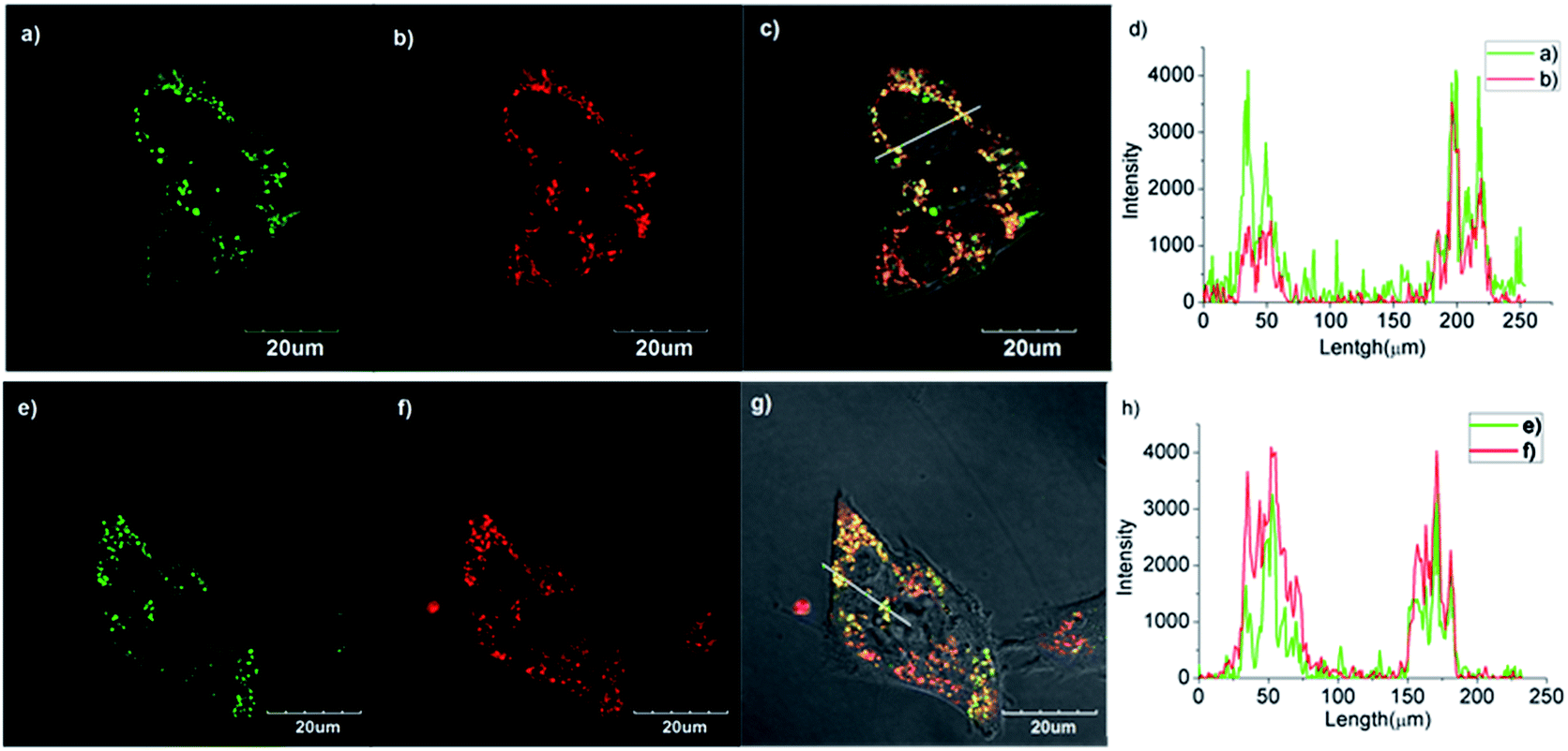 | ||
| Fig. 2 Colocalization imaging studies of the mitochondira within COS-7 cells transfected with pSNAPf-Cox8A. COS-7 cells are stained with (a) PYBG-TMR (channel 1: λex = 559 nm, λem = 575–620 nm), (e) PYBG-BODIPY (channel 1: λex = 488 nm, λem = 500–560 nm), and (b) or (f) MitoTracker Deep Red Fm (channel 2: λex = 635 nm, λem = 655–755 nm). (c) overlay of (a) and (b). (g) overlay of (e) and (f). (d) and (h) show the intensity profile of regions of interest across the cells. | ||
In addition, localization of the SNAP-tag to nucleus is achieved by fusion of SNAP-tag to the C-terminus of the nuclear histone H2B proteins (pSNAPf-H2B, NEB). After transfected with the plasmid, the nuclear histone H2B targeted SNAP-tag proteins are obtained in live cells. And then we use PYBG-TMR and PYBG-BODIPY to label nucleus. As shown in Fig. 3, the stable transfected cells stained with PYBG-TMR or PYBG-BODIPY show specific labeling of the nucleus to produce a high resolution fluorescence image without staining the area outside the nucleus. In contrast, the control cells without any transfection show no fluorescent labeling, as shown in Fig. 3b and d, which also reflect the specificity of the nucleus labeling described above.
 | ||
| Fig. 3 COS-7 cells transfected with pSNAPf-H2B (a) and (c) and non-transfected COS-7 cells (b) and (d). COS-7 cells are stained with (a) or (b) PYBG-TMR (λex = 559 nm, λem = 576–675 nm, pseudo red) and (c) or (d) PYBG-BODIPY (λex = 488 nm, λem = 495–560 nm, pseudo green). | ||
In conclusion, we have developed PYBG, a terminal alkyne substituted O6-benzylguanine, as a precursor for the efficient syntheses of new labels to SNAP-tags. PYBG can be facilely conjugated with functional molecules through ‘Click chemistry’ and Sonogashira coupling reactions. By the two ways, strong fluorescent labels, PYBG-TMR and PYBG-BODIPY, have been obtained with high yields. In the cells transfected with pSNAPf-H2B and pSNAPf-Cox8A, PYBG-TMR and PYBG-BODIPY can readily and specifically react with the SNAP-tags and label cells to produce the high resolution fluorescent images. The excellent fluorescent imaging indicates the usability of the conjugated dyes derived from PYBG. Now, the syntheses and applications of other PYBG-based labels are ongoing in our group.
Acknowledgements
This work is supported by National Natural Science Foundation of China (nos 21174022, 21376038 and 21421005), National Basic Research Program of China (no. 2013CB733702), the Program for Changjiang Scholars and Innovative Research Team in University (IRT1213) and the Fundamental Research Funds for the Central Universities (no. DUT14YQ103) and Specialized Research Fund for the Doctoral Program of Higher Education (no. 20110041110009).Notes and references
- J. W. Lichtman and J.-A. Conchello, Nat. Methods, 2005, 2, 910–919 CrossRef CAS PubMed.
- W.-S. Lo, W.-M. Kwok, G.-L. Law, C.-T. Yeung, C. T.-L. Chan, H.-L. Yeung, H.-K. Kong, C.-H. Chen, M. B. Murphy, K.-L. Wong and W.-T. Wong, Inorg. Chem., 2011, 50, 5309–5311 CrossRef CAS PubMed.
- H. Yu, Y. Xiao and L. Jin, J. Am. Chem. Soc., 2012, 134, 17486–17489 CrossRef CAS PubMed.
- B. A. D. Neto, J. R. Correa and R. G. Silva, RSC Adv., 2013, 3, 5291–5301 RSC.
- D.-S. Mei, Y. Qu, J.-X. He, L. Chen and Z.-J. Yao, Tetrahedron, 2011, 67, 2251–2259 CrossRef CAS PubMed.
- A. Keppler, S. Gendreizig, T. Gronemeyer, H. Pick, H. Vogel and K. Johnsson, Nat. Biotechnol., 2003, 21, 86 CrossRef CAS PubMed.
- A. Keppler, H. Pick, C. Arrivoli, H. Vogel and K. Johnsson, Proc. Natl. Acad. Sci. U. S. A., 2004, 101, 9955–9959 CrossRef CAS PubMed.
- E. Tomat, E. M. Nolan, J. Jaworski and S. J. Lippard, J. Am. Chem. Soc., 2008, 130, 15776–15777 CrossRef CAS PubMed.
- M. Bannwarth, I. R. Corrêa, M. Sztretye, S. Pouvreau, C. Fellay, A. Aebischer, L. Royer, E. Ríos and K. Johnsson, ACS Chem. Biol., 2009, 4, 179–190 CrossRef CAS PubMed.
- A. Keppler and J. Ellenberg, ACS Chem. Biol., 2009, 4, 127–138 CrossRef CAS PubMed.
- D. Srikun, A. E. Albers, C. I. Nam, A. T. Iavarone and C. J. Chang, J. Am. Chem. Soc., 2010, 132, 4455–4465 CrossRef CAS PubMed.
- T. Komatsu, K. Johnsson, H. Okuno, H. Bito, T. Inoue, T. Nagano and Y. Urano, J. Am. Chem. Soc., 2011, 133, 6745–6751 CrossRef CAS PubMed.
- C.-J. Zhang, L. Li, G. Y. J. Chen, Q.-H. Xu and S. Q. Yao, Org. Lett., 2011, 13, 4160–4163 CrossRef CAS PubMed.
- S. Banala, D. Maurel, S. Manley and K. Johnsson, ACS Chem. Biol., 2012, 7, 289–293 CrossRef CAS PubMed.
- H. Gong, J. L. Kovar, B. Baker, A. Zhang, L. Cheung, D. R. Draney, I. R. Correa Jr, M. Q. Xu and D. M. Olive, PLoS One, 2012, 7, e34003 CAS.
- T. Kobayashi, T. Komatsu, M. Kamiya, C. Campos, M. González-Gaitán, T. Terai, K. Hanaoka, T. Nagano and Y. Urano, J. Am. Chem. Soc., 2012, 134, 11153–11160 CrossRef CAS PubMed.
- H. Yu, Y. Xiao and H. Guo, Org. Lett., 2012, 14, 2014–2017 CrossRef CAS PubMed.
- M. Yuan, X. Yin, H. Zheng, C. Ouyang, Z. Zuo, H. Liu and Y. Li, Chem.–Asian J., 2009, 4, 707–713 CrossRef CAS PubMed.
Footnotes |
| † Electronic supplementary information (ESI) available: Details of the synthesis, NMR spectroscopic, 1H NMR, 13C NMR, HPLC, cell culture, protein expressing and labeling, and confocal imaging. See DOI: 10.1039/c4ra17072e |
| ‡ These two authors contribute equally. |
| This journal is © The Royal Society of Chemistry 2015 |