Filled and peptide-modified single-walled carbon nanotubes: synthesis, characterization, and in vitro test for cancer cell targeting

Zhiyuan Hu*ab, Jingjing Zhaoc, Zhaozheng Songa and Chunpeng Yanga
aState Key Laboratory of Heavy Oil Processing, China University of Petroleum, Beijing 102249, China. E-mail: zh209@cup.edu.cn
bDepartment of Applied Chemistry, China University of Petroleum, Beijing 102249, China
cDepartment of Chemical Engineering and Technology, Beijing University of Chemical Technology, Beijing, 100029, China

Received 25th December 2014 , Accepted 12th January 2015

First published on 12th January 2015


Abstract

Multi-functional single-walled carbon nanotubes (SWNTs) with metal endohedral filling and a high degree of polycarboxylation on the sidewalls were synthesized without affecting the SWNT σ-framework. The encapsulation of Zr4+ ions within SWNTs from aqueous solutions by the wet chemistry method was achieved using tubes “corked” with C70 at both ends to prevent the loss of the loaded metal. Carboxylic acid functionalities were introduced on the sidewalls of the tubes without introducing holes in the framework through a modified Birch-type reaction. Moreover, these metal-filled, surface polycarboxylated SWNTs can serve as versatile anchor points for coupling to specific peptides, which can target integrins for potential applications in diagnostic imaging and therapy. This directly leads to the construction of multi-functional SWNT conjugates, equipped with peptides on the surface for the targeted delivery of radionuclides to specific sites in vivo. The different SWNT adducts were characterized by Raman spectroscopy, ultraviolet-visible/near-infrared (UV-vis/NIR) spectroscopy, thermogravimetric analysis combined with mass spectrometry and also transmission electron microscopy (TEM). The aqueous solubility, cytotoxicity and cancer cell binding assay were studied for investigating the potential of these multi-functional SWNT conjugates in biomedical applications.

1. Introduction

Since the discovery of carbon nanotubes (CNT) by Iijima,1 many scientists from different groups all over the world have been attracted to these materials and a large number of studies have been carried out to promote potential applications in electricity, advanced materials, and medicine. However, the low solubility and rapid formation of bundles when dispersed in most common solvents have proved to be obstacles in the advancement of industrial applications. Normally, the direct method for increasing the CNTs' solubility is the introduction of carboxylic groups (–COOH) on the surface of the nanotube scaffold via oxidation. The most important factor of this modification is that some subsequent functionalities including polymers or biomolecules can be anchored on the nanotube surface via coupling through the carboxylic group.2–7 The issue with this method is that the σ bonds in the nanotube structure need to be broken for the subsequent oxidation reaction. This means that the nanotube structure is inevitably damaged by the introduction of holes on the surface during oxidation. As such, the unique properties and electronic network of the tubes are affected. Some non-covalent wrapping methods based on van der Waals forces and aromatic stacking have been reported for the modification of CNTs without affecting the characteristic of tubes.8,9

An electrophilic addition of CO2 to reduced nanotubes by Birch reduction is a selective and efficient method for the introduction of carboxylic acid functionalities without affecting the single-walled carbon nanotube (SWNT) σ-framework.10 The direct electrophilic addition of CO2 to the SWNT sidewalls without breaking σ bonds can maintain the structure of the nanotube scaffold. This approach can also achieve a very high degree of functionalization because CNTs are reduced to negatively charged intermediates (CNT−1) in liquid ammonia by lithium metal11–13 prior to the addition of CO2. This degree of functionalization tunable by varying different reaction conditions (reaction time, ultrasonic treatment and pressure).

If the nanotube structure is kept intact without introducing holes on the sidewall, endohedral filling of the surface functionalized SWNTs with some metals will build a complex construct with interior filling and functionalities on the sidewall simultaneously. High-yield filling of SWNTs with different kinds of halides including ThCl4,14 CdCl2,14 TbCl3, TiCl4, and PbI2, lanthanide halides LnCl3,15 KI,16,17 ZrCl4,18 and silver halides under harsh conditions (above 300 °C) leading to continuous crystalline filling was demonstrated by Professor Malcolm L. H. Green's group at Oxford. In this typical endohedral filling method via a molten salt route, the end of the nanotubes normally close under the reaction conditions, which involves the thermal annealing of the SWNTs under harsh conditions, e.g., samples are heated at temperatures at least 20 °C above the melting point of the filling material.19 The condition of this typical endohedral filling method via a molten salt route is very harsh (above 300 °C) and time-consuming (about 24 h). Considering the practical constraints of typical chemistry laboratories, in our experience the molten salt route is not compatible with the rapid generation of metal filling carbon nanotubes in aqueous media for the further radiochemistry. Therefore we developed and report here a new protocol for the rapid metal filling under mild conditions in aqueous media, which can be generalised to be applicable to water soluble radionuclide precursors in the future. A stopper molecule with a compatible outer diameter (C70) is encapsulated within the inner cavity of the tubes simultaneously with the metal to minimise the leaking of the payload in aqueous media.

In recent decades, synthetic peptide-based targeting fluorescence/PET probes have been developed for diagnostic imaging and therapy. This is because of their advantages compared with large-sized antibodies: specific peptides are easily synthesized and modified, and they are less likely to be immunogenic and have rapid blood clearance.20–24 The gastrin-releasing peptide receptor (GRP-R) targeting peptide such as Bombesin is very good candidate for visualizing tumors earlier in diagnosis, such as prostate, non-small cell lung carcinoma, breast, and gastrointestinal stromal tumors.25 Tumor progression, invasion, and metastasis of breast cancer, glioma, melanoma, and ovarian carcinoma are linked to a specific biomolecule αvβ3 integrin over-expression during tumor angiogenesis. Pentapeptide cyclo(-Arg-Gly-Asp-DPhe-Val-) was developed by Kessler and co-workers and showed high affinity and selectivity for αvβ3 integrin.26–28 Moreover, cyclo-(RGDfK) is one the most prominent structures for the development of molecular imaging agents for the assessment of αvβ3 expression.

The aim of this study was to build a complex construct with a receptor targeting peptide anchored on the surface of polycarboxylate-functionalized SWNTs and potential radionuclide metals in the interior of nanotube scaffold. SWNTs used here were purified by the “steam” method with open ends, clean surface first. Metal filling in the hollow cavity of the SWNTs was achieved by the aqueous method under mild conditions. C70 was encapsulated within the inner cavity of the tubes to prevent load loss. A modified Birch-type reaction was explored in order to extend a type selective polycarboxylation of endohedral filled CNTs via gaseous carbon dioxide. After constructing endohedral filled SWNTs–COOHx complexes, the entire surface of the CNTs were rendered biocompatible via covalent functionalization with two cancer-targeting peptides (RGDfK and Bombesin).

2. Materials and methods

2.1. Filling carbon nanotubes with zirconium acetate Zr(OAc)4

Open-ended and steaming purified (see ESI) SWNTs (1.6 mg) were added to 20 mL of ethanol and then ultrasonicated in a water bath for 20 min. SWNTs ethanol solution was splitted into two samples (A and B). 23.4 mL of zirconium acetate solution (30 mg) was added to each vial. 10 mg of sodium acetate was added into B sample. Zr(OAc)4 formed gel in both samples after two days room temperature stirring. H2SO4 was added into both samples in dropwise until the pH value was adjusted to 2.4 for avoiding the gel formation of Zr(OAc)4. Ethanol was removed by evaporation and then 10–15 mL double-distilled H2O was added into each sample without the gel forming by dropping H2SO4. Samples were kept under constant stirring at 50 °C for 48 h. The solution was filtered through a 0.2 μm polycarbonate membrane and washed with excess distilled water to remove the large excess of Zr(OAc)4 and sodium acetate. The sample was then dried under vacuum giving zirconium acetate filled SWNTs, designated as Zr(OAc)4@SWNTs. Also the unwashed Zr(OAc)4@SWNTs solution can be “corked” with C70. Prior to washing, C70 (0.4 mg) was added, stirred and heated at 50 °C overnight. After 24 h, the solution was filtered and washed several times with excess double-distilled H2O, followed by toluene wash to remove the C70 left in solution and attached the sidewall of SWNTs.

2.2. Reductive polycarboxylation functionalization of SWNTs

Steam purified SWNTs (20 mg, 1.66 mmol of carbon) were dispersed in 200 mL DMSO after 30 min ultrasonication in a heat-dried and argon purged under 30 min ultrasonication four-necked round-bottom flask (500 mL) equipped with two gas inlets and pressure compensation. The dispersion was cooled to −78 °C (acetone/dry ice) and ammonia (150 mL) was added to this flask and condensed by ice. Lithium metal (58.34 mg, 8.32 mmol) was added to the flask and the solution was stirred for 1 h yielding a blue-black colored, stable dispersion. After the reaction fully finished, the cooling was removed and the solution was kept stirring until the complete evaporation of the ammonia. The resulting lithium–bronze was removed by a syringe and the DMSO solution was ultrasonicated for 30 min prior to the addition of CO2. All solution was transferred into an autoclave reactor which was subsequently sealed. CO2 was purged to the reactor through a pressure valve (55 bar) keeping the stirring and constant pressure for 1 h. After reaction, the pressure was slowly released to atmospheric conditions. The resulting black solids were filtrated and dried in a vacuum oven at 75 °C overnight.

2.3. Postfunctionalization of Zr4+@SWNT–(COOH) with cyclo(-RGDfK) and Bombesin[7–13]

In a heat-dried and argon purged round-bottom flask (250 mL) equipped with two gas inlets and pressure compensation, Zr4+ filling and polycarboxylation functionalized SWNTs (Zr4+@SWNT–(COOH), 12 mg, 0.7 mmol of carbon) were dispersed in 100 mL THF by 30 min ultrasonication. Then the solution was cooled to 4 °C in ice bath. EDC·HCl(N-(3-dimethylaminopropyl)-N′-ethylcarbodiimide hydrochloride) (201.3 mg, 1.05 mmol, 1.5 eq.) and HOBt·H2O(1-hydroxybenzotriazole hydrate) (141.9 mg, 1.05 mmol) was added sequentially to functionalized SWNTs solution and allowed to stir for 40 min in ice bath and to this solution was added Cyclo(-RGDfK) peptide (319.8 mg, 0.53 mmol, 0.75 eq.) or Bombesin[7–13] (428.47 mg, 0.53 mmol, 0.75 eq.). The pH value of solution was adjusted to 9 with DIPEA. Then the ice bath was removed and the solution was stirred at room temperature for 96 h. Afterwards the reaction was transferred to a separation funnel with cyclohexane and water (each 100 mL). The water/THF phase was discarded, and the cyclohexane layer with the functionalized nanotubes was purged two times with water and THF (100 mL, each). The organic layer with the functionalized nanotubes was filtered through a 0.2 μm reinforced cellulose membrane filter (Sartorius) and thoroughly washed with THF, ethanol, water and saturated aqueous Na2CO3 Solution, 200 mL each. The resulting solid was dried in a vacuum oven at 75 °C overnight.

2.4. Characterization

TGA-MS was performed on a Perkin-Elmer Pyris I thermogravimetric analyzer equipped with a Hiden HPR20 mass spectrometer. The initial sample weights were about 1–3 mg, and the whole experiment was executed under inert gas atmosphere with a He gas flow (20 mL min−1) at a ramp rate of 10 °C min−1 to 700 °C after being held at 120 °C for 30 min to remove any residual solvent.

UV/Vis-NIR absorption spectra were recorded on a Perkin-Elmer Lambda 900 spectrometer. The samples were prepared by dispersing the nanotube material in H2O by sonication in an ultrasonic bath (Ultrawave U50, 30–40 kHz) for 5 min followed by filtration through a plug of cotton wool to remove particulates after allowing the solution to stand for 2 h. The concentrations of SWNTs in the supernatant solutions were calculated with the extinction coefficient reported elsewhere.

Raman spectroscopy was performed using a Renishaw upright microscope using 830 nm for excitation, LWD objective 2 cm, operated at 50× magnifications. Laser power magnification at the sample was 220 mW and spot size 100 micrometers. All spectra were recorded on solid samples over several regions and were referenced to the silicon line at 520 cm−1.

High resolution transmission electron microscopy (HR TEM) images were taken on a high resolution microscope Jeol 3000F coupled with EDX (point resolution, 0.16 nm) or a Jeol 4000F. Samples for HR TEM observation were ground and dispersed in ethanol and placed dropwise onto a holey carbon support grid.

ζ-Potential measurements were conducted on a Malvern Zetasizer NanoZS system with irradiation from a 632.8 nm He–Ne laser. The samples were filled in folded capillary cells and measured using a mixed mode method combining fast field reversal and slow field reversal, which eliminated electroosmotic effects. The ζ-potential was determined from the measured electrophoretic mobility (μ), using the Smoluchowski approximation:

image file: c4ra17047d-t1.tif
where V is the applied voltage, η is the viscosity of the solution, εm is the dielectric constant of the medium, and D is the electrode separation. This approximation is rigorously valid only for spherical particles, and may overestimate in some cases the actual zeta potential.29–31 Nevertheless it has been successfully applied to nanotube dispersions to qualitatively understand the influence of the surface charge on dispersion quality.32–35

2.5. Concentration calibration and solubility determination of SWNT–(COOH)n–peptide

A UV-vis-NIR absorption-based approach is used for a direct measurement of the unknown concentration of SWNT in aqueous supernatant via the Beer–Lambert law, according to the relation A = αcl, where A is the absorbance, I [m] is the light path length, c [g L−1] is the concentration of dispersed graphitic material, and α [L g−1 m−1 ] is the absorption coefficient. The stock SWNT solution was diluted to 10%, to avoid possible scattering losses at higher concentrations. For this purpose, peptide SWNT conjugates dispersions with different known concentrations ranging from 0.06 to 0.003 g L−1 are prepared for absorption measurements. The wavelength at 660 nm caused by the eh22 transition in the absorption spectra is chosen for determined for different SWNT concentrations.

2.6. Cell culture and receptor binding (IC50) assays

The RGP receptor binding affinity (IC50) of SWNT–(COOH)n–Bombesin was determined by a competitive cell-binding assay on PC-3 cells in cultures using 125I Tyr4-bombesin as the GRP-specific radioligand, while 125I-echistatin was used as the integrin-specific radioligand for assessment of IC50 of SWNT–(COOH)n–RGDfK with integrin αvβ3 on human glioblastoma U87MG cells.

PC-3 cells were cultured at 37 °C in a humidified atmosphere of 5% CO2 in air and split once confluence had been reached, using Dulbecco's modified Eagle medium (DMEM) medium with 10% fetal bovine serum (FBS), 200 U mL−1 L-glutamine and 100 U mL−1 penicillin. U87MG human glioblastoma cells were grown in Dulbecco's medium (Gibco) supplemented with 10% FBS, 200 U mL−1 L-glutamine and 100 U mL−1 penicillin.

During the cell-binding assay experiment, the human glioblastoma U87MG cells or PC-3 cells were harvested, washed twice with PBS, and resuspended (2 × 10⁶ cells per mL) in binding buffer (20 mmol L−1 Tris, pH 7.4, 150 mmol L−1 NaCl, 2 mmol L−1 CaCl2, 1 mmol L−1 MgCl2, 1 mmol L−1 MnCl2, 0.1% bovine serum albumin). 2 × 10⁵ cells per mL U87MG cells were seeded in 96-well plates and incubated with 125I-echistatin (30[thin space (1/6-em)]000 cpm per well) in the presence of increasing concentrations of SWNT–(COOH)n–RGDfK (0–100 μg mL−1). For SWNT–(COOH)n–Bombesin affinities test, 2 × 10⁵ cells per mL PC-3 cells were incubated at 37 °C for 40 min under 5% CO2 in the presence of 20[thin space (1/6-em)]000 cpm 125I-[Tyr4]Bombesin (2200 Ci mmol−1) and increasing concentration of SWNT–(COOH)n–Bombesin conjugates (0–100 μg mL−1). The total incubation volume was adjusted to 200 μL. After the cells were incubated for 2 h at room temperature, the medium was removed and washed twice with cold binding buffer. The radioactivity bound with cell was determined using a NaI(Tl) γ-counter (Packard Instruments). The 50% inhibitory concentration (IC50) values for the U87MG cells were calculated by fitting the data by nonlinear regression using Origin 8.0 software. Experiments were performed twice with triplicate samples.

3. Results and discussion

The steam method was used to remove the catalyst encapsulated carbon anion, amorphous carbon layers, and opening of SWNTs following a method first reported by the research group of Professor Malcolm Green, University of Oxford with Thomas Swann Ltd. As-made SWNTs were purified by introducing steam carried by an argon flow through the reactor. This method gives rise to highly purified, opened CNTs, containing only SWNTs (when as samples are used) with dimensions ranging between 50–500 nm and diameters between 0.9 and 2 nm.

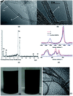
Although filling SWNTs with metal species is well established through the pioneering work of the Oxford Nanotube Group,14–16 the encapsulation of Zr4+ ions within SWNTs from aqueous solutions under rapid and mild conditions applicable for tracer preparation for PET radio-imaging has not been demonstrated. In this work, the Zr4+ filling experiment was performed from aqueous solutions using Zr(OAc)4 as the precursor as a model for the PET radioisotope filling with 89Zr.36–38 Gel formation occurred in the Zr(OAc)4 solution until the pH value was adjusted to 2.4 by the dropwise addition of H2SO4 to the gelatinous sample. Subsequently, the successful zirconium filling was achieved. However, the precise nature of the encapsulated Zr clusters is difficult to predict due to the complex aqueous chemistry of Zr(IV). The Zr4+@SWNT structures were analyzed using High Resolution TEM (HRTEM). By lowering the accelerating voltage, the knock-on damage of the carbon nonmaterial can be minimized. From HRTEM observations, crystal-filled CNTs were only found in sample 1 (C70@Zr4+@SWNT saturated with NaOAc, see Experiments section), indicating that the solution saturated with OAc is a key factor in Zr filling (Fig. 1a and b). Zirconium clusters were formed after the hydrolysis of precursor Zr(OAc)4 in acetic acid and the condensation of partially hydrolyzed species after 48 h is likely to form a three-dimensional gel network. The zirconium complex structure is likely to change after addition of sulfuric acid at pH 2.4 as the stoichiometry of Zr(IV) to the oxo-ions in solution is known to be strongly dependent on the pH. It is likely that the sulfate SO42− ions bind to Zr4+ and this counterion is expected to replace the AcO ions, but the precise nature of the zirconium clusters in this aqueous mixture could not be determined. Zirconium sulfate-based oxo-clusters are likely to be present in solution and may be introduced into the open-ended SWNTs by solution filling.39,40 The filling experiment at low pH values seemed to help convert the zirconium-aqueous gel-like material into discrete species, which in turn contributed to the successful filling of the SWNTs with Zr(IV) by the solution method.


image file: c4ra17047d-f1.tif
Fig. 1 Typical HRTEM images of (a) C70@Zr4+@SWNTs (1) observed in Zr-filled and C70 corked samples. (b) HRTEM of Zr4+encapsulated inside steam purified SWNTs. (c) The EDS spectrum of C70@Zr4+@SWNTs (1). (d) Solid state Raman spectrum of C70@Zr4+@SWNT (1 and 2) when filling was achieved in the presence or absence of NaOAc (λex = 830 nm) showing the intensity of G and D bands vs. Raman shift (cm−1) (up) RBM frequency modes (bottom).

The use of functionalized C60 was shown to allow the removal of the excess free salts in water.41 We found that the leaking of the encapsulated metallic species from open SWNTs may be prevented by the simultaneous encapsulation of C70 molecules (maximum van der Waals radii ca. 1.1 nm) rather than C60 (maximum external diameter = 0.7 nm), which remain stuck at the ends of the SWNTs. Several C70 molecules act as ideal-sized stoppers for the SWNTs carriers. HRTEM images show that the open ends of SWNTs are filled by several C70 molecules by standard endohedral encapsulation methods.41 We found that any free, un-bound C70 decorating the outside of the tubes can be removed via washing with toluene followed by the filtration/dispersion. After fullerene encapsulation, the mixture containing M@C70@SWNTs were then filtered and the solid residue was washed several times with distilled water to remove the Zr(IV) ions external to the SWNTs. Washing with water and toluene removed most of the free C70 adhering to the walls of the SWNTs, in addition to any other of bulk materials from the filling step that were external to the SWNT cavity.

Raman spectroscopy studies showed that sample 1 and 2 (sample 1: C70@Zr4+@SWNTs saturated with NaOAc; sample 2: C70@Zr4+@SWNTs without NaOAc, see Material and methods) exhibited characteristic radial breathing mode (RBM), G bands, and D bands (Fig. 1d). For sample 1 and 2, the D-band intensities decreased with respect to those of purified SWNTs, indicating that functionalization occurred. The ID/IG ratio of purified SWNTs, sample 1, and sample 2 were 0.121, 0.076 and 0.046, respectively. Clearly, there is a decrease of the band intensity ratio after filling with zirconium.

The sidewall carboxylation of CNTs was achieved by a modified Birch reaction which was reported before.42 This controllable and efficient carboxylation was based on the reduction of SWNTs in liquid ammonia to form the SWNTn intermediates which act as electron donors, transferring one electron to the CO2, yielding the respective carboxyl radical CO2˙. This radical species can subsequently attack the SWNT sidewall giving rise to carboxylated CO2–SWNT derivatives (Fig. 2a).


image file: c4ra17047d-f2.tif
Fig. 2 (a) Scheme of reductive polycarboxylation of zirconium filled SWNTs under modified Birch conditions. (b) Raman D- and G-band region of sidewall carboxylated Zr4+@SWNT: Zr4+@SWNT–(COOH)m (THF/ultrasound) (3), Zr4+@SWNT–(COOH)n (THF/ultrasound/55 bar) (4) compared with starting materials: Zr4+@SWNT (1). Excitation wavelength λex = 830 nm. (c) UV-vis/nIR absorption spectra of starting material Zr4+@SWNT and carboxylated SWNT derivatives Zr4+@SWNT–(COOH)m (THF/ultrasound) (3), Zr4+@SWNT–(COOH)n (THF/ultrasound/55 bar) (4) in DMF.

In this modified Birch reaction, the CNTs was dispersed via ultrasound in dry THF, and then the SWNTs were reduced with lithium metal in a liquid of ammonia condition. Afterwards, the ammonia was evaporated and remaining lithium–bronze can be removed forming a stable black solution with charged SWNTn species. Prior to the carboxylation, an ultrasonication step is essential for efficiently debundle CNTs, which were then reduced to charged SWNTn intermediates.

To this SWNTn intermediate solution, gaseous carbon dioxide was purged to yield the respective polycarboxylated CO2–SWNT derivatives after the final wash/workup. Raman spectroscopy method was explored for the study of polycarbonxylation of SWNTs. The direct sidewall carboxylation of CNTs can induce a prominent Raman spectra change with an increased intensity of the D-band (∼1300 cm−1) based on the rehybridization of the sp2 lattice carbon atoms into the sp3 configuration.

The resonant Raman spectra obtained with an excitation wavelength of 830 nm clearly showed a significant increase in the D-band intensity (Fig. 2b) of sidewall functionalized SWNTs. The normalized area ratio of the D-band to the G-band (1590 cm−1) (AD/AG) with respect to the ratio of the as-received starting material (AD0/AG0) can be regarded as a good measure for the degree of functionalization. The highest degree of sidewall functionalization was achieved when 50 bar pressure was applied (denoted as Zr4+@SWNT–(COOH)n n = 7.56 × 10⁵ [unit per CNT]), as compared to the Birch reduction without pressure. Specifically, the AD/AG ratio reached up to 20.2, while the AD/AG ratio of Zr4+@SWNT–(COOH) without pressure was only 6.3 (denoted as Zr4+@SWNT–(COOH)m m = 9.34 × 10⁴). This indicated that the polycarboxylation of CNTs was highly dependent on the pressure applied in the reaction.

The UV-Vis-NIR absorption spectra were studied for reveal the excitonic transitions of SWNTs after functionalization. The spectrum of 1 showed resolved absorption signals for the metallic M11 transitions from (350–600 nm), as well as from the S22 (600–1000 nm) and S11 (1100–600 nm) transitions (Fig. 2c). In contrast, the spectra of the functionalized SWNT derivatives only exhibited decreased metallic absorption signals in the M11 region between 500 and 600 nm. In the S22 and S11 regions, there were no semiconducting absorption signals detectable. Two potential explanations can be attributed to the nearly complete absence of the characteristic SWNT transitions in highly functionalized Zr4+@SWNT–(COOH)n (THF/ultrasound/55 bar) (4). One is a lack of debundle and another is covalent modification of the nanotube network. The morphology of sample 4 in TEM images clarified this issue. Representative TEM image of sample 4 (Fig. 3c) shows that the nanotubes were well dispersed and individualized. The loss of the transitions in the absorption spectra was predominantly attributed to the covalent functionalization.


image file: c4ra17047d-f3.tif
Fig. 3 Photographs showing (a) the precipitate of starting materials: C70@Zr4+@SWNTs (1) compared with (b) dissolution of Zr4+@SWNT–(COOH)n (THF/ultrasound/55 bar) (4) in DMF and (c) HR-TEM image of Zr4+@SWNT–(COOH)n (THF/ultrasound/55 bar) (4), scale bar 50 nm.

The prominent benefit of polycarboxylation of CNTs is the formation of functionalized SWNTs with highly accessible surfaces which can be post-functionalized by adding other adducts via EDC coupling (Scheme 1). Here, Zr4+@SWNT–(COOH)x–RGDfK and Zr4+@SWNT–(COOH)x–Bombesin (x = n, m) were successfully synthesized by coupling Zr4+@SWNT–(COOH)x (x = n, m) to the amino group of Bombesin's glutamine residue or the amino group of RGDfK. The synthesis of highly dispersed Zr4+@SWNT–(COOH)m (3) and Zr4+@SWNT–(COOH)n (4) is a fundamental step for subsequent functionalization of RGDfK and Bombesin to the surface of SWNT–(COOH)x through EDC coupling. Composites Zr4+@SWNT–(COOH)x–RGDfK and Zr4+@SWNT–(COOH)x–Bombesin have good solubility in DMSO, H2O and cell culture medium, which was necessary for the biomedical assessment.


image file: c4ra17047d-s1.tif
Scheme 1 EDC-mediated coupling of targeting peptides with zirconium filled, carboxylated SWNTs yielding the corresponding products: Zr4+@SWNT–(COOH)n–RGDfK (5), Zr4+@SWNT–(COOH)m–RGDfK (6), Zr4+@SWNT–(COOH)n–Bombesin (7) and Zr4+@SWNT–(COOH)m–Bombesin (8).

The respective TGA trace in combination with the ion currents of the detected peptides (RGDfK, Bombesin) for the peptide coupled SWNT derivatives Zr4+@SWNT–(COOH)n–RGDfK (5) and Zr4+@SWNT–(COOH)n–Bombesin (7) are depicted in Fig. 4. The major mass loss occurred in the temperature range between 300 and 500 °C (35.3% mass loss for 5 and 32.3% mass loss for 7) and was accompanied by the detection of the [RGDfK + H]+(m/z = 604.4) fragments as shown in Fig. 4(a), and [Bombesin + H]+(m/z = 809.4), [Bombesin/2 + H]+ (m/z = 405.38) fragments in Fig. 4(b). Assuming that the difference in weight between the functionalized derivatives Zr4+@SWNT–(COOH)n–RGDfK (5), Zr4+@SWNT–(COOH)n–Bombesin (7) and the pristine starting material can be correlated with the weight of the detached functional entities and that the residual weight can be attributed to the defunctionalized SWCNTs, the loading amount of RGDfK and Bombesin on the SWNT can be calculated. It revealed the presence of ∼1 RGDfK molecule for every 92 (1.09 atomic%) carbon atoms for Zr4+@SWNT–(COOH)n–RGDfK 5, ∼1 Bombesin molecule for every 141 (0.71 atomic%) carbon atoms for Zr4+@SWNT–(COOH)n–Bombesin 7.


image file: c4ra17047d-f4.tif
Fig. 4 (a) Thermogravimetric profile (weight loss, black) of TGA-MS for Zr4+@SWNT–(COOH)n–RGDfK (5) (heating rate: 10°C min−1), and the detected ion currents for the RGDfK fragments m/z = 604.4 ([M + H]+) (blue). (b) Thermogravimetric profile (weight loss) of Zr4+@SWNT–(COOH)n–Bombesin (7) (heating rate: 10 K min−1), vertical arrow corresponds to the weight loss (black) recorded within the temperature region of Bombesin peptide fragment detection (300–500 °C) – in combination of the detected ion currents for the two different potential Bombesin fragments m/z = 809.4(blue), m/z = 405.38(red) (corresponding to [M]2+ and [M]+).

It is important to investigate the solubility and dispersibility of peptide SWNT conjugates in aqueous media before we can move forward to biomedical experiment or application. To avoid unwanted precipitate of the SWNTs in solution, samples were mildly centrifuged (3 krpm, 5 min) after the wet chemical processing and the functionalization to remove the aggregate. SWNT concentrations (c) absorption calibration curves for SWNT–(COOH)n–Bombesin and SWNT–(COOH)n–RGDfK are shown in Fig. 5b. The slope of the curves gives the values of C value at wavelength of 660 nm. In this study the extinction coefficient corresponding to the eh22 transition (660 nm) is 3500 L g−1 m−1 for the SWNT–(COOH)n–peptide dispersions.


image file: c4ra17047d-f5.tif
Fig. 5 (a) Plot of the zeta potential of functionalized SWNTs as a function of the pH value. (b) Absorption calibration curves for SWNT obtained by monitoring the eh22 transition at wavelength of 660 nm.

The solubility and disperserbility data show that the dispersion efficiency in of Zr4+@SWNT–(COOH)n–RGDfK 5 and Zr4+@SWNT–(COOH)n–Bombesin 7 could reach 89% and 87% respectively after covalent attachment of the carboxylic acid groups and functionalized with peptides (Table 1). This data indicated that these functionalized SWNTs are highly soluble in aqueous media.

Table 1 Dispersability and receptor binding affinity of SWNT–COOH–peptide nano-constructsa
Sample 5 6 7 8
a The dispersibility is directly deduced from the ratio of the initial SWCNT concentrations 0.1 g L−1 to the actual concentrations in the supernatant.
Dispersibility (H2O) 89% 13% 85% 9%
Solubility (g L−1) 0.025 ± 0.001 0.013 ± 0.003 0.031 ± 0.003 0.011 ± 0.002
IC50 (μg mL−1) 4.21 ± 0.86 22.62 ± 3.97 5.80 ± 0.54 16.70 ± 2.24


Fig. 5a records the zeta potential of SWNT–(COOH)n, SWNT–(COOH)n–RGDfK and SWNT–(COOH)n–Bombesin as a function of the pH value. Aqueous suspensions of SWNT–(COOH)n 1, SWNT–(COOH)n–RGDfK 5 and SWNT–(COOH)n–Bombesin 7 are robust at pH conditions ranging from 3 to 10 without any observed precipitate. The zeta potential of SWNT–(COOH)n becomes more negative with increasing pH until to a neutral pH 6–7 is reached and then starts to increase again. The increase in the magnitude of the zeta potential with pH increasing from 3 to 7 can be explained by the ionization of the COOH groups on the SWNT. The peptide covalent functionalization has a dramatical influence in the reduction of the zeta potential of SWNT–(COOH)n, the zeta potential is shifted to a value range from 0 to −5 mV.

Before the binding affinity study of peptide SWNT conjugates, we evaluated the cytotoxicity of these multifunctional SWNTs to ensure the materials' concentration used for the next binding assay is safe to cells. Based the studies of SWNT's cytotoxity from literature,43–46 PC-3 cells were treated with 5 μg mL−1 to 800 μg mL−1 of SWNT–(COOH)n and SWNT–(COOH)n–Bombesin for 24 h, glioblastoma U87MG cells were treated with 5 μg mL−1 to 800 μg mL−1 of SWNT–(COOH)n and SWNT–(COOH)n–RGDfK for 24 h, respectively. The effect of three types of SWCNTs on cell viability was evaluated by MTT assay. As shown in Fig. 6, treatment of the U87MG cells and PC-3 cells with SWNT–(COOH)n decreased cell viability in a time and dose-dependent manner. The results showed that the EC50 values of was 454.1 ± 9.8 when PC-3 cells were treated with SWNT–(COOH)n for 24 h, and 454.1 ± 9.8 when U87MG cells were treated with SWNT–(COOH)n for 24 h. For SWNT–(COOH)n–RGDfK and SWNT–(COOH)n–Bombesin, both have maximum of 37.0% (in U87MG cells) and 34.6% (in PC-3 cells) inhibition at 800 μg mL−1 SWNT in comparison to the control. Therefore, no EC50 values can be determined from the MTT assay for both peptide functionalized SWNTs, as maximum cytotoxicity determined in both instances was less than 50%. Results from the MTT assay indicated the Bombesin or RGDfK functionalized SWNT to have very low acute toxicity to the PC-3 cells and U87MG cells respectively, as the 24 h EC50 values exceeded the limited concentration we can use (800 μg mL−1).


image file: c4ra17047d-f6.tif
Fig. 6 (a) Effect of SWNT–(COOH)n 1, SWNT–(COOH)n–RGDfK 5 on cell viability of U87MG cells, and (b) effect of SWNT–(COOH)n 1, SWNT–(COOH)n–Bombesin 7 on cell viability of PC-3 cells. Cells were treated with SWNTs for 24 at 37 °C. Cell viability was measured by the MTT method as described in materials and methods. Data are shown as mean ± SD values obtained from three separate experiments.

The binding affinities (IC50) of SWNT–COOH–peptides conjugates have been evaluated by competitively displacing radio-iodinated Bombesin 125I-[Tyr4]BBN by adding increasing amounts of SWNT–(COOH)m–Bombesin to the surface of PC-3 cells, and displacing the integrin-specific radioligand125I-echistatinwith the SWNT–(COOH)n–RGDfK conjugate from human glioblastoma U87MG cells. Plots of cell-bound radioactive peptides (Bombesin or RGDfK) versus increasing concentrations of SWNT–COOH–peptide conjugates have been used to determine IC50 values. It is evident from the data that the IC50 values or cell binding affinities of the conjugates depends on the degree of carboxylation on the CNT surface. Applying a high pressure (55 bar) during the reaction leads to a dramatic increase in the number of carboxyl groups covalently attached to the CNT surface. The functionalized CNTs used for EDC coupling with peptides can naturally load more bombesin/RGDfK peptides on the surface and exhibit a lower IC50 value (or a higher cell binding affinity) when compared with functionalized CNT synthesized without pressure (Table 1).

4. Conclusion

We demonstrated an aqueous solution method filling of SWNTs with Zr4+ by adjusting the pH to ca. 2 with H2SO4. Any significant leakage of metal ions from open SWNTs was avoided by simultaneous encapsulation of C70 molecules at the ends of SWNTs. The successful inner cavity filling with Zr4+ and both ends stacking with C70 molecules were imaged by HRTEM in this solution filling method. Then we explored the polycarboxylation of SWNTs by the reductive treatment of the tubes in sodium metal–ammonia solutions followed by use of gaseous carbon dioxide as electrophilic trapping molecules. Moreover, the initially introduced carboxylic acid functionalities served as anchoring points for subsequent coupling with peptides via an EDC-mediated reaction. The successful addition of specific peptides on SWNTS–(COOH)x was confirmed by the TGA-MS-based analysis of the functionalized SWNT derivatives. For both attached addends, the corresponding characteristic molecule fragments can be detected in the temperature region of a covalent bond cleavage. The solubility of functional SWNTs was precisely measured and confirms that both SWNT–peptide conjugates show excellent dispersibility in aqueous condition. The surface charge measurement reveals that the zeta potential of peptide covalent functionalized SWNTs shift to neutral compared to the higher negatively charged SWNT–(COOH)n, which is much more benefit for the cells' internalization and membrane transportation of SWNTs. The binding assay shows that higher carboxylated SWNT–COOH–peptides could load more addends on the surface and exhibited a lower IC50 value upon binding to PC-3 cells. The investigation of these polycarboxylated SWNT–COOH–peptides opens a new chapter for the application of carbon nanotube as targeting delivery therapy agent.

Acknowledgements

This work was supported by Science Foundation of China University of Petroleum, Beijing (no. 2462013YJRC029).

References

  1. S. Iijima, Nature, 1991, 354, 56 CrossRef CAS.
  2. A. M. Diez-Pascual and D. Gascon, ACS Appl. Mater. Interfaces, 2013, 5, 12107 CAS.
  3. Y. S. Park, J. Yi and J. Lee, Thin Solid Films, 2013, 546, 81 CrossRef CAS PubMed.
  4. S. Beg, M. Rizwan and A. M. Sheikh, et al., J. Pharm. Pharmacol., 2011, 63, 141 CrossRef CAS PubMed.
  5. X. Li, Y. Fan and F. Watari, Biomed. Mater., 2010, 5, 22001 CrossRef PubMed.
  6. F. Liang and B. Chen, Curr. Med. Chem., 2010, 17, 10 CrossRef CAS.
  7. H.-C. Wu, X. Chang and L. Liu, et al., J. Mater. Chem., 2010, 20, 1036 RSC.
  8. A. Ghosh, K. V. Rao and R. Voggu, et al., Chem. Phys. Lett., 2010, 488, 198 CrossRef CAS PubMed.
  9. Z. Yang, Z. Wang and X. Tian, et al., J. Chem. Phys., 2012, 136 Search PubMed.
  10. B. Gebhardt, F. Hof and C. Backes, et al., J. Am. Chem. Soc., 2011, 133, 19459 CrossRef CAS PubMed.
  11. B. Gebhardt, Z. Syrgiannis and C. Backes, et al., J. Am. Chem. Soc., 2011, 133, 7985 CrossRef CAS PubMed.
  12. D. Wunderlich, F. Hauke and A. Hirsch, J. Mater. Chem., 2008, 18, 1493 RSC.
  13. J. Chattopadhyay, A. K. Sadana and F. Liang, et al., Org. Lett., 2005, 7, 4067 CrossRef CAS PubMed.
  14. J. L. Hutchison, J. Sloan and A. I. Kirkland, et al., J. Electron Microsc., 2004, 53, 101 CrossRef CAS PubMed.
  15. C. G. Xu, J. Sloan and G. Brown, et al., Chem. Commun., 2000, 2427 RSC.
  16. R. R. Meyer, J. Sloan and R. E. Dunin-Borkowski, et al., Science, 2000, 289, 1324 CrossRef CAS.
  17. M. Wilson and P. A. Madden, J. Am. Chem. Soc., 2001, 123, 2101 CrossRef CAS.
  18. G. Brown, S. R. Bailey and J. Sloan, et al., Chem. Commun., 2001, 845 RSC.
  19. N. Thamavaranukup, H. A. Hoppe and L. Ruiz-Gonzalez, et al., Chem. Commun., 2004, 1686 RSC.
  20. P. Chowdhury, M. Gondry and R. Genet, et al., Photochem. Photobiol., 2003, 77, 151 CrossRef CAS.
  21. S. J. Miller, B. P. Joshi and Y. Feng, et al., Gastroenterology, 2011, 140, S104 Search PubMed.
  22. H. Sahoo and W. M. Nau, Chembiochem, 2007, 8, 567 CrossRef CAS PubMed.
  23. S. Scheffler, M. Sauer and H. Neuweiler, Z. Phys. Chem., 2005, 219, 665 CrossRef CAS.
  24. L. Voglino, S. A. Simon and T. J. McIntosh, Biochemistry, 1999, 38, 7509 CrossRef CAS PubMed.
  25. X. Wen, C. Chao and K. Ives, et al., BMC Mol. Biol., 2011, 12 CAS.
  26. M. A. Dechantsreiter, E. Planker and B. Matha, et al., J. Med. Chem., 1999, 42, 3033 CrossRef CAS PubMed.
  27. R. Haubner, H. J. Wester and W. A. Weber, et al., Cancer Res., 2001, 61, 1781 CAS.
  28. U. Hersel, C. Dahmen and H. Kessler, Biomaterials, 2003, 24, 4385 CrossRef CAS.
  29. A. A. Ameen, A. N. Giordano and J. R. Alston, et al., Phys. Chem. Chem. Phys., 2014, 16, 5855 RSC.
  30. H. Kato, A. Nakamura and M. Horie, RSC Adv., 2014, 4, 2129 RSC.
  31. R. W. O'Brien, J. K. Beattie and A. M. Djerdjev, J. Colloid Interface Sci., 2014, 420, 70 CrossRef PubMed.
  32. K. Min, J. Kim and K. Park, et al., J. Mol. Catal. B: Enzym., 2012, 83, 87 CrossRef CAS PubMed.
  33. Z. Sun, V. Nicolosi and D. Rickard, et al., J. Phys. Chem. C, 2008, 112, 10692 CAS.
  34. E. F. de la Cruz, Y. Zheng and E. Torres, et al., Int. J. Electrochem. Sci., 2012, 7, 3577 CAS.
  35. B. White, S. Banerjee and S. O'Brien, et al., J. Phys. Chem. C, 2007, 111, 13684 CAS.
  36. Y. Zhang, H. Hong and W. Cai, Curr. Radiopharm., 2011, 4, 131 CrossRef CAS.
  37. M. A. Deri, S. Ponnala and B. M. Zeglis, et al., J. Med. Chem., 2014, 57, 4849 CrossRef CAS PubMed.
  38. A. Kasbollah, P. Eu and S. Cowell, et al., J. Nucl. Med. Technol., 2013, 41, 35 CrossRef PubMed.
  39. A. C. Geiculescu and H. J. Rack, J. Non-Cryst. Solids, 2001, 289, 53 CrossRef CAS.
  40. A. C. Geiculescu and H. J. Rack, J. Non-Cryst. Solids, 2002, 306, 30 CrossRef.
  41. M. Yudasaka, K. Ajima and K. Suenaga, et al., Chem. Phys. Lett., 2003, 380, 42 CrossRef CAS PubMed.
  42. B. Gebhardt, F. Hof and C. Backes, et al., J. Am. Chem. Soc., 2011, 133, 19459 CrossRef CAS PubMed.
  43. L. M. Pasquini, S. M. Hashmi and T. J. Sommer, et al., Environ. Sci. Technol., 2012, 46, 6297 CrossRef CAS PubMed.
  44. R. Subbiah, S. Ramasundaram and P. Du, et al., J. R. Soc., Interface, 2013, 10, 20130694 CrossRef PubMed.
  45. H. N. Yehia, R. K. Draper and C. Mikoryak, et al., J. Nanobiotechnol., 2007, 5, 8 CrossRef PubMed.
  46. B. Farshid, G. Lalwani and B. Sitharaman, J. Biomed. Mater. Res., Part A, 2014, 4, 1 Search PubMed.

Footnote

Electronic supplementary information (ESI) available: Steaming purification of SWNTs; synthesis of bombesin [7–13] peptide (2) and cyclic deprotected RGDfK peptide; 1HNMR. See DOI: 10.1039/c4ra17047d

This journal is © The Royal Society of Chemistry 2015
Click here to see how this site uses Cookies. View our privacy policy here.