Zhao Chenab,
Xiaochi Wanga,
Hua Lic,
Chao Lid,
Qinghua Lub,
Guang Yangd,
Jiangang Longc and
Lingjie Meng*a
aSchool of Science, State Key Laboratory for Mechanical Behavior of Materials, Xi'an Jiaotong University, Xi'an, 710049, P. R. China. E-mail: menglingjie@mail.xjtu.edu.cn
bSchool of Chemistry and Chemical Technology, State Key Laboratory of Metal Matrix Composites, Shanghai Jiaotong University, Shanghai, 200240, P. R. China
cSchool of Life Science and Technology, Xi'an Jiaotong University, Xi'an, 710049, P. R. China
dInternational Center for Dielectric Research, Xi'an Jiaotong University, Xi'an, 710049, P. R. China
First published on 13th February 2015
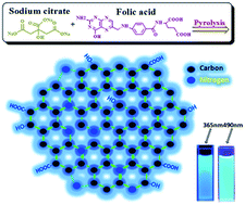
A facile but effective bottom-up method for the mass preparation of N-doped carbon dots (N–C-dots) was developed by the direct heating of a solid mixture of folic acid (FA) and sodium citrate (SC) for several minutes at 300 °C. The nitrogen content of the resulting N–C-dots could be easily and precisely tuned from 0% to 14% by adjusting the SC to FA mass ratio. Uniform N–C-dots with honeycomb-like ordered structures were obtained, with sizes ranging from 1 to 3 nm in diameter. The N-doping content affected not only the emission quantum yield but also the emission wavelength. The N–C-dots emitted strong blue-green fluorescence based on the excitation wavelength and N-doping content. Because of their excellent water solubility, low toxicity, powerful fluorescence, and high resistance to photobleaching, the N–C-dots can enter various cells and serve as ideal candidates for multicolour cell imaging.
For these reasons, it is imperative to develop effective methods for preparing C-dots and N–C-dots for various applications. Two basic strategies have been proposed: “top-down” methods by cutting the carbon resources22–27 and “bottom-up” approaches by chemical synthesis from small organic molecules.28–39 N–C-dots were typically synthesized by the “bottom-up” strategy, which involved carbonizing the mixtures of N-sources (urea,14 ammonia,15 hydroxylamine,30 amoxicillin,31 NaNH,32 dimethylamine,33 among others) and C-sources under hydrothermal or microwave-assisted hydrothermal conditions. However, these fabrication methods were often time-consuming; they also afforded low yield of products and were difficult to extend to mass production and to precisely control the N-doping content.
Herein, we developed a facile but effective synthesis to prepare N–C-dots in one-pot by directly heating a solid mixture of folic acid (FA) and sodium citrate (SC) for several minutes. The N-doping content of the resulting N–C-dots could be well tuned from 0% to 14% by adjusting the mass ratio of SC to FA. Through systematic investigations, we found that the N-doping content affected not only the QY but also the emission wavelength. This method provides a new method for the mass production (several grams or more) of N–C-dots with tunable emissions for various applications. Owing to their good water solubility, low toxicity, and strong fluorescence, the N–C-dots were directly applied in cell imaging to demonstrate one of their promising applications.
:
0, 40
:
1, 20
:
1, 10
:
1, 6.7
:
1, 5
:
1, 2.5
:
1, 2
:
1, 1
:
1, 1
:
2, 1
:
4, and 0
:
1) was fully grinded in an agate mortar. The mixed powders were placed into glass tubes and heated at 300 °C in an oil bath for 4 min. The resulting black or brown solid was dissolved in pure water (10 mL) and filtered through a PVDF membrane, resulting the C-dot or N–C-dot aqueous solution.
The PL property of N–C-dots is closely related to their concentration and the degree of thermopolymerization; thus, the reaction time and concentration were optimized through fluorescence tests. An aqueous N–C-dot solution emitted a bright fluorescence under light irradiation of 365 nm. The fluorescence intensity increased with increasing N–C-dot concentration and reached a maximum at about 0.5 mg mL−1 (see ESI, Fig. S1†). With further increase in the N–C-dot concentration, the fluorescence intensity decreased, probably because of the aggregation of the N–C-dots, thereby resulting in fluorescence quenching. Based on the result, we further investigated the fluorescence intensity of 0.5 mg mL−1 N–C-dots prepared at different reaction times. The N–C-dots prepared at 300 °C for 4 min exhibited the strongest fluorescence (see ESI, Fig. S2†), suggesting that the optimal reaction time is approximately 4 min.
The sizes and structures of the as-synthesized N–C-dots were investigated by HRTEM. Fig. 1 shows typical HRTEM images of N–C-dots derived from an SC
:
FA mixture of 20
:
1. As shown in Fig. 1a and c, relatively uniform N–C-dots were observed, with sizes ranging from 1 to 3 nm in diameter. The magnified image provides direct evidence that SC and FA react with each other to form N–C-dots with clear lattice fringes, which is similar to those reported previously.23,36 Along with amorphous ultrathin carbon-coated structures on TEM grids, honeycomb-like structures were observed in some regions of the N–C-dots, with each honeycomb-like hole being approximately 0.12 nm (Fig. 1d). The partially ordered structures in the dots might benefit the fluorescence property. All other samples exhibited size and morphology similar to those in Fig. 1.
![]() | ||
Fig. 1 (a and b) HRTEM images of N–C-dots (SC to FA mass ratio is 20 : 1). (c) Size distribution of N–C-dots in (a). (d) Amplified HRTEM of N–C-dots in (b). | ||
C bonds during the reaction. In the C-dot spectrum, the peaks at 1690, 2950, and 3300 cm−1 corresponded to the stretching vibrations of OH, CH2, and C
O groups, respectively, implying that C-dots still contain some COOH and CH2 groups and that carbonization is not complete. In the FTIR spectrum of N–C-dots prepared from pure FA, the peaks at 3200 and 1390 cm−1 corresponding to NH2 and NH groups, respectively, almost disappeared, probably because amides react with neighboring carboxylic groups by intra- and intermolecular dehydrolysis. However, the peaks at 1690 and 3300 cm−1 corresponding to the stretching vibrations of OH and C
O groups, respectively, were still observed, implying that the N–C-dots also contain some COOH. Of note, the C
N vibration peak at 1670 cm−1 gradually increased proportionally with the FA content (see ESI, Fig. S3†).
![]() | ||
Fig. 2 (a) FTIR spectra of the C-dots, N–C-dots and their raw materials. (b) Raman spectra of N–C-dots derived from different SC : FA mass ratios. | ||
Raman spectra provided additional evidence that all the obtained C-dots and N–C-dots have ordered microstructures (Fig. 2b). All samples exhibited an obvious peak at 1600 cm−1, which is the characteristic tangential displacement mode of the highly crystalline graphite. In addition, the D band at approximately 1350 cm−1 resulting from amorphous carbon and defects weakened with increasing FA content, probably because the rigid pteridine rings of FA might aid in the formation of ordered structures in N–C-dots.
The actual N-doping content in the N–C-dots was studied by XPS analysis (Fig. 3 and Table 1). In the XPS spectra of N–C-dot samples, the peaks at approximately 400 and 284 eV corresponded to N 1s and C 1s, respectively. The nitrous content of the C-dots was zero since the SC precursor did not contain any nitrogen element. Interestingly, the nitrous content in the N–C-dots increased proportionally to the FA
:
SC mass ratio. An increase of approximately 1.4% in the nitrous content for the N–C-dots corresponded to a 10% increase of FA in the precursor mixtures. Therefore, the N-doping content of the resulting C-dots can be tuned from 0% to 14% in a facile and quantitative manner by adjusting the SC
:
FA mass ratio. On the other hand, FA had a significantly lower content of C–O and C
O than SC. The oxygenated carbon decreased as a result of the increase of the FA
:
SC mass ratio. In addition, the zeta potentials of the N–C-dots increased with increasing nitrous content (Table 1), which is in good agreement with the results obtained from XPS.
![]() | ||
Fig. 3 XPS full-scan survey. Inset: high-resolution C 1s and N 1s XPS spectra. The mass ratio of FA to SC is (a) 0 : 1, (b) 1 : 10, (c) 1 : 2, and (d) 1 : 0. | ||
XPS is a standard technique for studying the nitrogen and carbon configurations through N 1s and C 1s spectra.37,38 In the C 1s spectra of N–C-dots, four peaks were deconvoluted: C
C peak at 284.66 eV, C–C peak at 285.39 eV, C–O/C–N peak at 286.1 eV, and C
O/C
N peak at 287.59 eV (see Fig. 3 and ESI, Table S1†). The C–C configuration constitutes the majority of carbon atoms in a C-dot sample, indicating that most of carbon atoms are sp3 hybridized, and only small sp2 clusters are isolated within the incompletely carbonized C-dots. With increasing FA
:
SC mass ratio, the content of C
C gradually increased because the pteridine and benzene rings of FA were maintained. In addition, three common bonding configurations may be obtained when doping nitrogen into the C-dots: pyridinic N (398.38 eV), pyrrolic N (399.08 eV), and quaternary N (400.14 eV). At a low FA
:
SC mass ratio, the amino and imino groups on FA were significantly lesser than the carboxyl groups on FA and SC; multiple carboxyl groups might react with one amino group and lead to high quaternary N content. With increasing FA
:
SC mass ratio, the amount of quaternary N gradually decreased, while that of pyridinic N increased correspondingly (see the inset in Fig. 3 and ESI, Table S1†).
C bonds. With increasing FA content, the aqueous solutions of N–C-dots gradually became darker and exhibited two dominant absorbance peaks at 270 and 365 nm, respectively. The absorption curves of N–C-dots were very similar to those of FA, except for slight peak broadening and a small blue shift, implying that the conjugated FA structure is mainly maintained during the reaction.
Based on the UV-Vis spectra, the fluorescent properties of C-dots and N–C-dots were compared under light irradiation of 254, 365, and 490 nm (Fig. 4c). All samples exhibited very weak or no PL under light excitation of 254 nm (Fig. 4a and c); however, they emitted strong luminescence under light irradiation of 365 and 490 nm. The maximum emission wavelength of C-dots and N–C-dots derived from pure SC and FA was 520 and 445 nm under light irradiation of 365 nm, respectively, while that of all other N–C-dots was 475 nm (Fig. 4b). Interestingly, all the samples emitted strong green fluorescence under exposure to 490 nm (Fig. 4c), and their maximum emission wavelength was about 550 nm with a half-peak width of approximately 70 nm (see ESI, Fig. S4†).
The exact fluorescence mechanism of C-dots and N–C-dots remains to be elucidated. We proposed that several sp2 clusters were isolated within the sp3 C–C, C–N, and C–O matrix to prevent the formation of large conjugated structures;39,40 therefore, only blue and green fluorescence can be observed. It is consistent with the results of XPS and UV-Vis spectra. Interestingly, the fluorescence QY is closely related to the SC
:
FA mass ratio. For example, the QY of C-dots under light irradiation of 365 nm was approximately 1.3%, and the QY of N–C-dots reached 4.56% when the FA
:
SC mass ratio increased to 1
:
10. With further increase in the FA content, the QY and fluorescence intensity of N–C-dots gradually decreased, and the emission almost disappeared at a FA
:
SC mass ratio of 1
:
1 or greater. Remarkably, the QY of N–C-dots prepared from pure FA dramatically increased to 28.39%, probably caused by the involvement of a totally different reaction mechanism and formation of a new structure. Therefore, the N content affected not only the maximum emission wavelength but also the fluorescence QY. The substitution of N atoms, which introduces the unpaired electron, might change the atomic charge distribution and electronic gap. This can be indirectly demonstrated by the fluorescence lifetime. With increasing N-doping content, the lifetime gradually increased from 4.57 ns to 12.5 ns (see ESI, Fig. S5†).
Photostability is a particularly important criterion in cases in which it is often desirable to observe markers for extended periods against the background of intrinsic cellular emissions. Remarkably, the N–C-dots were quite stable under light irradiation of 365 nm (2 W) and exhibited only negligible reduction in the observed intensities for 30 min (see ESI, Fig. S6†).
![]() | ||
| Fig. 5 Viability of (a) HeLa and (b) H9C2 cells incubated with N–C-dots at diverse concentrations for 24 h (black) and 48 h (gray) determined by the MTT assay. | ||
Based on their low toxicity, powerful fluorescence, and excellent resistance to photobleaching, the N–C-dots were directly used for cell imaging to demonstrate one of their promising applications. The HeLa and H9C2 cells were incubated with N–C-dots (50 μg mL−1) for 6 h, and fluorescence images were recorded by confocal microscopy. Bright blue and green images were observed for HeLa cells incubated with N–C-dots upon light irradiation of 405 and 488 nm, respectively, caused by the strong fluorescence emitted by N–C-dots (Fig. 6a and b). Surprisingly, the regions at both the cell wall and nucleus were bright, indicating that a large amount of C-dots has been internalized into the cells and even in the nucleus. When being merged into one picture, Fig. 6a and b perfectly overlap (Fig. 6c). A similar phenomenon was observed for H9C2 cells (Fig. 6d–f). However, the nucleus region in H9C2 was dark, implying that the N–C-dots cannot enter the H9C2 nucleus, probably because of the denser nuclear membrane and smaller nuclear pores of H9C2. Therefore, N–C-dots can act as ideal fluorescent nanomaterials for various cell imaging applications.
:
FA mass ratio. Uniform C-dots and N–C-dots with some honeycomb-like order structures were obtained, with sizes ranging from 1 to 3 nm in diameter. The C-dots and N–C-dots emitted blue and green fluorescence based on the excitation wavelength and N-doping content. The nitrogen content affected not only the emission wavelength but also the emission quantum yield. This study provides a new method to achieve the mass production of N–C-dots with tunable emissions for various applications. Because of their low toxicity, powerful fluorescence, and high photostability, the N–C-dots can enter various cells and serve as ideal candidates for multicolour cell imaging.
Footnote |
| † Electronic supplementary information (ESI) available: FL spectra of C-dots at different concentration, FTIR spectra of N–C-dots prepared with different mass ratio of SC to FA, FL spectra of N–C-dots (0.5 mg mL−1) prepared at different reaction time, FL spectra of C-dots and N–C-dots under irradiation of 490 nm, FL lifetime of the C-dots under irradiation of 365 nm, FL spectra of N–C-dots under 365 nm light (2 W) at different irradiation time, and XPS analysis of N–C-dots. See DOI: 10.1039/c4ra16990e |
| This journal is © The Royal Society of Chemistry 2015 |