Characterization of supports activated with divinyl sulfone as a tool to immobilize and stabilize enzymes via multipoint covalent attachment. Application to chymotrypsin

Jose C. S. dos Santosab, Nazzoly Ruedaac, Oveimar Barbosad, Jorge F. Fernández-Sáncheze, Antonio L. Medina-Castillof, Teresa Ramón-Márqueze, María C. Arias-Martosf, Ma Carmen Millán-Linaresg, Justo Pedrocheg, María del Mar Yustg, Luciana R. B. Gonçalvesb and Roberto Fernandez-Lafuente*a
aICP-CSIC, Departamento de Biocatálisis, Instituto de Catálisis-CSIC, C/ Marie Curie 2, Campus UAM-CSIC, Cantoblanco, 28049 Madrid, Spain. E-mail: rfl@icp.csic.es
bDepartamento de Engenharia Química, Universidade Federal do Ceará, Campus do Pici, CEP 60455-760, Fortaleza, CE, Brazil
cEscuela de Química, Grupo de Investigación en Bioquímica y Microbiología (GIBIM), Edificio Camilo Torres 210, Universidad Industrial de Santander, Bucaramanga, Colombia
dFacultad de Ciencias, Departamento de Química, Universidad del Tolima, Ibagué, Colombia
eDepartment of Analytical Chemistry, University of Granada, Avd. Fuentenueva s/n, 18071 Granada, Spain
fNanoMyP, Nanomateriales y Polimeros S.L., Spin-Off company of the UGR, BIC building, Avd. Innovacion 1, 18100, Granada, Spain
gInstituto de la Grasa-CSIC, Av Padre García Tejero 4, 41012 Sevilla, Spain

Received 23rd December 2014 , Accepted 16th February 2015

First published on 16th February 2015


Abstract

Divinyl sulfone (DVS) has been used to activate agarose beads. The DVS activated agarose resulted quite stable in the pH range 5–10 at 25 °C under wet conditions, and can react rapidly with α-amides of Cys and His, at pH 5–10, with Lys mainly at pH 10 and with Tyr in a much slower fashion. After blocking with different nucleophiles, the support lost all reactivity, confirming that this protocol could be useful as an enzyme–support reaction end point. Then, chymotrypsin was immobilized on this support at pH 5, 7 and 10. Even though the enzyme was immobilized at all pH values, the immobilization rate decreased with the pH value. The effect of the immobilization on the activity depended on the immobilization pH, at pH 7 the activity decreased (to 50%) more than at pH 10 (by a 25%), while at pH 5 the immobilization has no effect. Then, the effect of blocking with different reagents was analyzed. It was found that blocking with ethylenediamine improved the enzyme activity by 70% and gave the best stability. The stability of all enzyme preparations improved when 24 h incubation was performed at pH 10, but the qualitative stabilization depended on the inactivation conditions. The analysis of the amino acids of the preparation immobilized at pH 10 showed that Lys, Tyr and Cys residues were involved in the immobilization, involving a minimum of 10 residues (glyoxyl agarose gave 4 Lys involved in the immobilization). The new preparation was 4–5 fold more stable than glyoxyl agarose preparation, considered a very stable one, and in some instances was more active than the free enzyme (170% for the enzyme immobilized at pH 10). Thus, DVS activated supports are very promising to permit the multipoint covalent attachment of enzymes, and that way to improve their stability.

1. Introduction

Immobilization is in many instances a compulsory step in the design of a biocatalyst, as it is the simplest solution to the problems generated by the solubility of the proteins in aqueous media, enabling the enzyme recovery and its separation from the reaction medium.1–6 Immobilization also simplifies bioreactor design and control over the reaction.7–10

Thus, many researchers have focused their efforts on the use of the “compulsory” immobilization step to improve other enzymes properties, mainly the enzyme stability,9,11,12 but also activity, specificity or selectivity.13–15

Enzyme stabilization via immobilization may be achieved via different phenomena.16,17 Any enzyme molecule that is immobilized and dispersed in the surface of a porous support cannot suffer any intermolecular inactivation process (precipitation, proteolysis, interaction with external hydrophobic interfaces).16,17 Moreover, a proper immobilization system may permit to improve the enzyme stability by generating a favorable enzyme environment,17–19 by avoiding the subunit dissociation of multimeric proteins11 or by increasing the enzyme rigidity via multipoint covalent attachment.6,10,12,14,20–22

Multipoint covalent attachment has revealed itself as one of the most potent tools to improve enzyme stability.12,14,20,22 The selection of the support, the immobilization conditions and the reactive groups on enzyme and support are key points in the preparation of enzyme biocatalysts stabilized via multipoint covalent attachment.23,24

The support must offer flat surfaces to the reaction with the enzyme (e.g., agarose) and must present many reactive groups on that surface.12 The immobilization conditions must favor the enzyme mobility and the reactivity of the supports and enzyme groups (moderate temperatures, alkaline pH values, long reaction times).23,24 Finally, the reactive group on the support needs to be able to react with enzyme moieties that are generally abundant in the enzyme surface.12 Moreover, it must offer low steric hindrances to the reaction with the enzyme groups, good stability under immobilization conditions and be placed at a moderate distance from the support surface to really transmit the rigidity of the support to the enzyme.12 The number of support groups suitable for producing very intense multipoint covalent attachment is not very high. The supports activated with glyoxyl,25,26 epoxyde27–29 and the versatile glutaraldehyde30,31 have offered good results in this topic and some industrial enzymatic biocatalysts have been prepared using these chemistries. However, each of these active groups has some problems which avoid their universal use, making very interesting to find some new protocols.

Glyoxyl supports have been described as very suitable to get an intense multipoint covalent attachment.25 This good result occurs even though the immobilization on this support only involves the primary amino groups of the protein.25 However, immobilization needs to be performed at alkaline pH value, even proteins with low density of Lys cannot be immobilized26 and the end point of the reaction requires the use of borohydride.32 Epoxy-supports may react with a wide range of protein groups (amino, thiol, phenol, imidazole, carboxy),33 but they have very low reactivity, even making a first adsorption of the enzyme on the support necessary to get a first covalent immobilization34–37 (this has been useful to develop epoxy heterofunctional supports).38 Due to this low reactivity, a very intense multipoint covalent attachment is hard to achieve compared to glyoxyl supports.39 Glutaraldehyde activated supports have low stability, and very low stability at alkaline pH,31 usually the best results are achieved by modifying anionically exchanged enzymes on amino supports (and that is not always positive for the enzyme activity because it means a global enzyme surface modification).30 Thus, the search of new reactive groups on the support potentially useful to stabilize enzymes via multipoint covalent attachment is still a demand in the biocatalyst design.

In this regard, supports activated with divinyl sulfone (DVS) have been successfully used to immobilize some enzymes,40–47 but its potential use to stabilize the immobilized proteins and some of the relevant features to determine their prospects as a support for industrial immobilization of enzymes have not been even analyzed.

Activation with DVS may be achieved in supports bearing in its surface a very wide range of groups, like amino, thiol or hydroxy groups.42,47,48 The reactive vinyl sulfone groups placed in the support can react with amino, phenol, imidazol or thiol groups of the proteins,47,49 moieties that are frequently placed in its surface, therefore useful to get many enzyme–supports linkages.50

In this paper, we have analyzed the prospects of DVS-agarose beads as support not just to immobilize enzyme, but to stabilize enzymes via multipoint covalent attachment. In literature there are a lack of information on some key features of a support to be considered a good support to produce an intense multipoint covalent attachment. For example, the reactivity of the different moieties of the proteins at different pH values and the stability of the groups under immobilization conditions are very important characteristic and have been analyzed for first tome ion this paper, also some alternatives as enzyme–support reaction end-point have been compared. Then, a model enzyme, bovine alpha-chymotrypsin, has been immobilized on the DVS-support and the variables that determine the final stabilization have been studied. This enzyme has been selected because it may be highly stabilized via multipoint covalent attachment; in fact it has been highly stabilized after immobilization on glyoxyl agarose beads.51 The protocol of enzyme immobilization is usually critical to take the maximum profit of the characteristics of the support to achieve an intense multipoint covalent attachment, it may be stated that good results may be achieved only if a good support and a good immobilization protocol is utilized.

2. Materials and methods

2.1. Materials

Divinyl sulfone, α-chymotrypsin from bovine pancreas, benzoyl-L-tyrosine-p-nitroanilide (BTNA), ethylenediamine, ethanolamine, glycine, aspartic acid, cysteine and 2-mercaptoethanol were purchased from Sigma Chemical Co. (St. Louis, MO). The α-amides of Lys, His, Tyr and Cys were purchased from Bachem. Agarose beads 6% (w/v) were purchase from Agarose Bead Technologies (ABT, Spain). All other reagents were of analytical grade.

2.2. Methods

All experiments were performed by triplicate and the results are reported as the mean of this value and the standard deviation (under 10%).
2.2.1. Enzymatic assays. The activity of the soluble or immobilized enzyme (30 mg mL−1) was assayed by determination of the increase in absorbance at 405 nm which accompanies the hydrolysis of the synthetic substrate BTNA (100 or 200 μL of soluble or suspended enzyme were added to 2.5 mL 50 mM sodium phosphate 40% ethanol, pH 7.0, containing 30 μL of 40 mM BTNA in hexane[thin space (1/6-em)]:[thin space (1/6-em)]dioxane 1[thin space (1/6-em)]:[thin space (1/6-em)]1 (v/v) at room temperature).23

In the determination of the effects of the pH value on the enzyme activity, the followed protocol was similar but the buffer in the measurements was changed according to the pH value studied: 100 mM of sodium acetate at pH 5–0, sodium phosphate at pH 6.0–8.0 and sodium borate at pH 9–0 and pH 10.0. At 25 °C, all the preparations remained fully stable after incubation for several hours at any of these pH values. The ε values suffered very slight variations with the change in the pH value.

2.2.2. Preparation of glyoxyl-agarose-support. The activation of agarose gels was done according to the procedure previous described.25,26 The agarose beads were suspended in 1 M NaOH and 0.5 M NaBH4 (3 mL of solution per g of support). This suspension was maintained in an ice bucket under mechanical stirring, and glycidol was added drop wise in order to reach a 2 M final concentration. The resulting suspension was gently stirred overnight at room temperature. The activated gel was then washed with abundant distilled water.

Them, the glycidol activated support was incubated in a solution of water containing 80 μmol of NaIO4 per wet gram of beads (10 mL of oxidation solution per g of wet support). This oxidative reaction was allowed to proceed for 2 h under mild stirring at room temperature.52 Then, the glyoxyl support was washed with an excess of distilled water and stored at 4 °C under wet conditions.

2.2.3. Preparation of divinyl sulfone-support. A volume of 7.5 mL divinyl sulfone was added to 200 mL of 333 mM sodium carbonate buffer at pH 12.5 and vigorously stirred until the solution became homogeneous, then 10 g of agarose beads was added and left under gentle agitation for 35 minutes. Then, the support was washed with an excess of distilled water and stored at 4 °C.
2.2.4. Determination of the reaction rates between DVS-support and different amino acids. The pH of 10 mL of 2 mM of amides with the α-amino acid of the amino acids His, Tyr, Lys and Cys, was adjusted at pH 5.0 (100 mM, sodium acetate), pH 7.0 (100 mM, sodium phosphate) or pH 10.0 (100 mM, sodium bicarbonate). Then, 1 g of DVS-support was added. Inert agarose was used as a reference. The remaining amide in the supernatant was measured periodically using an UV spectra (wavelength was 220 nm) (Jasco V-630) and in some cases the concentration was confirmed by HPLC (Spectra Physic SP 100) coupled to an UV detector (Spectra Physic SP 8450).
2.2.5. Immobilization of the enzymes.
2.2.5.1. Immobilization on glyoxyl-support. The immobilization was performed suspending 10 g of wet support in 100 mL of chymotrypsin solution (maximum protein concentration was 1 mg mL−1), prepared in 50 mM sodium carbonate at pH 10.0–10.1 at 25 °C for 3 h under continuous stirring.51 As a reaction end point, derivatives were reduced by addition of solid NaBH4 (to reach a concentration of 1 mg mL−1). After gentle stirring for 30 min at room temperature, the resulting derivatives were washed with abundant distilled water to eliminate residual sodium borohydride.
2.2.5.2. Immobilization on divinyl sulfone-support. The immobilization was performed suspending 10 g of wet support in 100 mL of proteins solutions (maximum protein concentration was 1 mg mL−1), prepared in 50 mM sodium acetate at pH 5.0, sodium phosphate at pH 7.0 or sodium carbonate at pH 10.0, always at 25 °C. In some cases, the immobilized enzyme preparations were incubated in 100 mL of 100 mM bicarbonate at pH 10.0 and 25 °C for different times before stopping the enzyme–support reaction by blocking the support. As a reaction end point, all the immobilized biocatalysts were incubated for 24 hours at room temperature in 1 M of different nucleophiles (ethylenediamine, ethanolamine, glycine, aspartic acid, cysteine or mercaptoethanol) dissolved in 100 mM sodium carbonate at pH 10.0. Finally, the immobilized enzyme preparations were washed with an excess of distilled water and stored at 4 °C.
2.2.6. Thermal inactivations. To check the stability of the enzyme derivatives, 1 g of immobilized enzyme was suspended in 5 mL of 50 mM sodium acetate at pH 5.0, sodium phosphate at pH 7.0 or sodium carbonate at pH 9.0 at different temperatures. Periodically, samples were withdrawn and the activity was measured using BTNA. Half-lives were calculated from the observed inactivation courses.
2.2.7. Determination of the amino acids involved on the enzyme–support multipoint covalent attachment. The bonds formed by the enzymes and the glyoxyl (after reduction), or the DVS supports (after blocking) are highly stable and may stand acid hydrolysis of proteins. This strategy has been used previously with very good results on different glyoxyl-immobilized enzymes.23,24 The number of free amino acids of the different biocatalysts was obtained by determining the amino acids of each biocatalyst that could be released to the medium by the method previously described by Alaiz et al.53 Briefly, samples of each derivative, containing 2–4 mg of enzyme, were hydrolyzed with 6 N HCl at 120 °C and subsequently analyzed by high-performance liquid chromatography (HPLC) after derivatization with ethoxymethylenmalonate, using D,L-α-aminobutyric acid as internal standard, and a 300 × 3.9 mm i.d. reverse-phase column (Novapack C18, 4 μm; waters). Likewise, amino acid composition of soluble chymotrypsin was also determined, in the presence and absence of blocked DVS support to ensure the lack of artifacts caused by the support. Concentrations (mol per g protein) of each amino acid were determined and the number of residues was calculated as followed:
Number of amino acid/molecule of chymotrypsin × amino acid concentration in sample/amino acid concentration in chymotrypsin.
2.2.8. Studies of enzyme structure and amino acid accessibility. Protein structures were modeled using PyMol software version 0.99rc6.54 Surface accessibility (ASA) values of residues from 1TCA were calculated by the web-based program ASA-view.55 Solvent accessibility was divided into three classes: buried, partially exposed and exposed, indicating, respectively, the least, moderate and high accessibility of the amino acid residues to the solvent.56,57

3. Results

3.1. Characterization of the DVS-agarose as a matrix to immobilize proteins and stabilize them via multipoint covalent attachment

3.1.1. DVS reactivity versus different amino acids under different pH values. The scheme of the reaction between the DVS and the agarose is shown in Scheme 1. A support to be used in immobilization and stabilization of enzymes via multipoint covalent attachment must have a good reactivity with groups frequently placed in the enzyme surface. DVS has been reported to react with hydroxyl, imidazol, amino or thiol groups.40–47,58 Thus, Lys, Tyr, His and Cys are the amino acids that can exhibit a higher reactivity with the DVS-support.
image file: c4ra16926c-s1.tif
Scheme 1 Activation of agarose with DVS and reaction of DVS activated supports with proteins.

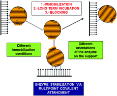
Moreover, it may be expected that the reactivity of the different amino acids with DVS may differ at different pH value. This may open the opportunity to control the enzyme orientation by using conditions that can favor the reactivity of the support with one group kind or another, changing the amino acids involved in the first covalent attachment.

To this purpose, the amides (to eliminate the reactivity of the amino acid alpha-amino group) of Lys, Cys, His and Tyr were offered to DVS-activated supports at pH 5.0, 7.0 and 10.0. Table 1 shows the results. As expected, the highest immobilization rates for all amino acids were observed at pH 10.0 and the lowest at pH 5.0.

Table 1 Reaction rates of the α-amides of different amino acids. The experiments have been performed as described in Section 2. The immobilization rates are given as μmol of immobilized amide per h per g
Amino acid Immobilization rates
pH 10 pH 7 pH 5
Lysine 14.20 ± 0.5 1.09 ± 0.2 0.04 ± 0.01
Cysteine 24.80 ± 1 5.60 ± 0.4 2.60 ± 0.1
Tyrosine 0.73 ± 0.1 0.40 ± 0.1 0.27 ± 0.05
Histidine 21.00 ± 1 7.33 ± 0.8 1.67 ± 0.2


Lys, the most abundant residues among the studied ones had a good reactivity at pH 10.0, which drastically decreased at pH 7.0 and even more at pH 5.0. Cys and His are the most reactive groups at all studied pH values, decreasing the reactivity when the pH value was lowered in a slower fashion than Lys, while Tyr is the less reactive group, except at pH 5.0 where it is more reactive than Lys. Thus, it seems that at pH 5.0 the Lys residues will play an irrelevant role in the first immobilization of an enzyme on DVS activated supports even being the most abundant group, because the ionization degree of its amino group. At lower pH value, it is expected that the terminal amino group may be more relevant in the immobilization of the enzyme. The terminal amino group should have a pK value 2–3 unit below that of the Lys (10.7), thus its reactivity at low pH value should be much more significant than that of the Lys residues, mainly at pH 5.0.

3.1.2. DVS stability under different experimental conditions. The usefulness of a support to immobilize enzymes at industrial level may be marked by the stability of the active groups under different conditions. This may affect the storage (i.e., it is simpler if the support may be stored under wet conditions at 25 °C that if it requires to be conserved under dry conditions at −20 °C). Stability of the support groups also determines the range of conditions where the support can be used in the enzyme immobilization. Another point to be considered is that only if the support reactive groups are stable enough, the support surface may be fully covered of enzyme molecules because this may require a relative long time. On the contrary, if the support groups are instable, it is possible that they become inactivated before the full support surface is coated with enzyme. Multipoint covalent attachment usually requires a quite long time, as the support and the enzyme are quite rigid structures that need to get the correct alignment to get an intense multipoint covalent attachment under conditions where the support and enzyme have a good reactivity attachment.23,24 This makes that only supports having good stability may be used in the long term incubation necessary to get an intense multipoint covalent.

In order to check the stability of the DVS groups under different conditions that could be useful to immobilize different enzymes, the supports were incubated at pH values ranging from pH 4.0 to pH 10.5 at 25 °C.

Periodically, samples were extracted and their reactivity versus N-alpha-amide-Lys at pH 10.0 was evaluated. After 24 h of incubations at 25 °C, there was no difference in the reaction rate between the Lys-amide and the DVS support in all conditions, which means that the support remained fully reactive in this range of pH values. At pH 7.0 and 25 °C, after 60 days, the reactivity of the support did not suffer any relevant decrement (less than 5%). The stability was really high even at 36 °C, over 90% of the reaction rate versus Lys was observed after 60 days of incubation at pH 7.

Thus, storage seems to be possible even under wet conditions at pH 7.0. Moreover, DVS supports seem to be useful to immobilize enzymes at 25 °C in a broad range of pH values, also useful to get multipoint covalent attachment (even at pH 10.0 in 24 h the reactivity of the support is maintained).

3.1.3. Reaction end point. The usefulness of a support to immobilize enzymes, and more if the final goal is to have a highly stabilized biocatalyst, is favored if there is some simple protocol to eliminate the chemical reactivity of the support with the enzyme when the desired degree of enzyme–support reaction has been achieved. This will permit the full control of the enzyme–support reaction, otherwise during operation the enzyme and the support can produce new covalent bonds that can drive to the inactivation of the enzyme by stabilizing inactive enzyme structures. To this goal, the DVS support was blocked by incubation in the presence of different compounds for 24 h, and also was reduced with 1 mg mL−1 sodium borohydride (compatible with many enzymes stability),32 or submitted to incubation overnight in 1 M NaOH at 40 °C to destroy the support reactivity. The incubation in NaOH left a support fully unreactive with the α-amide of Lys. Similar results were obtained by the blocking with all the studied compounds. However, using NaBH4, the reactivity of the support decreased to 20%, but we were not able to fully eliminate the support reactivity even using 5 mg NaBH4 mL−1, although this very high concentration was not compatible with the stability of many enzymes.32 Thus, as in the case of the epoxyde-activated supports, the proposed end point to the enzyme support reaction is the blocking of the support with different nucleophiles.59 The selection of the blocking reagent will depend on the specific enzyme properties, and may be used also as a tool to further tuning the enzyme properties.60

3.2. Immobilization of chymotrypsin in DVS supports at different pH values

From the previous results, the DVS-support seems very adequate to immobilize enzymes at industrial level. It may be handled or stored even in wet conditions and room temperature for weeks without decreasing its reactivity, and it permits its use at pH values from 5.0 to 10.0 for 24 h without any significant decrease in support reactivity. Coupling this stability results with the data on reactivity versus different amino acids, and the possibility of blocking by incubation with different nucleophiles, DVS supports seemed to have very good prospects to immobilize proteins under a broad range of pH values and to produce an intense support/enzyme multi-reaction.

Next, the immobilization of chymotrypsin has been tried in DVS-agarose at pH ranging from 5.0 to 10.0.

3.2.1. Analysis of the accessibility of the different amino acids to the medium. First of all, the number of reactive groups of the enzyme and their accessibilities in the enzyme surface (ASA) were studied. Table 2 shows the amount of likely reactive groups of chymotrypsin and their medium exposition degree. From Table 2, the 14 Lys residues that this enzyme has, are reasonably exposed to the medium, while from the 4 Tyr, only Tyr-171 have good exposition, while Tyr-228 is not exposed. Both His have a very low exposition. Regarding the 10 Cys all of them are involved in disulfide bridges (1–122; 42–58; 136–201; 168–182; 191–220) and therefore with low reactivity via the thiol, group. Moreover, the amino terminal Cys has a very high exposition (and can react via its amino group), while the other have moderate or even null exposition to the medium.
Table 2 List of reactive groups of chymotrypsin and their medium accessibilities (ASA). Calculations have been performed as described in Section 2. Surface accessibility (ASA) values of residues from 1TCA were calculated by the web-based program ASA-view
Amino acid Tyr 94 Tyr-146 Tyr-171 Tyr-228 Lys-36 Lys-79 Lys-82 Lys-84
%ASA 24.3 15.9 59 0.5 74.4 99.2 73.4 73.4
Amino acid Lys-87 Lys-90 Lys-93 Lys-107 Lys-169 Lys-170 Lys-175 Lys-177
%ASA 55.9 62.7 70 19.9 43.3 89.9 46.7 37.4
Amino acid Lys-202 Lys-203 His-40 His-57 Ile-16 Ala-149 Cys-1 Cys-42
%ASA 62.2 37.9 12.1 2.7 0.5 27.2 77 2.8
Amino acid Cys-58 Cys-122 Cys-136 Cys-168 Cys-182 Cys-191 Cys-201 Cys-220
%ASA 11.1 7.6 0 0 0 4.9 0.7 16


The terminal amino groups Ile-16 and Ala-149 have a moderate or very low exposition respectively. The requirement for the medium exposition is higher when we intend to achieve the reaction between the enzyme and a flat surface (e.g., groups inside pockets will hardly react with the support, at least in a first step, before the enzyme is distorted by the enzyme–support interactions). However, the enzyme may suffer some distortions during immobilization involving some new groups in the immobilization. Moreover, once the enzyme is immobilized, only groups located in that face of the protein can react with the support. To better visualize, this, Fig. 1 shows the structure of the chymotrypsin with the reactive groups marked, it seems that many groups on the enzyme may take part on the immobilization step and on the further multipoint covalent attachment, mainly in the upper area to the active center, while in the other face the number of reactive groups decreased. The possibilities of getting an intense multipoint attachment seems to be not limited by the number and distribution of groups located on the enzyme surface.


image file: c4ra16926c-f1.tif
Fig. 1 3D surface structure model of chymotrypsin. The 3D surface structure model of Chymotrypsin indicates lysine, tyrosine and histidine residues and the N-terminal amino acids. (a) N-terminal face, (b) back face. The 3D surface structure was obtained using PyMol versus 0.99. The 3D structure of chymotrypsin was obtained from the Protein Data Bank (PDB). For chymotrypsin pdb code is 5CHA.
3.2.2. Chymotrypsin immobilization. As expected from the data on amino acids reactivity, immobilization proceeded far more rapid at pH 10.0 than at pH 7.0 or 5.0 (Fig. 2). In fact, at pH 10.0 immobilization in DVS-agarose (90% enzyme immobilized in 2 h) is even more rapid than the immobilization in the same support activated with glyoxyl groups (80% immobilization after 2 h), a support described as very suitable for the immobilization/stabilization of this enzyme.51 At pH 7.0, the enzyme immobilization was slower, with a yield of 75% after 24 h, and at pH 5.0 was even slower, with immobilization yield of only 10% after 24 h.
image file: c4ra16926c-f2.tif
Fig. 2 Immobilization courses of chymotrypsin on DVS activated agarose at different pH values experiments have been performed at 25 °C, other specifications are described in Section 2. Panel A: (pH 5.0), Panel B: (pH 7.0), Panel C: (pH 10.0): circles (suspension), square (supernatant), triangle (soluble enzyme).

Regarding the activity (Fig. 2), the immobilization on DVS at pH 10.0 produced a decrease in enzyme activity of 25% after 24 h. At pH 7.0, the decrease in activity is more significant, the expressed activity of the immobilized enzyme is around 50% of that of the free enzyme. At pH 5.0, there are no significant changes in enzyme activity. The more drastic decrease in activity after immobilization at pH 7.0 cannot be related to a more intense reaction between enzyme and support, as show in the analysis of the reactivity of the different amino acids with this support showed in point 3.1.1, but may be explained if a different orientation of the enzyme in the support is produced. Fig. 1 shows that many of the groups relevant for enzyme immobilization are quite close to the active center of the enzyme.

3.3. Effect of the blocking step in the stability of chymotrypsin immobilized on DVS supports

Next, the enzyme that had been immobilized at pH 10.0 was subject to incubation in the presence of different nucleophiles to check the effects on enzyme activity and stability of the blocking of the reactive groups on the support. Fig. 3 shows that the incubation of the enzyme that had been immobilized at pH 10.0 in the presence of EDA permitted to increase the enzyme activity by a 75% that decreases to 70% after 24 h.
image file: c4ra16926c-f3.tif
Fig. 3 Effect on enzyme activity of the incubation of the immobilized enzyme in the presence of different blocking agents. Experiments have been carried out at 25 °C and at pH 10.0 using the enzyme immobilized at pH 10.0. Other specifications are described in Section 2. Circles, solid black line: (EDA); squares, solid black line: (ethanolamine); triangles, solid black line: (Gly); rhombus, solid black line: (Asp); stars, solid black line: (Cys); gray circles, solid gray line: (mercaptoethanol).

In opposition, the blocking with Cys produced a severe decrease of the enzyme activity, perhaps by their effect on the disulfide bonds that have chymotrypsin, while the incubation in the presence of the other blocking reagents did not produce a significant effect on enzyme activity. The biocatalyst blocked with EDA presented an activity slight higher than the activity of the free enzyme, reverting the slight decrease on enzyme activity observed during the immobilization step.

Fig. 4 shows the thermal inactivation courses of the enzyme blocked with the different reagents.


image file: c4ra16926c-f4.tif
Fig. 4 Thermal inactivation courses of the enzyme blocked with the different blocking agents. Experiments have been performed at 60 °C and pH 8.0, using the enzyme immobilized at pH 10.0. Other specifications as described in Section 2. Circles, solid black line: (EDA); squares, solid black line: (ethanolamine); triangles, solid black line: (glycine); rhombus, solid black line: (aspartic acid); stars, solid black line: (cysteine); gray circles, solid gray line: (pH 10.0).

The less stable preparation was that unblocked, as the support still has the possibility to react with the distorted enzyme structure induced by heat. The most stable one was that blocked with EDA, while the less stable ones were those blocked with Gly or Asp, suggesting that the ionic nature of the support surface plays an important role on the stability of the immobilized chymotrypsin. Considering the high isoelectric point of chymotrypsin (9.2), the support with EDA may produce a lower number of ionic bridges with the enzyme than the Asp.

Thus, the EDA was selected as blocking reagent in further experiments.

3.4. Effect of the long term alkaline incubation on the activity/stability of DVS-chymotrypsin biocatalysts

In order to improve the enzyme stability, the enzyme that had been immobilized at pH 10.0 was further incubated at pH 10.0 for different times before the EDA blocking step. It should be considered that is not possible to ensure that during the blocking step there is not some enzyme–support reaction, and that can somehow minimize the effect of the long term incubation.

In this experiment, the biocatalyst was blocked with EDA just after immobilization (2 h), after 24 h, and after 72 h. The effect of this treatment on enzyme activity and stability has been analyzed (Fig. 5). The long term incubation produced a certain decrement in enzyme activity (near to 50% activity was lost after 72 h, Fig. 5a), while improving the enzyme stability, more clearly for the comparison between 2 and 24 h and in an almost negligible way if the incubation was prolonged from 24 to 72 h (Fig. 5b). Thus, incubation for 24 h at pH 10.0 seemed adequate to get optimal activity/stability parameters.


image file: c4ra16926c-f5.tif
Fig. 5 Effect of the long incubation time on the activity/stability of DVS-chymotrypsin biocatalysts. Panel (A) evolution of the activity of the chymotrypsin immobilized at pH 10.0 and 25 °C. Other features are described in Section 2. Panel (B) inactivation course of the different enzyme preparation at pH 10.0 and 25 °C. Other features are described in Section 2. Circles, solid black line: DVS-chymotrypsin-6 h; squares, solid black line: DVS-chymotrypsin-24 h; triangles, solid black line: DVS-chymotrypsin-72 h.

3.5. Effect of the immobilization pH on the final activity/stability of the DVS-chymotrypsin preparation

As previously discussed, the immobilization pH value may alter the orientation of the enzyme on the support, and that way this may alter the final enzyme stability. This may be caused by the different amount of protein nucleophiles in each area (giving more or less possibilities of establishing many enzyme–support linkages) or by the relative importance of a specific area on enzyme stability. Thus, the activities and stabilities of enzymes immobilized at pH 5.0, 7.0 and 10.0 and blocked directly after immobilization have been compared to that of all these immobilized enzymes incubated for 24 h at pH 10.0 before the blocking step, giving similar possibilities at all enzyme preparations of producing an intense multipoint covalent attachment. Again, it must be considered that during the blocking step at alkaline pH, that may favor the enzyme–support reactivity, it is likely that some enzyme–support reaction may occur, and that may reduce the impact of the long term alkaline incubation. Table 3 shows the recovered activities of the different preparations and the half-lives obtained in thermal inactivations performed at pH 5.0, 7.0 and 9.0.
Table 3 Thermal stability of the different enzyme preparations is given as half-lives in minutes. Temperatures were 55 °C at pH 5.0, 65 °C at pH 7.0 and 60 °C at pH 9.0. Other specifications are described in Section 2
Biocatalysts Recovered activitya (%) Half-lives
a 100 is the activity of the soluble enzyme activity recovered after the blocking step.
Soluble 100 pH 5 pH 7 pH 9
pH 5 (24 h) 170 ± 10 38 ± 3 17 ± 2 60 ± 4
pH 5 (24 h) + pH 10 (24 h) 140 ± 15 150 ± 10 60 ± 4 125 ± 8
pH 7 (24 h) 148 ± 14 47 ± 3 11 ± 2 23 ± 2
pH 7 (24 h) + pH 10 (24 h) 110 ± 14 180 ± 12 90 ± 4 87 ± 5
pH 10 (2 h) 175 ± 12 60 ± 3 120 ± 7 33 ± 4
pH 10 (24 h) 140 ± 13 195 ± 8 200 ± 9 117 ± 8
Glyoxyl 80 ± 5 31 ± 3 10 ± 2 11 ± 1


Regarding the activity, in many instances the positive effects of the blocking with EDA compensated the decrease of activity produced by immobilization. In fact, all preparations were finally more active than the free enzyme. The positive effect of the blocking was similar in all preparations, and also the decrease caused by the alkaline pH incubation (even though the immobilization presented a higher negative effect on the enzyme activity at pH 7.0).

Regarding the effect on stability, the results offer a complex picture. At all three pH values of inactivation and immobilization, the enzymes incubated at pH 10.0 for 24 h showed an improved stability when compared to the enzyme blocked just after immobilization, the stabilization caused by the incubation ranged from less than 2 to more than 4 folds depending on the biocatalyst pH immobilization value and inactivation pH value. As the only possible difference between the enzymes just immobilized and those incubated for longtime at alkaline pH value is an increase in the number of enzyme–support linkages, it seems that the alkaline incubation favored the enzyme–support reaction.

In inactivations at pH 5.0 of the just immobilized biocatalysts, the most stable one was that immobilized at pH 10.0 and the least stable was that immobilized at pH 5.0. After alkaline incubation, stability of all preparations increased, and the least stable preparations are the most stabilized. Thus, finally the stabilities of all of them became quite similar (half-lives from 150 min for preparation immobilized at pH 5.0 to 195 minutes when immobilized at pH 10.0).

When the inactivations were performed at pH 7.0, the enzyme immobilized at pH 10 is again clearly the most stable one just after immobilization (120 minutes versus half-live of 17 minutes for the biocatalyst prepared at pH 5.0 or 11 minutes if the biocatalyst is prepared at pH 7.0), and remains in this relative position after alkaline incubation even though it is the less stabilized by this treatment (half live of 200 minutes versus 90 for the derivative immobilized at pH 7.0 or 60 if the immobilization is at pH 5.0).

However, in inactivations at pH 9.0, the most stable preparation just after immobilization is that immobilized at pH 5.0 (half-live of 60 minutes), doubling the stability of the other preparations. After incubation at pH 10.0, stability of the enzyme immobilized at pH 5.0 and 10.0 become similar, while that of the enzyme immobilized at pH 7.0 is clearly inferior. These qualitative differences on the stabilities of the different preparations suggest that the relevance of the different areas involved in the immobilization is not identical under any inactivation cause.61–63

Thus, the immobilization pH seems to really alter the enzyme orientation on the support, as that should be the only relevant difference between the immobilized enzymes after incubation at pH 10.0 for 24 h.

3.6. Comparison of glyoxyl-chymotrypsin, BrCN-agarose and glyoxyl-chymotrypsin

Fig. 6 shows an inactivation course of the enzyme immobilized on DVS-agarose at pH 10.0 and incubated for 24 h before the blocking step and the chymotrypsin immobilized on glyoxyl under optimal conditions51 or BrCN agarose. BrCN-chymotrypsin is by far the least stable preparation, with full inactivation in the first measure in all the inactivation pH values. Glyoxyl-chymotrypsin was a much more stable preparation, as has been previously reported. Nevertheless, DVS-agarose-chymotrypsin is more stable than the very stable glyoxyl preparation at all studied pH values. In fact, even the biocatalysts prepared without the alkaline long term incubation were quite more stable than the glyoxyl support. This result suggested the good prospects of this support to give an important stabilization of enzymes via immobilization.
image file: c4ra16926c-f6.tif
Fig. 6 Inactivation courses of the chymotrypsin immobilized on DVS-agarose, glyoxyl-agarose or BrCN-agarose. Experiments have been performed at 60 °C and pH 8.0. Other features are described in Section 2. Triangles, solid black line: CNBr; square, solid black line: glyoxyl; circles, solid black line: DVS.

3.7. Determination of the number of enzyme–support linkages

To confirm that the enzyme was attached to the support via several attachments, the number of amino acids that can be released from the DVS and glyoxyl supports had been compared (Table 4). As reference, Arg, Ala and Pro were selected. The implication of the Ile-16 and Ala-149 (amino terminal groups) in the immobilization was not evaluated, as just one amino acid is not detected by this method.
Table 4 Free amino acids of different immobilized chymotrypsin preparations. Experiments have been performed as described in Section 2 (CT is chymotrypsin)
  CT CT experimental DVS/CT DVS-CT (pH 10.0) Glyoxyl-CT
His 2 2.1 ± 0.3 1.8 ± 0.2 1.5 ± 0.3 1.8 ± 0.3
Arg 3 3.9 ± 0.1 3.6 ± 0.1 2.6 ± 0.1 3.3 ± 0.6
Ala 22 22.3 ± 1.5 18.7 ± 1 14.4 ± 1 17.9 ± 1
Pro 9 9.3 ± 0.5 8.9 ± 0.8 9.8 ± 0.7 9.0 ± 0.3
Tyr 4 4.0 ± 0.3 3.9 ± 0.2 2.1 ± 0.2 3.3 ± 0.2
Cys 10 9.8 ± 0.5 10.3 ± 0.9 6.5 ± 0.3 9.5 ± 0.5
Lys 14 14.2 ± 0.6 13.6 ± 0.6 8.0 ± 0.4 10.6 ± 0.5


The amounts of the target groups in the presence or absence of blocked DVS were quite coincident; suggesting that the support did not alter the results (that is, can not react with any amino acid). Glyoxyl support involved in the immobilization around 4 Lys groups, while DVS-involved at least 6 Lys, 1–2 Tyr and even several Cys (3–4) seemed to be involved in the immobilization that means that the disulfide bridge has been broken, as only the Cys 1 (that is the amino terminal) can react by its amino group, suggesting some enzyme distortion while multipoint covalent attachment was established. There were no clear indications on the involvement of any of the His on the immobilization. These results show two points: first, a very intense multipoint covalent attachment has been achieved (at least 10–12 groups involved), and second, the implication of at least Tyr, Lys and Cys on the multipoint covalent attachment has been shown. This occurred even though the reactivity of the free Tyr seemed to be very low even at pH 10.0, perhaps because the reaction is now “intramolecular”. Thus, it is evident that DVS-supports are very efficient to produce an intense multipoint covalent attachment, even using the only moderately favorable agarose 4BCL, that is an agarose with not very thick agarose fibers.23

3.8. Activity/pH profile of different immobilized chymotrypsin preparations

Fig. 7 shows the pH/Activity curve. The free enzyme had a clear optimum at pH 8.0, and the activity decrease is relatively marked around this optimum value. All immobilized preparations presented a different optimum pH value, and the curve is less narrow than using the free enzyme. The pH of immobilization on the DVS-support produced significant changes on the activity/pH curve, while the enzyme immobilized at pH 5.0 had the highest activity at pH 9.0, the other two preparations have the highest activity at pH 10.0, the maximum value used in this study to prevent chemical hydrolysis of the substrate.
image file: c4ra16926c-f7.tif
Fig. 7 Effect of the pH on the activity versus BTNA of the different α-chymotrypsin preparations. Experiments have been performed as described in Section 2. Circles, solid black line: (pH 5.0); circles, solid gray line: (pH 5.0 + pH 10.0); square, solid black line: (pH 7.0); square, solid gray line: (pH 7.0 + 10.0); triangle, solid black line: (pH 10.0); triangle, solid gray line: (pH 10.0 + pH 10.0); stars, solid black line: (soluble).

The 24 h incubation at alkaline pH produced also changes. The enzyme immobilized at pH 5.0 is the one that suffered the greatest alterations in the pH/activity curve, changing a clear optimal at pH 9.0 to a flat plateau in the range 7–10. The enzyme immobilized at pH 7 also improved the percentage of activity at pH values from 7.0 to 9.0, while the enzyme immobilized at pH 10.0 almost did not suffer any change after the long term alkaline incubation.

4. Conclusion

This paper shows the great potential of DVS-activated agarose not to immobilize enzymes, but to stabilize them via multipoint covalent attachment. First, the main features of the active groups to this goal has been analyzed. The support is very stable, maintaining its reactivity after storage for two months even at 36 °C in wet condition, also retained full reactivity after 24 h of incubation at pH 4.0 to 10.5 at 25 °C. This support is able to react with Lys, His, Cys and Tyr, with a rate that depends on the pH value. Regarding reactivity with groups of proteins, DVS and epoxide are capable to react with different nucleophiles,42 while glyoxyl only can react with primary amino groups.25 However, DVS is much more reactive than epoxy groups, being able to covalently immobilize enzymes without requiring the previous adsorption of the enzyme. Moreover, DVS supports can be used in a wide range of pH values, in opposition to glyoxyl agarose that generally require the immobilization at alkaline pH value.26 DVS can directly yield stable enzyme–support linkages, being no necessary any treatment to stabilize these bonds (e.g., imine bonds obtained using glyoxyl require reduction). However, to avoid uncontrolled enzyme–support reaction, the support may be blocked using different nucleophiles. This may become a tool to further tailoring immobilized enzyme features.60

Using it as a support to obtain a stabilized preparation of chymotrypsin, the results have been really good. To take advantages of the support properties, a proper immobilization protocol needs to be utilized, as multipoint covalent attachment is a quite complex process. The first enzyme immobilization may be performed at different pH values, obtaining preparations with different activity/stability properties, but still multipoint covalent attachment has not be maximized. Further incubation at pH 10.0 produced an increase in enzyme stability with some costs in terms of activity. The blocking of the remaining sulfone groups is another critical variable, as show in this paper and expected from the higher stability of DVS groups. In this case, the blocking with EDA permitted to avoid undesired covalent enzyme–support reactions, and improved the enzyme activity, that become even slightly higher than that of the free enzyme (175% measured at pH 7.0) and stability. The results after the long term incubation at alkaline pH are different depending on the immobilization pH, considering that the support is full stable in the used conditions, and that the blocking is identical, the only likely explanation is that the enzyme orientation may be different depending on the immobilization pH value and that determine the number of enzyme groups that can react with the support, or affecting regions of the enzyme with different relevance for the enzyme stability. Thus, the immobilization protocol to have an optimized enzyme stabilization via an intense multipoint covalent attachment is a first immobilization of the enzyme on DVS-agarose at different pH values (to involve different areas of the enzyme in the immobilization), an incubation under alkaline conditions to improve the enzyme reactivity and have an intense multipoint covalent attachment, and an optimization of the blocking step (assaying different blocking reagents) to have the best activity/stability features.

Acknowledgements

We gratefully recognize the support from the Spanish Government, grant CTQ2013-41507-R. The predoctoral fellowships for Mr dos Santos (CNPq, Brazil) and Ms Rueda (Colciencias, Colombia) are also recognized. The authors wish to thank Mr Ramiro Martínez (Novozymes, Spain) for kindly supplying the enzymes used in this research. The help and comments from Dr Ángel Berenguer (Instituto de Materiales, Universidad de Alicante) are kindly acknowledged.

References

  1. I. Chibata, T. Tosa and T. Sato, J. Mol. Catal., 1986, 37, 1–24 CrossRef CAS.
  2. W. Hartmeier, Trends Biotechnol., 1985, 3, 149–153 CrossRef CAS.
  3. E. Katchalski-Katzir, Trends Biotechnol., 1993, 11, 471–478 CrossRef CAS.
  4. J. F. Kennedy, E. H. M. Melo and K. Jumel, Chem. Eng. Prog., 1990, 86, 81–89 CAS.
  5. A. M. Klibanov, Science, 1983, 219, 722–727 CAS.
  6. D. Brady and J. Jordaan, Biotechnol. Lett., 2009, 31, 1639–1650 CrossRef CAS PubMed.
  7. L. Cao and F. I. Innovation, in Comprehensive Biotechnology, 2011, vol. 1, pp. 461–476 Search PubMed.
  8. P. Adlercreutz, Chem. Soc. Rev., 2013, 42, 6406–6436 RSC.
  9. M. Petkar, A. Lali, P. Caimi and M. Daminati, J. Mol. Catal. B: Enzym., 2006, 39, 83–90 CrossRef CAS PubMed.
  10. R. K. Singh, M. K. Tiwari, R. Singh and J. K. Lee, Int. J. Mol. Sci., 2013, 14, 1232–1277 CrossRef CAS PubMed.
  11. R. Fernandez-Lafuente, Enzyme Microb. Technol., 2009, 45, 405–418 CrossRef CAS PubMed.
  12. C. Mateo, J. M. Palomo, G. Fernandez-Lorente, J. M. Guisan and R. Fernandez-Lafuente, Enzyme Microb. Technol., 2007, 40, 1451–1463 CrossRef CAS PubMed.
  13. R. C. Rodrigues, C. Ortiz, Á. Berenguer-Murcia, R. Torres and R. Fernández-Lafuente, Chem. Soc. Rev., 2013, 42, 6290–6307 RSC.
  14. K. Hernandez and R. Fernandez-Lafuente, Enzyme Microb. Technol., 2011, 48, 107–122 CrossRef CAS PubMed.
  15. U. Guzik, K. Hupert-Kocurek and D. Wojcieszyńska, Molecules, 2014, 19, 8995–9018 CrossRef CAS PubMed.
  16. A. Illanes, Comprehensive Biotechnology, Elsevier, 2011 Search PubMed.
  17. R. C. Rodrigues, Á. Berenguer-Murcia and R. Fernandez-Lafuente, Adv. Synth. Catal., 2011, 353, 2216–2238 CrossRef CAS.
  18. C. Mateo, B. Fernandes, F. van Rantwijk, A. Stolz and R. A. Sheldon, J. Mol. Catal. B: Enzym., 2006, 38, 154–157 CrossRef CAS PubMed.
  19. K. Hernandez, A. Berenguer-Murcia, R. C. Rodrigues and R. Fernandez-Lafuente, Curr. Org. Chem., 2012, 16, 2652–2672 CrossRef CAS.
  20. A. M. Klibanov, Anal. Biochem., 1979, 93, 1–25 CrossRef CAS.
  21. L. Gianfreda and M. R. Scarfi, Mol. Cell. Biochem., 1991, 100, 97–128 CrossRef CAS.
  22. C. Garcia-Galan, Á. Berenguer-Murcia, R. Fernandez-Lafuente and R. C. Rodrigues, Adv. Synth. Catal., 2011, 353, 2885–2904 CrossRef CAS.
  23. J. Pedroche, M. del Mar Yust, C. Mateo, R. Fernández-Lafuente, J. Girón-Calle, M. Alaiz, J. Vioque, J. M. Guisán and F. Millán, Enzyme Microb. Technol., 2007, 40, 1160–1166 CrossRef CAS PubMed.
  24. R. M. Blanco, J. J. Calvete and J. Guisán, Enzyme Microb. Technol., 1989, 11, 353–359 CrossRef CAS.
  25. C. Mateo, J. M. Palomo, M. Fuentes, L. Betancor, V. Grazu, F. López-Gallego, B. C. C. Pessela, A. Hidalgo, G. Fernández-Lorente, R. Fernández-Lafuente and J. M. Guisán, Enzyme Microb. Technol., 2006, 39, 274–280 CrossRef CAS PubMed.
  26. C. Mateo, O. Abian, M. Bernedo, E. Cuenca, M. Fuentes, G. Fernandez-Lorente, J. M. Palomo, V. Grazu, B. C. C. Pessela, C. Giacomini, G. Irazoqui, A. Villarino, K. Ovsejevi, F. Batista-Viera, R. Fernandez-Lafuente and J. M. Guisán, Enzyme Microb. Technol., 2005, 37, 456–462 CrossRef CAS PubMed.
  27. E. Katchalski-Katzir and D. M. Kraemer, J. Mol. Catal. B: Enzym., 2000, 10, 157–176 CrossRef CAS.
  28. T. Boller, C. Meier and S. Menzler, Org. Process Res. Dev., 2002, 6, 509–519 CrossRef CAS.
  29. L. Hilterhaus, B. Minow, J. Müller, M. Berheide, H. Quitmann, M. Katzer, O. Thum, G. Antranikian, A. P. Zeng and A. Liese, Bioprocess Biosyst. Eng., 2008, 31, 163–171 CrossRef CAS PubMed.
  30. O. Barbosa, C. Ortiz, Á. Berenguer-Murcia, R. Torres, R. C. Rodrigues and R. Fernandez-Lafuente, RSC Adv., 2014, 4, 1583 RSC.
  31. O. Barbosa, R. Torres, C. Ortiz and R. Fernandez-Lafuente, Process Biochem., 2012, 47, 1220–1227 CrossRef CAS PubMed.
  32. R. M. Blanco and J. Guisán, Enzyme Microb. Technol., 1989, 11, 360–366 CrossRef CAS.
  33. J. Turková, K. Bláha, M. Malaníková, D. Vancurová, F. Svec and J. Kálal, Biochim. Biophys. Acta, 1978, 524, 162–169 CrossRef.
  34. W. R. Melander, D. Corradini and C. Horváth, J. Chromatogr., 1984, 317, 67–85 CrossRef CAS.
  35. K. Smalla, J. Turkova, J. Coupek and P. Hermann, Biotechnol. Appl. Biochem., 1988, 10, 21–31 CAS.
  36. J. B. Wheatley and D. E. Schmidt, J. Chromatogr., 1993, 644, 11–16 CrossRef CAS.
  37. J. B. Wheatley and D. E. Schmidt, J. Chromatogr. A, 1999, 849, 1–12 CrossRef CAS.
  38. O. Barbosa, R. Torres, C. Ortiz, A. Berenguer-Murcia, R. C. Rodrigues and R. Fernandez-Lafuente, Biomacromolecules, 2013, 14, 2433–2462 CrossRef CAS PubMed.
  39. J. M. Bolivar, C. Mateo, V. Grazu, A. V. Carrascosa, B. C. Pessela and J. M. Guisan, Process Biochem., 2010, 45, 1692–1698 CrossRef CAS PubMed.
  40. F. J. Lopez-Jaramillo, M. Ortega-Munõz, A. Megia-Fernandez, F. Hernandez-Mateo and F. Santoyo-Gonzalez, Bioconjugate Chem., 2012, 23, 846–855 CrossRef CAS PubMed.
  41. P. Prikryl, J. Lenfeld, D. Horak, M. Ticha and Z. Kucerova, Appl. Biochem. Biotechnol., 2012, 168, 295–305 CrossRef CAS PubMed.
  42. M. Ortega-Muñoz, J. Morales-Sanfrutos, A. Megia-Fernandez, F. J. Lopez-Jaramillo, F. Hernandez-Mateo and F. Santoyo-Gonzalez, J. Mater. Chem., 2010, 20, 7189 RSC.
  43. A. L. Medina-Castillo, J. Morales-Sanfrutos, A. Megia-Fernandez, J. F. Fernandez-Sanchez, F. Santoyo-Gonzalez and A. Fernandez-Gutierrez, J. Polym. Sci., Part A: Polym. Chem., 2012, 50, 3944–3953 CrossRef CAS.
  44. K. Labus, A. Turek, J. Liesiene and J. Bryjak, Biochem. Eng. J., 2011, 56, 232–240 CrossRef CAS PubMed.
  45. J. Bryjak, J. Liesiene and B. N. Kolarz, Colloids Surf., B, 2008, 61, 66–74 CrossRef CAS PubMed.
  46. M. D. Bale Oenick, S. J. Danielson, J. L. Daiss, M. W. Sunderberg and R. C. Sutton, in Annales de Biologie Clinique, 1990, vol. 48, pp. 651–654 Search PubMed.
  47. J. Morales-Sanfrutos, J. Lopez-Jaramillo, M. Ortega-Muñoz, A. Megia-Fernandez, F. Perez-Balderas, F. Hernandez-Mateo and F. Santoyo-Gonzalez, Org. Biomol. Chem., 2010, 8, 667–675 CAS.
  48. J. Porath and R. Axén, Immobilized Enzymes, Elsevier, 1976, vol. 44 Search PubMed.
  49. E. Ilan, P. G. Morton and T. M. S. Chang, Biochim. Biophys. Acta, Protein Struct. Mol. Enzymol., 1993, 1163, 257–265 CrossRef CAS.
  50. P. A. Srere and K. Uyeda, Immobilized Enzymes, Elsevier, 1976, vol. 44 Search PubMed.
  51. J. M. Guisán, A. Bastida, C. Cuesta, R. Fernandez-Lufuente and C. M. Rosell, Biotechnol. Bioeng., 1991, 38, 1144–1152 CrossRef PubMed.
  52. T. P. Nevell, Methods in Carbohydrate Chemistry, R. L. Whistler, Academic Press, NY, 1963, vol. 3 Search PubMed.
  53. M. Alaiz, J. L. Navarro, J. Giron and E. Vioque, J. Chromatogr., 1992, 591, 181–186 CrossRef CAS.
  54. W. DeLano, CCP4 Newsl. Protein Crystallogr., 2002 Search PubMed.
  55. S. Ahmad, M. Gromiha, H. Fawareh and A. Sarai, BMC Bioinf., 2004, 5, 51 CrossRef PubMed.
  56. D. Gilis and M. Rooman, J. Mol. Biol., 1996, 257, 1112–1126 CrossRef CAS PubMed.
  57. D. Gilis and M. Rooman, J. Mol. Biol., 1997, 272, 276–290 CrossRef CAS PubMed.
  58. J. C. Begara-Morales, F. J. López-Jaramillo, B. Sánchez-Calvo, A. Carreras, M. Ortega-Muñoz, F. Santoyo-González, F. J. Corpas and J. B. Barroso, BMC Plant Biol., 2013, 13, 61 CrossRef CAS PubMed.
  59. C. Mateo, O. Abian, G. Fernández-Lorente, J. Pedroche, R. Fernández-Lafuente, J. M. Guisan, A. Tam and M. Daminati, Biotechnol. Prog., 2002, 18, 629–634 CrossRef CAS PubMed.
  60. P. Bonomi, T. Bavaro, I. Serra, A. Tagliani, M. Terreni and D. Ubiali, Molecules, 2013, 18, 14349–14365 CrossRef PubMed.
  61. V. Grazú, F. López-Gallego, T. Montes, O. Abian, R. González, J. A. Hermoso, J. L. García, C. Mateo and J. M. Guisán, Process Biochem., 2010, 45, 390–398 CrossRef PubMed.
  62. J. Mansfeld and R. Ulbrich-Hofmann, Biotechnol. Appl. Biochem., 2000, 32(Pt 3), 189–195 CrossRef CAS.
  63. J. Mansfeld, G. Vriend, B. Van Den Burg, V. G. H. Eijsink and R. Ulbrich-Hofmann, Biochemistry, 1999, 38, 8240–8245 CrossRef CAS PubMed.

This journal is © The Royal Society of Chemistry 2015
Click here to see how this site uses Cookies. View our privacy policy here.