DOI:
10.1039/C4RA16663A
(Paper)
RSC Adv., 2015,
5, 28554-28569
Rational design, synthesis, and 2D-QSAR study of anti-oncological alkaloids against hepatoma and cervical carcinoma†
Received
18th December 2014
, Accepted 2nd March 2015
First published on 5th March 2015
Abstract
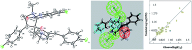
Antitumor active dispiro[3H-indole-3,2′-pyrrolidine-3′,3′′-piperidine]-2(1H),4′′-diones 11–19 were regioselectively synthesized via azomethine ylide cycloaddition reactions with 3E,5E-1-alkyl-3,5-bis(arylmethylidene)-4-piperidones 3–7. Compounds 13, 14, and 16 reveal higher potency against the HeLa (cervical) tumor cell line than the standard reference cisplatin, while 11, and 12 seem more potent against the HepG2 (liver) carcinoma cell line relative to the standard reference doxorubicin hydrochloride as determined by in vitro Sulfo-Rhodamine-B bio-assay. 3D-Pharmacophores of the HeLa comprise five chemical features viz., two hydrogen bond acceptors, two hydrophobic centers and one positive ionizable center and HepG2 contains three chemical features viz., a hydrogen bond acceptor, a hydrophobic center and a positive ionizable center. These features of the tumor cell lines explain the variation of bioactivity relative to chemical structure. Statistically significant QSAR models describing the spiro-alkaloid bio-properties were obtained employing CODESSA-Pro software validating the observed pharmacological observations and identifying the most important parameters governing activity.
Introduction
Although there has been progress in diagnosing, treating and managing cancer, the disease still results in the death of a significant number of patients. It remains the second leading cause of death worldwide after cardiovascular disease. The worldwide cancer burden is expected to increase by as much as 15 million new cases per year by 2020, according to the World Health Organization, unless further preventive measures are put into practice.1,2 Generally, cancers of the breast, lung, colorectal, and prostate are the most frequent types in developed countries and cancers of the stomach, liver, oral cavity, and cervix the most frequent in developing countries, although these patterns are changing, especially due to population aging and life style changes.3,4 The preliminary cancer treatment options usually remain a combination of radiotherapy, surgery and chemotherapy. Chemotherapy is considered one of the effective approaches in suppressing tumor growth and eradication of tumors. However, many patients undergoing chemotherapy suffer from side effects such as nausea, vomiting, cachexia, lethargy and poor oral intake.5 In spite of availability of a large number of anticancer drugs, the development of new chemotherapeutics is one of the most noteworthy challenges to non-selectivity and emergence of resistance by cancerous cells towards existing anticancer agents. A constant need to develop better alternatives to such problems is therefore in demand.6
Recently we described regioselective synthesis of fluoro-substituted dispiro-oxindole and the structure was investigated by X-ray and theoretical studies.7 We now report full details of the synthesis of spiro-indoles and their anti-oncological properties against human cervical (HeLa), and hepatoma (HepG2) cancer cell lines. The rationale for this is as follows. Cervical carcinoma is the third most common cancer and the fourth leading cause of cancer-related death in women worldwide; every year, approximately 529
800 new cases are diagnosed, and approximately 275
000 women die from this disease.8,9 More than 80% of cervical cancer cases occur in developing countries, while incidence and mortality have substantially declined in developed countries.10,11 Persistent infections with oncogenic types of human papillomavirus (HPV) are the main risk factors for cervical cancer development.12 From almost 160 HPV types that have been characterized,13 close to 30 infect the anogenital epithelium and 14 of them have been classified as oncogenic types.14,15 The biological behavior of HPV infections is influenced by viral cellular and host factors and varies in different lesions, even when the same viral type is involved.16 Human cervical cancer cells can be categorized according to HPV type as HeLa (HPV-18+), ME-180 (HPV-68+), SiHa (HPV-16+), and SW756 (HPV-18+) cells.17–19 Although several advances in screening, diagnostic and treatment modalities have been made, the overall prognosis of cervical cancer has not changed dramatically, and the mortality rate still approaches 50%. The treatment of choice is by radiotherapy or surgery for early stage disease and concurrent chemoradiation for advanced stage patients.20 Concurrent chemotherapy and radiotherapy (CCRT) is the standard of care for locally advanced cervical cancer, able to achieve a 6% improvement in a 5 years survival compared to radiotherapy alone.21–23 A larger survival advantage occurs when adjuvant chemotherapy is administered after CCRT.23–25 Cisplatin is the drug of choice either alone or in combination with topotecan.26 The combination of cisplatin with 5-fluorouracil has also been reported.27,28 However, severe side-effects like bone-marrow depression, neutropenia, thrombocytopenia and anaemia due to haematological toxicity along with nephrotoxicity and neurotoxicity29 and acquired chemoresistance30 throughout the course of treatment have limited the usage of cisplatin. Other reports describe the severe renal toxicity and gastrointestinal side effects of cisplatin that limits its clinical application.31
Liver malignancies including hepatocellular carcinoma, cholangiocarcinoma and hepatoblastoma are jointly the fifth most prevalent form of cancer and globally the third leading cause of cancer related death, after mortality due to lung cancer and colon cancer.32 In addition, the liver is a favorite site for metastasis of other cancers, in particular colorectal cancer, esophageal cancer and pancreatic cancer. The five-year natural mortality rate for hepatocellular carcinoma is more than 95%, and it affects more than 500
000 people worldwide per year.33 The major risk factors for liver cancer are persistent infection with hepatitis B virus (HBV) and hepatitis C virus (HCV), both of which increase the risk of liver cancer some 20-fold.34 Other well-established liver cancer risk factors include cirrhosis, aflatoxin exposure, heavy alcohol drinking, tobacco smoking and some rare monogenic syndromes such as hereditary hemochromatosis and α-1 antitrypsin deficiency.35 Doxorubicin (also named as adriamycin, ADR) has a broad antitumor effect as a typical DNA intercalating agent, and is often used as the first-line anticancer drug in treatment of acute leukemia, malignant lymphoma, multiple myeloma, breast cancer, osteosarcoma, soft tissue sarcoma and liver cancer.36 The clinical application of doxorubicin is limited by its toxicity to normal tissues and cells, especially its cardiotoxicity.37 The only clinically approved chemotherapy drug for advanced hepatocellular carcinoma is sorafenib, which shows only modest efficacy, improving survival of patients by just 3 months.38,39
3D-QSAR pharmacophoric generation and 2D-QSAR (quantitative structure–activity relationship) study is considered in the present work utilizing Discovery Studio 2.5 and Comprehensive Descriptors for Structural and Statistical Analysis (CODESSA-Pro) software. This allows a better understanding of the observed pharmacological activity and determines the most important structural parameters controlling bio-activity. These studies are also used to validate the observed bio-data.
Results and discussion
Chemistry
3E,5E-1-Alkyl-3,5-bis(arylmethylidene)-4-piperidones 3–7, the starting compounds for constructing the targeted spiro-alkaloids, were synthesized via base-catalyzed condensation of 1-alkyl-4-piperidones 1,2 with aromatic aldehydes (Scheme 1). Due to the huge melting point difference (≈30 °C) between the literature reported40 3E,5E-3,5-bis[(2,4-dichlorophenyl)methylidene]-1-ethyl-4-piperidones 3 and our synthesized analogue, the structure of the compound was investigated using a variety of spectroscopic techniques. The IR spectrum of 3 reveals a strong stretching vibration band at ν = 1674 cm−1 assignable to the α,β-unsaturated ketonic function. The 1H-NMR spectrum of 3 exhibits the exocyclic olefinic methine protons as a sharp singlet signal at δ = 7.91 confirming the formation of a single geometrical isomer (3E,5E).41 The 13C-NMR spectrum of 3 shows the olefinic methine carbon at δ = 136.1, and the carbonyl carbon at δ = 186.3. A single crystal X-ray study of 3 shows a half chair conformation for the piperidinyl ring. The exocyclic olefinic bonds at C-3 and C-5 of the piperidinyl ring possess E,E′-configurations (Fig. 1).
 |
| Scheme 1 Synthetic route towards 3E,5E-1-alkyl-3,5-bis(arylmethylidene)-4-piperidones 3–7. | |
 |
| Fig. 1 X-ray crystal structure of compound 3. | |
[3 + 2]-Cycloaddition of non-stabilized azomethine ylides (generated in situ via decarboxylative condensation of isatins 8–9 with sarcosine 10) and 3E,5E-1-alkyl-3,5-bis(arylmethylidene)-4-piperidones 3–7 in refluxing ethanol, proceeds regioselectively to afford single products (silica gel TLC) in good to excellent yields (62–98%) obeying the reported procedure.42 The structure of the isolated products was assigned as 1′′-alkyl-4′-aryl-5′′-(arylmethylidene)-1′-methyl-dispiro[3H-indole-3,2′-pyrrolidine-3′,3′′-piperidine]-2(1H),4′′-diones 11–19 based on spectroscopic (IR, 1H, 13C-NMR, 1H, 1H–COSY, HSQC, HRMS) data and elemental analysis (Fig. S1–S32 of ESI†). The reaction commences with nucleophilic attack of the amino group of sarcosine 10 on the 3-carbonyl function of isatins 8–9, followed by dehydration to form a spiro-oxazalidinone system. This, expels carbon dioxide to generate a reactive, non-stabilized azomethine ylide, that undergoes in situ 1,3-dipolar cycloaddition to the exocyclic olefinic linkage of piperidones 3–7 affording eventually spiro-alkaloids 11–19 (Scheme 2).
 |
| Scheme 2 Synthetic route towards dispiro[3H-indole-3,2′-pyrrolidine-3′,3′′-piperidines] 11–19. | |
The IR spectrum of 11, representative of the family, exhibits an indolyl amidic NH stretching vibration band at ν = 3285 cm−1, and strong stretching vibration bands at ν = 1701 and 1678 cm−1 corresponding to the carbonyl of ketonic and amidic functions, respectively. The methylene protons H2C-5′, H2C-2′′ and H2C-6′′ of 11 are diastereotopic. The methylene protons of the ethyl group attached at the piperidinyl N-1′′ appear as a multiplet at δ = 1.93–2.11, due to mutual coupling with each other and in turn with the vicinal methyl protons (diastereotopic protons). The 13C-NMR spectrum of 11 reveals the methylene carbons H2C-6′′, H2C-5′ and H2C-2′′ at δ = 53.4, 57.0, 56.1, respectively. The methine HC-4′ is observed at δ = 41.3 and the spiro-carbons C-3′ (C-3′′) and C-3 (C-2′) are exhibited at δ = 62.1, 76.2, respectively. The carbonyl carbons C-2 and C-4′′ appear at δ = 176.3, 196.8, respectively. 1H, 1H–COSY spectrum (Fig. S31 of ESI†) and 1H,13C-heteronuclear single quantum coherence (HSQC) spectrum of compound 14 (Fig. S32A and S32B of ESI†) support these interpretations.
A single crystal X-ray study of 14 (Fig. 2), supports the stereochemical structure. The indolyl as well as the 4-fluorophenyl rings have planar configurations and the exocyclic olefinic double bond has the E-configuration. The pyrrolidine ring has an envelope conformation with the flap atom being the ring nitrogen which is out of the plane of the remaining four atoms.
 |
| Fig. 2 X-ray crystal structure of compound 14. | |
Antitumor properties
Antitumor properties of the synthesized dispiro[3H-indole-3,2′-pyrrolidine-3′,3′′-piperidines] 11–19 were screened against HeLa (cervical), and HepG2 (liver) human tumor cell lines utilizing the reported in vitro Sulfo-Rhodamine-B standard method.43–49 The results in Table 1 (Fig. S33 and S34 of ESI†) show that, all the synthesized compounds have considerable antitumor activity against the tested cell lines with variable potencies. Compounds 13, 14, and 16 reveal higher potency (IC50 = 4.87, 5.75, and 7.25 μM, respectively) against HeLa (cervical) cell line than the standard reference cisplatin (IC50 = 7.71 μM) (clinically used against cervical carcinoma26). On the other hand, compounds 11 and 12 seem more potent (IC50 = 3.53, and 7.20 μM, respectively) than doxorubicin hydrochloride (IC50 = 8.05 μM) (clinically applicable agent against liver carcinoma36).
Table 1 Antitumor properties of the synthesized compounds 11–19
Entry |
Compd |
R |
R′ |
X |
IC50a, μg ml−1 (μM) |
HeLa |
HepG2 |
IC50 = Concentration required to produce 50% inhibition of cell growth compared to control experimental. |
1 |
11 |
2,4-Cl2C6H3 |
Et |
H |
10.27 (16.69) |
2.17 (3.53) |
2 |
12 |
2,4-Cl2C6H3 |
Et |
Cl |
8.26 (12.71) |
4.68 (7.20) |
3 |
13 |
4-FC6H4 |
Et |
H |
2.50 (4.87) |
5.60 (10.90) |
4 |
14 |
4-FC6H4 |
Et |
Cl |
3.15 (5.75) |
6.85 (12.50) |
5 |
15 |
2-Thienyl |
Et |
H |
5.33 (10.89) |
9.02 (18.42) |
6 |
16 |
2-Thienyl |
Et |
Cl |
3.80 (7.25) |
9.09 (17.34) |
7 |
17 |
3-Pyridinyl |
Me |
H |
9.35 (20.08) |
10.27 (22.06) |
8 |
18 |
3-Pyridinyl |
Et |
H |
5.16 (10.76) |
8.17 (17.04) |
9 |
19 |
3-Pyridinyl |
Et |
Cl |
11.58 (22.53) |
5.91 (11.50) |
10 |
Doxorubicin hydrochloride |
— |
— |
— |
4.19 (7.22) |
4.67 (8.05) |
11 |
Cisplatin |
— |
— |
— |
4.19 (7.71) |
3.58 (11.89) |
Structure–activity relationships (SAR) based on the observed antitumor activity data against HeLa (cervical carcinoma) reveal that the nature of the substituent attached to the phenyl group at C-4′ and consequently the exocyclic olefinic linkage, seems to be a controlling factor governing the antitumor properties. Substitution of this phenyl group by fluorine atom enhances the observed antitumor properties more than two chlorine atoms, as exhibited in pairs 11, 13 (IC50 = 16.69, 4.87 μM, respectively), and 12, 14 (IC50 = 12.71, 5.75 μM, respectively).
SAR due to the observed antitumor activity data against HepG2 (liver carcinoma) cell line describes a contrast behavior than the aforementioned cervical cell line. 2,4-Dichlorophenyl substituent at C-4′ and consequently the exocyclic olefinic linkage, seems the best choice for constructing antitumor active agent against hepatocellular carcinoma compared with the 4-fluorophenyl group as exhibited in pairs 11, 13 (IC50 = 3.53, 10.90 μM, respectively), and 12, 14 (IC50 = 7.20, 12.50 μM, respectively). Additionally, the 3-pyridinyl group at C-4′ and consequently the exocyclic olefinic linkage, seem to optimize antitumor activity against HepG2 (liver carcinoma) when compared with the thienyl group as exhibited in pairs 15, 18 (IC50 = 18.42, 17.04 μM, respectively), and 16, 19 (IC50 = 17.34, 11.50 μM, respectively). This can be attributed to the π-deficient heterocyclic properties of the 3-pyridinyl group compared with the electron-donating properties of thienyl group.
In order to better understand the observed antitumor properties and determine the most important structural parameters controlling bio-activity, computational chemistry studies were undertaken. Additionally, validation of the observed antitumor properties was established via these studies.
Computational chemistry
3D-Pharmacophore modeling. The pharmacophore modeling technique has been widely used in lead discovery and optimization as a key tool in computer aided drug design. The 3D-pharmacophore study was performed using Discovery Studio 2.5 software (Accelrys Inc., San Diego, CA, USA) which permits 3D-pharmacophore generation, structural alignment, activity prediction and 3D-database creation.45,50–52 A 3D-QSAR pharmacophore protocol was used to generate predictive pharmacophores via aligning different conformations in which the molecules are likely to bind with the receptor. A given hypothesis may be combined with known activity data to create a 3D-pharmacophore model that identifies overall aspects of molecular structure governing activity. 3D-QSAR pharmacophore was constructed using collections of molecules with activities ranging over a number of orders of magnitude. Pharmacophores explain the variability of bioactivity with respect to the geometric localization of the chemical features present in the molecules. The observed HYPOGEN identifies a 3D-array of five chemical features in the case of HeLa (cervical) and 3 features in case of HepG2 (liver) tumor cell lines which are common to the bio-active set compounds 11–19 that are consistent with binding to a proposed common receptor site.The five chemical features of the HeLa pharmacophore are two hydrogen bond acceptors (HBA-1, HB-2), two hydrophobic centers (H-1, H-2), and one positive ionizable (PosIon) (Fig. 3, Table 2 exhibits constraint distances and angles between features of the generated 3D-pharmacophore). On the other hand, the HepG2 pharmacophore contains three chemical features, a hydrogen bond acceptor (HBA), a hydrophobic (H) and a positive ionizable (PosIon) (Fig. 4, Table 2). Table 3 exhibits fit values and estimated/predicted activities of the synthesized compounds 11–19 due to the generated 3D-pharmacophore models. Through the pharmacophore mapping study (Fig. S35 and S36 of ESI†) it has been found that the major structural factors affecting the potency of the synthesized compounds are related to their basic skeleton. Additionally, most of the estimated activity as well as the fit values derived from the generated pharmacophores correlate with the experimentally observed potency. For example, the most potent analogue 13 (R = 4-FC6H4, R′ = Et, X = H; IC50 = 4.87 μM) among all the synthesized compounds, shows an estimated potency (IC50 = 5.75 μM) preserving its lead behavior and bio-potency compared to the standard reference used.
 |
| Fig. 3 (A) Constraint distances and (B) constraint angles of the generated 3D-pharmacophore for the synthesized compounds 11–19 against HeLa (cervical) cell line which contains two hydrogen bonding acceptor (HBA-1, HBA-2, green), two hydrophobics (H-1, H-2, light blue), and one positive ionizable (PosIon, red). | |
Table 2 Constraint distances (Å) and angles (°) between features of the generated pharmacophores
Cancer cell line |
Constraint distances (Å) |
Constraint angles (°) |
HeLa (cervical) |
(HBA-1)–(PosIon) = 3.146, (PosIon)–(H-1) = 4.551, (PosIon)–(HBA-2) = 4.494, (H-1)–(HBA-2) = 4.374, (HBA-2)–(H-2) = 6.746; (H-2)–(HBA-1) = 4.581, (H-1)–(H-2) = 6.148 |
(HBA-1)–(H-1)–(PosIon) = 39.22, (HBA-1)–(HBA-2)–(PosIon) = 38.88, (HBA-1)–(H-1)–(H-2) = 47.54, (HBA-1)–(H-2)–(PosIon) = 13.42 |
HepG2 (liver) |
(H)–(PosIon) = 8.239, (PosIon)–(HBA) = 4.245, (HBA)–(H) = 7.672 |
(PosIon)–(H)–(HBA) = 30.68 |
|
|
|
 |
| Fig. 4 (A) Constraint distances and (B) constraint angles of the generated 3D-pharmacophore for the synthesized compounds 11–19 against HepG2 (liver) cell line which contains a hydrogen bonding acceptor (HBA, green), a hydrophobics (H, light blue), and a positive ionizable (PosIon, red). | |
Table 3 Best fit values and estimated/predicted activities for the synthesized compounds 11–19 mapped with the generated 3D-pharmacophore models due to HeLa (cervical) and HepG2 (liver) cancer cell lines
Entry |
Compd |
R |
R′ |
X |
HeLa (cervical) cell line |
HepG2 (liver) cell line |
Observed IC50, μM |
Estimated IC50, μM |
Fit value |
Observed IC50, μM |
Estimated IC50, μM |
Fit value |
1 |
11 |
2,4-Cl2C6H3 |
Et |
H |
16.69 |
15.00 |
9.74 |
3.53 |
4.26 |
5.75 |
2 |
12 |
2,4-Cl2C6H3 |
Et |
Cl |
12.71 |
10.65 |
9.88 |
7.20 |
4.34 |
5.74 |
3 |
13 |
4-FC6H4 |
Et |
H |
4.87 |
5.75 |
10.15 |
10.90 |
11.51 |
5.31 |
4 |
14 |
4-FC6H4 |
Et |
Cl |
5.75 |
7.02 |
10.07 |
12.50 |
13.55 |
5.24 |
5 |
15 |
2-Thienyl |
Et |
H |
10.89 |
11.05 |
9.87 |
18.42 |
19.15 |
5.09 |
6 |
16 |
2-Thienyl |
Et |
Cl |
7.25 |
10.42 |
9.89 |
17.34 |
17.59 |
5.13 |
7 |
17 |
3-Pyridinyl |
Me |
H |
20.08 |
14.69 |
9.74 |
22.06 |
21.71 |
5.04 |
8 |
18 |
3-Pyridinyl |
Et |
H |
10.76 |
9.47 |
9.94 |
17.04 |
18.90 |
5.10 |
9 |
19 |
3-Pyridinyl |
Et |
Cl |
22.53 |
22.04 |
9.57 |
11.50 |
12.26 |
5.29 |
Mapping of the HeLa 3D-pharmacophore with compound 13 (Fig. S35 of ESI†) describes the correlation of the pyrrolidinyl nitrogen with the positive ionizable feature. The same alignment was also observed for compound 11 (R = 2,4-Cl2C6H3, R′ = Et, X = H; IC50 = 16.69, 15.00 μM corresponding to the observed and estimated potency, respectively). The high potency difference between the observed/estimated activity of these compounds (11, 13) explains the role of the substituent attached to the phenyl group linked at the C-4′ as explained previously in SAR due to the observed bio-data. The 2,4-dichlorophenyl group attached to C-4′ position deactivates the positive ionizable properties of the pyrrolidinyl nitrogen much more than the p-fluorophenyl group (although the −I effect of fluorine is higher than chlorine, the two chlorine substituents combine to give a higher −I effect). The same applies for compound 16 (R = 2-thienyl, R′ = Et, X = Cl; IC50 = 7.25, 10.42 μM corresponding to the observed and estimated potency, respectively) when compared with compound 19 (R = 3-pyridinyl, R′ = Et, X = Cl; IC50 = 22.53, 22.04 μM corresponding to the observed and estimated potency, respectively). This latter observation correlates well with the electron donating properties of the thienyl group (five-membered heterocycle with one hetero atom) strengthen the positive ionizable property of pyrrolidinyl nitrogen upon compared with the effect of a 3-pyridinyl group (π-deficient heterocycle).
Mapping of the HepG2 3D-pharmacophore with compound 11 (R = 2,4-Cl2C6H3, R′ = Et, X = H; IC50 = 3.53, 4.26 μM corresponding to the observed and estimated potency, respectively), the most potent analogue among all the synthesized spiro-alkaloids (Fig. S36 of ESI†), describes the alignment of the 2,4-dichlorophenyl group attached to the pyrrolidinyl C-4′ with the pharmacophoric hydrophobic feature and the pyrrolidinyl nitrogen with the pharmacophoric positive ionizable while the piperidinyl carbonyl is aligned with the hydrogen bond acceptor. A relatively similar mapping is exhibited by compound 13 (R = 4-FC6H4, R′ = Et, X = H; IC50 = 10.90, 11.51 μM corresponding to the observed and estimated potency, respectively), where the 4-fluorophenyl group attached to the exocyclic olefinic linkage is aligned with the pharmacophoric hydrophobic feature, the pyrrolidinyl nitrogen with the positive ionizable, and the piperidinyl carbonyl is aligned with the hydrogen bond acceptor feature. The potency difference of compounds 11 and 13 can be attributed to the slight difference in mode of alignment and high hydrophobic properties of the 2,4-dichlorophenyl group aligned with the hydrophobic feature than the corresponding p-fluorophenyl group aligned with the same pharmacophoric feature. This observation is the same mentioned for SAR rules governing HepG2 bio-data. The potency decrease of compounds 12 (R = 2,4-Cl2C6H3, R′ = Et, X = Cl; IC50 = 7.20, 4.34 μM corresponding to the observed and estimated potency, respectively) and 14 (R = 4-FC6H4, R′ = Et, X = Cl; IC50 = 12.50, 13.55 μM corresponding to the observed and estimated potency, respectively) “which exhibit typical alignment to compounds 11, 13, respectively” compared to their similar analogues 11 and 13 can be attributed to the effect of chloro substitution attached to the indolyl group which decreases the positive ionizable properties of the pyrrolidinyl nitrogen. Mapping of the HepG2 3D-pharmacophore with compound 18 (R = 3-pyridinyl, R′ = Et, X = H; IC50 = 17.04, 18.90 μM corresponding to the observed and estimated potency, respectively), describes the alignment of the 3-pyridinyl group attached to the exocyclic olefinic linkage with the pharmacophoric hydrophobic, the pyrrolidinyl nitrogen with the positive ionizable, and the piperidinyl carbonyl with the hydrogen bond acceptor feature. A slightly modified mapping is observed for compound 15 (R = 2-thienyl, R′ = Et, X = H; IC50 = 18.42, 19.15 μM corresponding to the observed and estimated potency, respectively) where, the 2-thienyl group linked to the exocyclic olefinic linkage is aligned with the pharmacophoric hydrophobic, the pyrrolidinyl nitrogen with the positive ionizable, and the indolyl carbonyl with the hydrogen bond acceptor feature. The enhanced potency effect of compound 18 relative to compound 15 can be attributed not only to the slight difference in mode of alignment but also to the higher hydrophobic properties of the 3-pyridinyl group than the 2-thienyl function. Another reason for the observed pharmacological potency enhancement is extracted from the differences in mode of alignment where, the hydrogen bond acceptor property of the piperidinyl ketonic carbonyl is higher than that of indolyl carbonyl. Meanwhile, potency enhancement of compound 19 (R = 3-pyridinyl, R′ = Et, X = Cl; IC50 = 11.50, 12.26 μM corresponding to the observed and estimated potency, respectively) compared with its similar analogue 18 is explained by the HepG2 pharmacophoric mapping of the former analogue in a completely different mode of alignment where, the chlorine substituent of the indolyl group is aligned with the pharmacophoric hydrophobic, the piperidinyl nitrogen with the positive ionizable, and the nitrogen of the 3-pyridinyl group linked at the C-4′ is aligned with the hydrogen bond acceptor feature. The same applies also for compound 16 (R = 2-thienyl, R′ = Et, X = Cl; IC50 = 17.34, 17.59 μM corresponding to the observed and estimated potency, respectively) that reveals a completely different mode of alignment in the hypothesized pharmacophore. Where the chlorine substituent of the indolyl group is aligned with the pharmacophoric hydrophobic, the piperidinyl nitrogen with the positive ionizable, and the indolyl carbonyl is aligned with the hydrogen bond acceptor feature. This is could be the reason for observed antitumor properties enhanced for compound 16 relative to its similar analogue 15.
2D-QSAR study
Data set. The basic idea behind QSAR is to generate a relationship between the chemical structure of an organic compound and its physico-chemical properties. Due to the limited pharmacologically active data set mentioned in the present study, external data points were considered. The external data points are derived from spiro-alkaloids having the same chemical scaffold (homogeneous/non-diverse data set protocol) and their bio-properties were determined by the same standard technique adopted in the present study. The QSAR study was undertaken using comprehensive descriptors for structural and statistical analysis (CODESSA-Pro) software employing the synthesized compounds of the present study 11,13,15–17,19 in addition to compounds 20–44 which are recently reported by our group53 forming 31 spiro-alkaloids used as a training set for constructing QSAR models (Table 4). Compounds 12, 14, and 18 (about one third of the synthesized analogues) representing high, and low potent antitumor active agents, were used as external data set for validating the attained QSAR models (Table 5).
Table 4 Observed and predicated values of training set compounds 11,13,15–17, and 19–44 according to the multi-linear QSAR models
Entry |
Compd |
R |
R′ |
X |
HeLa (cervical) cell line |
HepG2 (liver) cell line |
Observed IC50, μM |
Estimated IC50, μM |
Error |
Observed IC50, μM |
Estimated IC50, μM |
Error |
1 |
11 |
2,4-Cl2C6H3 |
Et |
H |
16.69 |
12.26 |
4.43 |
3.53 |
3.75 |
−0.22 |
2 |
13 |
4-FC6H4 |
Et |
H |
4.87 |
5.94 |
−1.07 |
10.90 |
7.63 |
3.27 |
3 |
15 |
2-Thienyl |
Et |
H |
10.89 |
10.48 |
0.41 |
18.42 |
15.69 |
2.73 |
4 |
16 |
2-Thienyl |
Et |
Cl |
7.25 |
7.86 |
−0.61 |
17.34 |
16.97 |
0.37 |
5 |
17 |
3-Pyridinyl |
Me |
H |
20.08 |
26.07 |
−5.99 |
22.06 |
21.29 |
0.77 |
6 |
19 |
3-Pyridinyl |
Et |
Cl |
22.53 |
20.89 |
1.64 |
11.50 |
16.32 |
−4.82 |
7 |
20 |
Ph |
Me |
H |
6.21 |
5.92 |
0.29 |
7.46 |
8.34 |
−0.88 |
8 |
21 |
Ph |
Me |
Cl |
5.92 |
5.41 |
0.51 |
5.66 |
6.16 |
−0.50 |
9 |
22 |
4-ClC6H4 |
Me |
H |
6.74 |
6.30 |
0.44 |
6.03 |
6.13 |
−0.10 |
10 |
23 |
4-ClC6H4 |
Me |
Cl |
5.08 |
5.72 |
−0.64 |
6.26 |
6.34 |
−0.08 |
11 |
24 |
4-ClC6H4 |
Et |
Cl |
4.96 |
5.28 |
−0.32 |
5.73 |
5.81 |
−0.08 |
12 |
25 |
4-ClC6H4 |
Me |
OMe |
5.78 |
5.90 |
−0.12 |
8.89 |
6.32 |
2.57 |
13 |
26 |
4-ClC6H4 |
Et |
OMe |
5.20 |
5.43 |
−0.23 |
5.43 |
5.33 |
0.10 |
14 |
27 |
4-FC6H4 |
Me |
H |
6.51 |
5.95 |
0.56 |
8.73 |
8.90 |
−0.17 |
15 |
28 |
4-FC6H4 |
Me |
Cl |
5.15 |
5.71 |
−0.56 |
5.77 |
7.41 |
−1.64 |
16 |
29 |
4-FC6H4 |
Me |
OMe |
5.44 |
6.21 |
−0.77 |
5.82 |
6.31 |
−0.49 |
17 |
30 |
4-H3CC6H4 |
Me |
H |
8.64 |
7.09 |
1.55 |
14.18 |
8.87 |
5.31 |
18 |
31 |
4-H3CC6H4 |
Me |
Cl |
6.65 |
6.71 |
−0.06 |
7.32 |
6.67 |
0.65 |
19 |
32 |
4-H3CC6H4 |
Et |
Cl |
5.55 |
7.78 |
−2.23 |
5.46 |
7.14 |
−1.68 |
20 |
33 |
4-H3CC6H4 |
Me |
OMe |
6.96 |
7.68 |
−0.72 |
6.15 |
6.36 |
−0.21 |
21 |
34 |
4-H3COC6H4 |
Me |
H |
6.45 |
7.17 |
−0.72 |
6.68 |
8.60 |
−1.92 |
22 |
35 |
4-H3COC6H4 |
Et |
H |
7.22 |
6.54 |
0.68 |
6.68 |
7.21 |
−0.53 |
23 |
36 |
4-H3COC6H4 |
Me |
Cl |
11.20 |
6.53 |
4.67 |
13.67 |
8.36 |
5.31 |
24 |
37 |
4-H3COC6H4 |
Et |
Cl |
8.74 |
6.27 |
2.47 |
5.91 |
8.05 |
−2.14 |
25 |
38 |
4-H3COC6H4 |
Me |
OMe |
6.10 |
6.94 |
−0.84 |
6.95 |
7.35 |
−0.40 |
26 |
39 |
4-H3COC6H4 |
Et |
OMe |
5.51 |
7.84 |
−2.33 |
7.49 |
7.35 |
0.14 |
27 |
40 |
4-Me2NC6H4 |
Me |
Cl |
24.36 |
20.24 |
4.12 |
23.71 |
21.13 |
2.58 |
28 |
41 |
2-Thienyl |
Me |
H |
8.94 |
8.18 |
0.76 |
14.02 |
13.76 |
0.26 |
29 |
42 |
2-Thienyl |
Me |
Cl |
6.86 |
7.98 |
−1.12 |
9.29 |
11.04 |
−1.75 |
30 |
43 |
2-Thienyl |
Me |
OMe |
9.65 |
10.77 |
−1.12 |
8.40 |
7.07 |
1.33 |
31 |
44 |
5-Methyl-2-furanyl |
Me |
Cl |
9.88 |
8.46 |
1.42 |
9.37 |
10.81 |
−1.44 |
Table 5 Observed and predicated values of external test set compounds 12, 14, and 18 according to the multi-linear QSAR models
Entry |
Compd |
R |
R′ |
X |
HeLa (cervical) cell line |
HepG2 (liver) cell line |
Observed IC50, μM |
Estimated IC50, μM |
Error |
Observed IC50, μM |
Estimated IC50, μM |
Error |
1 |
12 |
2,4-Cl2C6H3 |
Et |
Cl |
12.71 |
8.99 |
3.72 |
7.20 |
4.03 |
3.17 |
2 |
14 |
4-FC6H4 |
Et |
Cl |
5.75 |
5.64 |
0.11 |
12.50 |
8.00 |
4.50 |
3 |
18 |
3-Pyridinyl |
Et |
H |
10.76 |
23.70 |
−12.94 |
17.04 |
16.97 |
0.07 |
Methodology. Geometry of the compounds was optimized using molecular mechanics force field (MM+) followed by the semi-empirical AM1 method implemented in the HyperChem 8.0 package. The structures were fully optimized without fixing any parameters, thus bringing all geometric variables to their equilibrium values. The energy minimization protocol employed the Polake–Ribiere conjugated gradient algorithm. Convergence to a local minimum was achieved when the energy gradient was ≤0.01 kcal mol−1. The RHF method was used in spin pairing for the two semi-empirical tools.45,50,54,55 The resulting output files were exported to CODESSA-Pro that includes MOPAC capability for final geometry optimization. CODESSA-Pro software includes the following: (a) a calculation engine for more than 500 descriptors and (b) an engine for the development of the statistically most important linear and nonlinear QSAR regression models. CODESSA-Pro calculated 728 molecular descriptors including constitutional, topological, geometrical, charge-related, semi-empirical, molecular-type, atomic-type and bond-type descriptors for the exported 31 bio-active spiro-alkaloids 11,13,15–17,19, and 20–44 which were used as a training set in the present study. Different mathematical transformations of the experimentally observed property/activity (IC50, μM which is the concentration required to produce 50% inhibition of cell growth compared to control experimental) against HeLa (cervical) and HepG2 (liver) tumor cell lines of the training set compounds were utilized for the present QSAR modeling determination including property (IC50, μM), 1/property, log(property) and 1/log(property) values in searching for the best QSAR models.
QSAR modeling. Best multi-linear regression (BMLR) was utilized which is a stepwise search for the best n-parameter regression equations (where, n stands for the number of descriptors used), based on the highest R2 (squared correlation coefficient), Rcv2OO (squared cross-validation “leave one-out, LOO” coefficient), Rcv2MO (squared cross-validation “leave many-out, LMO” coefficient), F (Fisher statistical significance criteria) values, and s2 (standard deviation). The QSAR models up to 3 and 4 descriptor models describing bio-activity of the antitumor active agents against HeLa (cervical) and HepG2 (liver) cell line, respectively were generated (obeying the thumb rule of 5
:
1, which is the ratio between the data points and the number of QSAR descriptor models). Statistical characteristics of the QSAR models are presented in Tables 6 and 7. The established QSAR models are statistically significant. The descriptors are sorted in descending order of the respective values of the Student's t-criterion, which is a widely accepted measure of statistical significance of individual parameters in multiple linear regressions. Fig. 5, and 6 exhibit the QSAR multi-linear model plot of correlations representing the observed vs. predicted IC50 values for HeLa and HepG2 tumor cell line active agents, respectively. The scattered plots are uniformly distributed, covering ranges, observed 0.688–1.387, 0.548–1.375; predicted 0.723–1.416, 0.574–1.328 logarithmic units for HeLa and HepG2 cell lines, respectively.
Table 6 Descriptor of the best multi-linear QSAR model for the HeLa (cervical) tumor cell line active agents
Entry |
N = 31, n = 3, R2 = 0.815, Rcv2OO = 0.738, Rcv2MO = 0.776, F = 39.615, s2 = 0.008 |
ID |
Coefficient |
s |
t |
Descriptor |
1 |
0 |
0.141 |
0.185 |
0.763 |
Intercept |
2 |
D1 |
0.247 |
0.027 |
9.200 |
Min.(#HA, #HD) (MOPAC PC) |
3 |
D2 |
0.596 |
0.107 |
5.546 |
FNSA-2 fractional PNSA (PNSA-2/TMSA) (MOPAC PC) |
4 |
D3 |
0.426 |
0.096 |
4.424 |
HASA-2/SQRT(TMSA) (Zefirov PC) (all) |
log(IC50) = 0.141 + (0.247 × D1) + (0.596 × D2) + (0.426 × D3) |
|
Table 7 Descriptor of the best multi-linear QSAR model for the HepG2 (liver) tumor cell line active agents
Entry |
N = 31, n = 4, R2 = 0.799, Rcv2OO = 0.729, Rcv2MO = 0.741, F = 25.768, s2 = 0.009 |
ID |
coefficient |
s |
t |
Descriptor |
1 |
0 |
5.211 |
1.493 |
3.491 |
Intercept |
2 |
D1 |
0.152 |
0.021 |
7.157 |
Min.(#HA, #HD) (Zefirov PC) |
3 |
D2 |
83.943 |
17.380 |
4.830 |
Partial charged surface area for atom H |
4 |
D3 |
−41.614 |
8.822 |
−4.717 |
Partial charged surface area for atom O |
5 |
D4 |
−60.210 |
14.595 |
−4.125 |
Min. (>0.1) bond order for atom C |
log(IC50) = 5.211 + (0.152 × D1) + (83.943 × D2) − (41.614 × D3) – (60.210 × D4) |
|
 |
| Fig. 5 QSAR best multi-linear model plot of correlations representing the observed vs. predicted IC50 values for HeLa (cervical) tumor cell line active agents (compound 36 is an outlier). | |
 |
| Fig. 6 QSAR best multi-linear model plot of correlations representing the observed vs. predicted IC50 values for HepG2 (liver) tumor cell line active agents (compounds 30, and 36 are outliers). | |
Molecular descriptors. Molecular descriptors are the physico–chemical parameters used to correlate chemical structure and property value expressed as log(IC50). The descriptors were obtained based on BMLR method. The descriptors controlling the bio-activity (property) by the established multi-linear QSAR models are presented in Tables 6 and 7 and are arranged, based on their level of significance (t-criterion).
HeLa (cervical) tumor cell line. The descriptors controlling bio-activity of the synthesized compounds against HeLa (cervical) tumor cell line according to the attained 3 descriptor QSAR model (Table 6) are: min. (#HA, #HD) (MOPAC PC), FNSA-2 fractional PNSA (PNSA-2/TMSA) (MOPAC PC), and HASA-2/SQRT(TMSA) (Zefirov PC) (all). The first descriptor controlling the HeLa QSAR model is minimum (#HA, #HD), which is a molecular type descriptor explaining the capability of the bio-active agent as hydrogen donor/acceptor. Although this descriptor is considered the most important one governing the attained QSAR model based on its t-criterion (9.200), its coefficient in the QSAR is the minimum among all the other observed QSAR descriptor (0.247). The second most important descriptor controlling the HeLa QSAR model based on the t-criterion (5.546) is FNSA-2 fractional PNSA (PNSA-2/TMSA), which is a charge related descriptor. The fractional total charge weighted partial negative surface area (FNSA2) is determined by eqn (1).56 |
 | (1) |
where, PNSA2 stands for total charge weighted partial negatively charged molecular surface area, and TMSA for total molecular surface area. This descriptor has the highest coefficient (0.596) among the other descriptors controlling the HeLa QSAR model. This observation coincide with our mentioned SAR rules governing bio-activity concerning type of substituent (halogen) attached to the phenyl group linked at the C-4′ and consequently the exocyclic olefinic linkage, affecting greatly the observed potency of the bio-active agent. The same phenomenon is also extracted from the HeLa pharmacophoric hypothesis. The last descriptor of the HeLa QSAR model (t-criterion = 4.424), is HASA-2/SQRT(TMSA), which is also a charge related descriptor. The area-weighted surface charge of hydrogen bonding acceptor atoms (HASA2) is determined by eqn (2).56 |
 | (2) |
where, SA stands for solvent-accessible surface area of H-bonding acceptor atoms, qA for partial charge on H-bonding acceptor atoms, and Stot for total solvent-accessible molecular surface area. This descriptor is considered the second most effective parameter controlling the QSAR model based on its coefficient (0.426). The present descriptor also supported the attained SAR rules as explained in the previous descriptor (FNSA2).
HepGe (liver) tumor cell line. The attained HepG2 QSAR model exhibits 4 controlling descriptors (Table 7) which are: min. (#HA, #HD) (Zefirov PC), partial charged surface area for atom H, partial charged surface area for atom O, and min. (>0.1) bond order for atom C. The most important descript controlling the HepG2 QSAR model based on its t-criterion (7.157) is minimum (#HA, #HD) (Zefirov PC). This descriptor is a common for both HeLa and HepG2 QSAR models explaining its importance for the observed bio-properties in variable tumor cell lines. Although this descriptor is the most important one governing the HepG2 QSAR model, its coefficient is the minimum among all the other observed QSAR descriptor (0.152). The second and the third important descriptors of the HepG2 QSAR model are, partial charged surface area for atom H, and partial charged surface area for atom O, which are charge related descriptor. The partial positively or negatively charged surface area is determined by eqn (3).56 |
 | (3) |
where, SA stands for positively or negatively charged solvent-accessible atomic surface area. The partial charged surface area for atom H descriptor participates in the HepG2 QSAR model with the greatest share among all the other descriptors (coefficient = 83.943). However, the partial charged surface area for atom O descriptor participated negatively in the HepG2 QSAR model (coefficient = −41.614), explaining that the higher of the latter descriptor value, the lower of total log(IC50) observed, hence the higher potency of the constructed agent against HepG2 tumor cell line. This descriptor in particular can explain the potency of compounds 11 (R = 2,4-Cl2C6H3, R′ = Et, X = H; IC50 = 3.53 μM) relative to 13 (R = 4-FC6H4, R′ = Et, X = H; IC50 = 10.90 μM). This can be correlated with the halogen type/number substituted the phenyl group at C-4′ and consequently the exocyclic olefinic linkage. The same observation was mentioned in the SAR rules governing bio-activity and 3D-pharmacophore modeling. The last descriptor of the HepG2 QSAR model is min. (>0.1) bond order for atom C, which is an atomic type descriptor.
Validation of QSAR model
Internal validation. The reliability and statistical relevance of the QSAR models are examined by internal and external validation procedures. Internal validation is applied by the CODESSA-Pro technique employing both Leave One Out (LOO), which involves developing a number of models with one example omitted at a time, and Leave Many Out (LMO), which involves developing a number of models with many data points omitted at a time (up to 20% of the total data points). The observed correlations due to the internal validation techniques are Rcv2OO = 0.738, 0.729; Rcv2MO = 0.776, 0.741, for HeLa and HepG2 QSAR models, respectively. Both of them are significantly correlated with the squared correlation coefficient of the attained QSAR models (R2 = 0.815, 0.799 for HeLa and HepG2 QSAR models, respectively). Standard deviation of the regressions (s2 = 0.008, 0.009 for HeLa and HepG2 QSAR models, respectively) is also a measurable value for the attained model together with the Fisher test value (F = 39.615, 25.768 for HeLa and HepG2 QSAR models, respectively) that reflects the ratio of the variance explained by the model and the variance due to their errors. A high value of F-test compared with the s2 is a validation of the model. A randomization test was also performed in the present study which adds good support for the present QSAR models.The predicted/estimated IC50 value of compound 13 (the most potent synthesized analogue among all the training set compounds) is 5.94 μM based on the HeLa QSAR model, matched with the experimentally observed value (4.87 μM, error “difference between observed and predicted values” = 1.07). All the other potent training set analogues (compounds 16,20–29,31,33–35,38, and 42) relative to cisplatin (standard reference clinically used against cervical carcinoma, IC50 = 7.71 μM) exhibit predicted IC50 values matched with their experimentally observed potency (error range = 0.06–1.12). Compounds 32, and 39 which are also considered potent analogues against cervical carcinoma (IC50 = 5.55, 5.51 μM corresponding to compounds 32, and 39, respectively) reveal relatively higher predicated potency beyond the mentioned error range (predicted IC50 = 7.78, 7.84 μM; error = 2.23, 2.33 corresponding to compounds 32, and 39, respectively). The mild antitumor active agents against HeLa cell line, compounds 15,30,37,41,43, and 44 (IC50 range = 8.64–10.89 μM), reveal predicted potency (IC50 range = 6.27–10.77 μM) with relatively higher error range (0.41–2.47) than the high potent analogues. Additionally, the low potent analogues against HeLa cell lines, compounds 11,17,19,36, and 40 (IC50 range = 11.20–24.36 μM) reveal higher deviated predicted potency (IC50 range = 6.53–26.07 μM) with error range = 1.64–5.99. From all the above statistical observations, the attained HeLa QSAR model can be considered a good predicative model with a powerful ability to produce high potent HeLa antitumor hits compared to those of mild or low potency. Actually, this is an acceptable observation where most of the training set compounds belong to the high potent HeLa antitumor agents (20 compounds out of 31, i.e. two thirds of the entire training set).
The predicted IC50 values (IC50 = 3.75–8.60 μM) of the potent HepG2 analogues 11,20–24,26,28,29,31–35, and 37–39 (IC50 = 3.53–7.49 μM) relative to doxorubicin hydrochloride (IC50 = 8.05 μM standard reference used in the present study “clinically used as liver antitumor agent”) exhibit error range = 0.08–1.92. However, wilder error range was observed for the mild HepG2 antitumor agents, compounds 25,27, and 42–44 (IC50 = 8.73–9.37, 6.32–11.04 μM, corresponding to the observed and predicted values, respectively, error range = 0.17–2.57) and low HepG2 antitumor agents, compounds 13,15–17,19,30,36,40, and 41 (IC50 = 10.90–23.71, 7.63–21.29 μM, corresponding to the observed and predicted values, respectively, error range = 0.37–5.31). This is an indication for the ability of utilization of the attained HepG2 QSAR model for optimizing high potent HepG2 (liver) antitumor agents than either mild or low effective hits. The reason for this observation can be attributed as mentioned in the former HeLa QSAR model, due to participation of high number of potent agents compared to mild and low potent analogues (14 out of 31 analogous of the training set i.e. 45%).
External validation. Compounds 12, 14, and 18 were used as an external test set not only for validating the attained QSAR models but also for examining their predicative ability. The test set analogues experimentally exhibit high and low potency against the tested cell lines. The variation in potency can indicate the predication capabilities of the QSAR models. Table 5 reveals the observed and predicted IC50 values of the test set compounds. From the observed data, it has been noticed that compound 14 which is considered a high potent agent against HeLa cell line (IC50 = 5.75 μM), relative to cisplatin, standard reference used, reveals a predicted IC50 = 5.64 μM with minimum error value = 0.11. Compounds 12, and 18 that considered low potent analogues (IC50 = 12.71, 10.76 μM, respectively), afforded predicted IC50 = 8.99, 23.70 μM, respectively (error values = 3.72, 12.94, respectively).These observations give good sign for the predictive capability of the attained HeLa QSAR model and support the previous statement concerning its predictive power due to the statistical values.
Predicted value for compound 12, IC50 = 4.03 μM, is correlated to its high potency against HepG2 cell line (observed IC50 = 7.20 μM). Although the error value due to difference between the observed and predicted IC50 values is considered a relatively high (error value = 3.17), the observed potency of the analogue is still preserved relative to doxorubicin hydrochloride, standard reference used (IC50 = 8.05 μM). Compounds 14, and 18 which are considered low potent HepG2 antitumor agents (observed IC50 = 12.50, 17.04 μM, respectively) reveal predicted IC50 values = 8.00, 16.97 μM, respectively (error values = 4.50, 0.07, respectively). The overall observations due to test set predication values give a good indication for the predictive power of the attained QSAR models for optimizing high potent hits having spiro-alkaloid scaffold.
Conclusion
[3 + 2]-Cycloaddition reactions of non-stabilized azomethine ylide (generated in situ via decarboxylative condensation of isatins 8,9 with sarcosine 10) and 3E,5E-1-alkyl-3,5-bis(arylmethylidene)-4-piperidones 3–7 afforded 1′′-alkyl-4′-aryl-5′′-(arylmethylidene)-1′-methyl-dispiro[3H-indole-3,2′-pyrrolidine-3′,3′′-piperidine]-2(1H),4′′-diones 11–19 in regioselective manner. Compounds 13, 14, and 16 reveal higher potency against HeLa (cervical) cell line than the standard reference cisplatin, while compounds 11 and 12 seem more potent than doxorubicin hydrochloride (clinically applicable agent against liver carcinoma) through in vitro Sulfo-Rhodamine-B bio-assay. 3D-Pharmacophore study utilizing Discovery Studio 2.5 software explained the antitumor variability of the tested compounds based on chemical structural features. The HeLa pharmacophore contains five chemical features; two hydrogen bond acceptors, two hydrophobics, and one positive ionizable. The HepG2 pharmacophore comprises three chemical features; a hydrogen bond acceptor, a hydrophobic, and a positive ionizable. 2D-QSAR study was undertaken utilizing CODESSA-Pro software in order to validate the antitumor observed bio-data and determine the most important parameters controlling bio-activity. Statistically significant robust QSAR models describing the spiro-alkaloids bio-properties were obtained. External validation technique utilizing high and low potent synthesized agents, support the predictive power of the attained QSAR models. Homogeneity of the training set analogues (the same chemical scaffold) may be the main factor for the success of the QSAR models.
Experimental
Melting points were determined on a capillary tube digital Stuart SMP3 melting point apparatus. IR spectra (KBr) were recorded on a JASCO 6100 FT-IR spectrophotometer. NMR spectra were recorded on Mercury or Gemini NMR spectrometers operating at 300 MHz for 1H (with TMS as an internal standard) and 75 MHz for 13C, except 13C-NMR spectra of compounds 17–19 which were recorded on a Bruker Ascend 400/R (100 MHz) spectrometer. High-resolution mass spectra (HRMS) were recorded on an Agilent Technologies 6210 Time of Flight LC/MS instrument operating in the ESI mode. The starting compounds 4–7 were prepared according to the reported procedures.57–59
Synthesis of 3E,5E-3,5-bis[(2,4-dichlorophenyl)methylidene]-1-ethyl-4-piperidone (3)
A mixture of 1-ethyl-4-piperidone 2 (0.64 ml, 5 mmol), and 2,4-dichlorobenzaldehyde (1.75 g, 10 mmol) in 10 ml methanol containing KOH (0.56 g, 10 mmol), was stirred at room temperature (25 °C) for 3 h. The separated solid after storing the reaction mixture overnight at room temperature, was collected, washed with water, and crystallized from n-butanol affording 3 as pale yellow microcrystals, mp 136–138 °C (lit. mp 100.4–103.7 °C),50 yield 1.95 g (88%). IR: νmax/cm−1 1674 (C
O), 1616, 1584. 1H-NMR (CDCl3) (300 MHz): δ 0.99 (t, J = 7.2 Hz, 3H, NCH2CH3), 2.53 (q, J = 7.2 Hz, 2H, NCH2CH3), 3.64 (s, 4H, piperidinyl 2NCH2), 7.17 (d, J = 8.3 Hz, 2H, arom. H-6/6′), 7.29 (td, J = 8.3, 3.4, 1.7 Hz, Hz, 2H, arom. H-5/5′), 7.47 (t, J = 1.7 Hz, 2H, arom. H-3/3′), 7.91 (s, 2H, olefinic H's). 13C-NMR (CDCl3) (75 MHz): δ 12.4 (piperidinyl NCH2CH3), 51.0 (piperidinyl NCH2CH3), 54.0 [piperidinyl NCH2 (C-2/6)], 127.1 (arom. C-5′), 130.1 (arom. C-3′), 131.1 (arom. C-6′), 132.2 (arom. C-4′), 133.2 (arom. C-1′), 134.8 (arom. C-2′), 135.5 (piperidinyl C-3/5), 136.1 (olefinic CH), 186.3 [piperidinyl C-4 (C
O)]. Anal. calcd for C21H17Cl4NO (441.19): C, 57.17; H, 3.88; N, 3.17. Found: C, 57.33; H, 3.56; N, 2.97.
Synthesis of 1′′-alkyl-4′-aryl-5′′-(arylmethylidene)-1′-methyl-dispiro[3H-indole-3,2′-pyrrolidine-3′,3′′-piperidine]-2(1H),4′′-diones 11–19 (general procedure)
A mixture of equimolar amounts of the appropriate 1-alkyl-3,5-bis(arylmethylidene)-4-piperidones 3–7 (2 mmol), the corresponding isatins 8,9 and sarcosine 10 in absolute ethanol (10 ml) was heated under reflux for the appropriate time. The separated solid while refluxing was collected and crystallized from suitable solvent affording compounds 11–13,15,16, and 19. In the case of 14, the clear reaction mixture was stored at room temperature overnight. The separated solid was collected and crystallized from a suitable solvent. In case of 17, and 18, the reaction mixture was evaporated till dryness. The separated solid upon triturating the residue with methanol (5 ml) was collected and crystallized from a suitable solvent.
4′-(2,4-Dichlorophenyl)-5′′-[(2,4-dichlorophenyl)methylidene]-1′′-ethyl-1′-methyl-dispiro[3H-indole-3,2′-pyrrolidine-3′,3′′-piperidine]-2(1H),4′′-dione (11)
Obtained from reaction of 3, 8 and 10. Reaction time 12 h, pale yellow microcrystals from n-butanol, mp 232–234 °C, yield 1.07 g (87%). IR: νmax/cm−1 3285 (NH), 1701, 1678 (C
O), 1611, 1601. 1H-NMR (DMSO-d6) (300 MHz): δ 0.67 (t, J = 7.1 Hz, 3H, NCH2CH3), 1.71 (d, J = 12.5 Hz, 1H, upfield H of piperidinyl H2C-2′′), 1.92 (s, 3H, pyrrolidinyl NCH3), 1.93–2.11 (m, 2H, piperidinyl NCH2CH3), 2.91 (d, J = 12.8 Hz, 1H, downfield H of piperidinyl H2C-2′′), 3.10 (d, J = 15.4 Hz, 1H, upfield H of piperidinyl H2C-6′′), 3.28–3.40 (m, 2H, downfield H of piperidinyl H2C-6′′ + upfield H of pyrrolidinyl H2C-5), 3.77 (t, J = 9.0 Hz, 1H, downfield H of pyrrolidinyl H2C-5′), 4.89 (t, J = 8.7 Hz, 1H, pyrrolidinyl HC-4′), 6.65 (d, J = 7.6 Hz, 1H, arom. H), 6.82–6.84 (m, 2H, arom. H), 7.04–7.08 (m, 1H, arom. H), 7.11 (d, J = 8.3 Hz, 1H, arom. H), 7.34 (dd, J = 8.4, 2.1 Hz, 1H, arom. H), 7.42 (s, 1H, olefinic CH), 7.47–7.51 (m, 2H, arom. H), 7.61 (d, J = 2.2 Hz, 1H, arom. H), 7.98 (d, J = 8.5 Hz, 1H, arom. H), 10.57 (s, 1H, NH). 13C-NMR (DMSO-d6) (75 MHz): δ 11.0 (piperidinyl NCH2CH3), 33.9 (pyrrolidinyl NCH3), 41.3 (pyrrolidinyl HC-4′), 51.4 (piperidinyl NCH2CH3), 53.4 (piperidinyl H2C-6′′), 56.1 (piperidinyl H2C-2′′), 57.0 (pyrrolidinyl H2C-5′), 62.1[spiro C-3′ (C-3′′)], 76.2 [spiro C-3 (C-2′)], 108.8, 121.2, 125.9, 127.1, 127.2, 128.4, 129.1, 131.2, 131.5, 132.0, 132.1, 134.1, 134.7, 134.8, 135.6, 135.8, 143.6 (arom. C + olefinic C), 176.3 [indolyl C
O (C-2)], 196.8 [piperidinyl C
O (C-4′′)]. Anal. calcd for C31H27Cl4N3O2 (615.39): C, 60.50; H, 4.42; N, 6.83. Found: C, 60.89; H, 4.50; N, 6.46.
5-Chloro-4′-(2,4-dichlorophenyl)-5′′-[(2,4-dichlorophenyl)methylidene]-1′′-ethyl-1′-methyl-dispiro[3H-indole-3,2′-pyrrolidine-3′,3′′-piperidine]-2(1H),4′′-dione (12)
Obtained from reaction of 3, 9 and 10. Reaction time 12 h, pale yellow microcrystals from methanol, mp 203–204 °C, yield 0.81 g (62%). IR: νmax/cm−1 3414 (NH), 1721, 1694 (C
O), 1607, 1585. 1H-NMR (DMSO-d6) (300 MHz): δ 0.68 (t, J = 7.1 Hz, 3H, piperidinyl NCH2CH3), 1.72 (d, J = 12.5 Hz, 1H, upfield H of piperidinyl H2C-2′′), 1.94 (s, 3H, pyrrolidinyl NCH3), 1.98–2.13 (m, 2H, piperidinyl NCH2CH3), 2.95 (d, J = 12.6 Hz, 1H, downfield H of piperidinyl H2C-2′′), 3.15 (d, J = 13.6 Hz, 1H, upfield H of piperidinyl H2C-6′′), 3.29–3.35 (m, 2H, downfield H of piperidinyl H2C-6′′ + upfield H of pyrrolidinyl H2C-5′), 3.75 (t, J = 9.1 Hz, 1H, downfield H of pyrrolidinyl H2C-5′), 4.83 (t, J = 8.6 Hz, 1H, pyrrolidinyl HC-4′), 6.69 (d, J = 8.3 Hz, 1H, arom. H), 6.78 (d, J = 2.0 Hz, 1H, arom. H), 7.16 (dd, J = 8.2, 2.2 Hz, 1H, arom, H), 7.18 (s, 1H, arom. H), 7.37 (dd, J = 8.4, 1.9 Hz, 1H, arom. H), 7.43 (s, 1H, olefinic CH), 7.49–7.53 (m, 2H, arom. H), 7.63 (d, J = 2.1 Hz, 1H, arom. H), 7.91 (d, J = 8.5 Hz, 1H, arom. H), 10.73 (s, 1H, NH). 13C-NMR (DMSO-d6) (75 MHz): δ 11.0 (piperidinyl NCH2CH3), 34.0 (pyrrolidinyl NCH3), 41.6 (pyrrolidinyl HC-4′), 51.4 (piperidinyl NCH2CH3), 53.3 (piperidinyl H2C-6′′), 56.1 (piperidinyl H2C-2′′), 57.0 (pyrrolidinyl H2C-5′), 62.7 [spiro C-3′ (C-3′′)], 76.1 [spiro C-3 (C-2′)], 110.4, 125.4, 126.7, 127.2, 127.3, 128.2, 128.5, 128.6, 129.2, 131.2, 131.5, 131.9, 132.2, 134.3, 134.5, 134.7, 135.5, 135.6, 142.5 (arom. C + olefinic C), 176.0 [indolyl C
O (C-2)], 196.8 [piperidinyl C
O (C-4′′)]. Anal. calcd for C31H26Cl5N3O2 (649.84): C, 57.30; H, 4.03; N, 6.47. Found: C, 57.67; H, 3.77; N, 6.30.
1′′-Ethyl-4′-(4-fluorophenyl)-5′′-[(4-fluorophenyl)methylidene]-1′-methyl-dispiro[3H-indole-3,2′-pyrrolidine-3′,3′′-piperidine]-2(1H),4′′-dione (13)
Obtained from reaction of 4, 8 and 10. Reaction time 6 h, colorless microcrystals from n-butanol, mp 219–221 °C, yield 1.01 g (98%). IR: νmax/cm−1 3198 (NH), 1686 (C
O), 1611, 1601. 1H-NMR (DMSO-d6) (300 MHz): δ 0.74 (t, J = 7.1 Hz, 3H, piperidinyl NCH2CH3), 1.63 (d, J = 12.4 Hz, 1H, upfield H of piperidinyl H2C-2′′), 1.94 (s, 3H, pyrrolidinyl NCH3), 2.02–2.13 (m, 1H, upfield H of piperidinyl NCH2CH3), 2.17–2.28 (m, 1H, downfield H of piperidinyl NCH2CH3), 2.90 (dd, J = 14.9, 2.5 Hz, 1H, upfield H of piperidinyl H2C-6′′), 3.09–3.32 (m, 3H, downfield H of piperidinyl H2C-2′′ + downfield H of piperidinyl H2C-6′′ + upfield H of pyrrolidinyl H2C-5′), 3.75 (t, J = 9.6 Hz, 1H, downfield H of pyrrolidinyl H2C-5′), 4.61 (dd, J = 10.7, 7.4 Hz, 1H, pyrrolidinyl HC-4′), 6.56–7.36 (m, 13H, 12 arom. H + olefinic CH), 10.35 (s, 1H, NH). 13C-NMR (DMSO-d6) (75 MHz): δ 10.9 (piperidinyl NCH2CH3), 34.0 (pyrrolidinyl NCH3), 44.4 (pyrrolidinyl HC-4′), 51.1 (piperidinyl NCH2CH3), 53.2 (piperidinyl H2C-6′′), 56.0 (piperidinyl H2C-2′′), 56.6 (pyrrolidinyl H2C-5′), 64.1 [spiro C-3′ (C-3′′)], 75.3 [spiro C-3 (C-2′)], 108.6, 114.8, 115.1, 115.4, 115.6, 120.6, 126.7, 126.9, 128.5, 130.9, 131.0, 131.1, 131.12, 132.3, 132.4, 132.9, 134.6, 134.63, 135.5, 143.4, 159.5, 160.4, 162.7, 163.7 (arom. C + olefinic C), 176.5 [indolyl C
O (C-2)], 198.2 [piperidinyl C
O (C-4′′)]. HRMS (ESI): m/z [M + H]+ calcd for C31H29F2N3O2: 514.2301,found: 514.2326.
5-Chloro-1′′-ethyl-4′-(4-fluorophenyl)-5′′-[(4-fluorophenyl)methylidene]-1′-methyl-dispiro[3H-indole-3,2′-pyrrolidine-3′,3′′-piperidine]-2(1H),4′′-dione (14)
Obtained from reaction of 4, 9 and 10. Reaction time 10 h, pale yellow microcrystals from methanol, mp 210–212 °C, yield 0.80 g (73%). IR: νmax/cm−1 3157 (NH), 1701, 1678 (C
O), 1599, 1585. 1H-NMR (DMSO-d6) (300 MHz): δ 0.76 (t, J = 7.1 Hz, 3H, piperidinyl NCH2CH3), 1.64 (d, J = 12.5 Hz, 1H, upfield H of piperidinyl H2C-2′′), 1.98 (s, 3H, pyrrolidinyl NCH3), 2.06–2.14 (m, 1H, upfield H of piperidinyl NCH2CH3), 2.18–2.30 (m, 1H, downfield H of piperidinyl NCH2CH3), 2.95 (d, J = 13.9 Hz, 1H, upfield H of piperidinyl H2C-6′′), 3.10–3.34 (m, 3H, downfield H of piperidinyl H2C-2′′ + downfield H of piperidinyl H2C-6′′ + upfield H of pyrrolidinyl H2C-5′), 3.74 (t, J = 9.7 Hz, 1H, downfield H of pyrrolidinyl H2C-5′), 4.62 (dd, J = 10.7, 7.5 Hz, 1H, pyrrolidinyl HC-4′), 6.59–7.37 (m, 12H, 11 arom. H + olefinic CH), 10.55 (s, 1H, NH). 13C-NMR (DMSO-d6) (75 MHz): δ 10.9 (piperidinyl NCH2CH3), 34.1 (pyrrolidinyl NCH3), 44.2 (pyrrolidinyl HC-4′), 51.1 (piperidinyl NCH2CH3), 53.2 (piperidinyl H2C-6′′), 56.1 (piperidinyl H2C-2′′), 56.7 (pyrrolidinyl H2C-5′), 64.5 [spiro C-3′ (C-3′′)], 75.3 [spiro C-3 (C-2′)], 110.1, 114.9, 115.1, 115.5, 115.8, 124.8, 126.7, 128.4, 128.9, 130.8, 130.9, 131.0, 132.3, 132.4, 132.92, 132.94, 134.3, 134.31, 135.9, 142.4, 159.5, 160.5, 162.7, 163.8 (arom. C + olefinic C), 176.1 [indolyl C
O (C-2)], 198.0 [piperidinyl C
O (C-4′′)]. HRMS (ESI): m/z [M + H]+ calcd for C31H29ClF2N3O2: 548.1911, found: 548.1902.
1′′-Ethyl-1′-methyl-4′-(2-thienyl)-5′′-[(2-thienyl)methylidene]-dispiro[3H-indole-3,2′-pyrrolidine-3′,3′′-piperidine]-2(1H),4′′-dione (15)
Obtained from reaction of 5, 8 and 10. Reaction time 9 h, yellow microcrystals from N,N-dimethylformamide, mp 236–237 °C, yield 0.84 g (86%). IR: νmax/cm−1 3198 (NH), 1694, 1676 (C
O), 1614, 1576. 1H-NMR (DMSO-d6) (300 MHz): δ 0.86 (t, J = 6.9 Hz, 3H, piperidinyl NCH2CH3), 1.86 (d, J = 12.3 Hz, 1H, upfield H of piperidinyl H2C-2′′), 1.94 (s, 3H, pyrrolidinyl NCH3), 2.12–2.19 (m, 1H, upfield H of piperidinyl NCH2CH3), 2.34–2.42 (m, 1H, downfield H of piperidinyl NCH2CH3), 2.83 (d, J = 15.1 Hz, 1H, upfield H of piperidinyl H2C-6′′), 3.23–3.33 (m, 3H, downfield H of piperidinyl H2C-2′′ + downfield H of piperidinyl H2C-6′′ + upfield H of pyrrolidinyl H2C-5′), 3.77 (t, J = 9.7 Hz, 1H, downfield H of pyrrolidinyl H2C-5′), 4.87 (t, J = 8.9 Hz, 1H, pyrrolidinyl HC-4′), 6.62 (d, J = 7.8 Hz, 1H, arom. H), 6.70 (t, J = 7.4 Hz, 1H, arom. H), 6.79–7.61 (m, 8H, 7 arom H + olefinic CH), 7.81 (d, J = 4.5 Hz, 1H, arom. H), 10.38 (s, 1H, NH). 13C-NMR (DMSO-d6) (75 MHz): δ 11.0 (piperidinyl NCH2CH3), 33.8 (pyrrolidinyl NCH3), 40.2 (pyrrolidinyl HC-4′), 51.4 (piperidinyl NCH2CH3), 53.1 (piperidinyl H2C-6′′), 54.8 (piperidinyl H2C-2′′), 57.5 (pyrrolidinyl H2C-5′), 63.7 [spiro C-3′ (C-3′′)], 75.3 [spiro C-3 (C-2′)], 108.5, 120.4, 124.4, 126.0, 126.6, 126.9, 128.3, 128.8, 129.3, 132.2, 134.0, 137.6, 141.0, 143.6 (arom. C + olefinic C), 176.3 [indolyl C
O (C-2)], 196.5 [piperidinyl C
O (C-4′′)]. HRMS (ESI): m/z [M + H]+ calcd for C27H27N3O2S2: 490.1617,found: 490.1640.
5-Chloro-1′′-ethyl-1′-methyl-4′-(2-thienyl)-5′′-[(2-thienyl)methylidene]-dispiro[3H-indole-3,2′-pyrrolidine-3′,3′′-piperidine]-2(1H),4′′-dione (16)
Obtained from reaction of 5, 9 and 10. Reaction time 11 h, pale yellow microcrystals from n-butanol, mp 226–228 °C, yield 0.88 g (84%). IR: νmax/cm−1 3183 (NH), 1695, 1672 (C
O), 1618, 1574. 1H-NMR (DMSO-d6) (300 MHz): δ 0.86 (t, J = 7.2 Hz, 3H, piperidinyl NCH2CH3), 1.87 (d, J = 12.5 Hz, 1H, upfield H of piperidinyl H2C-2′′), 1.97 (s, 3H, pyrrolidinyl NCH3), 2.14–2.23 (m, 1H, upfield H of piperidinyl NCH2CH3), 2.31–2.40 (m, 1H, downfield H of piperidinyl NCH2CH3), 2.85 (d, J = 14.9 Hz, 1H, upfield H of piperidinyl H2C-6′′), 3.23 (d, J = 12.7 Hz, 1H, downfield H of piperidinyl H2C-2′′), 3.28–3.41 (m, 2H, downfield H of piperidinyl H2C-6′′ + upfield H of pyrrolidinyl H2C-5′), 3.74 (t, J = 9.7 Hz, 1H, downfield H of pyrrolidinyl H2C-5′), 4.85 (dd, J = 10.4, 7.4 Hz, 1H, pyrrolidinyl HC-4′), 6.63 (d, J = 8.3 Hz, 1H, arom. H), 6.74 (d, J = 2.0 Hz, 1H, arom. H), 6.96–7.61 (m, 7H, arom. H + olefinic CH), 7.85 (d, J = 5.0 Hz, 1H, arom. H), 10.55 (s, 1H, NH). 13C-NMR (DMSO-d6) (75 MHz): δ 11.0 (piperidinyl NCH2CH3), 33.9 (pyrrolidinyl NCH3), 34.7 (pyrrolidinyl HC-4′), 51.3 (piperidinyl NCH2CH3), 53.1 (piperidinyl H2C-6′′), 54.8 (piperidinyl H2C-2′′), 57.7 (pyrrolidinyl H2C-5′), 64.0 [spiro C-3′ (C-3′′)], 75.3 [spiro C-3 (C-2′)], 109.9, 124.5, 124.7, 126.1, 126.6, 126.9, 128.4, 128.7, 128.9, 129.6, 132.3, 134.0, 137.5, 140.7, 142.5 (arom. C + olefinic C), 175.9 [indolyl C
O (C-2)], 196.6 [piperidinyl C
O (C-4′′)]. HRMS (ESI): m/z [M + H]+ calcd for C27H26ClN3O2S2: 524.1228,found: 524.1250.
1′,1′′-Dimethyl-4′-(3-pyridinyl)-5′′-[(3-pyridinyl)methylidene]-dispiro[3H-indole-3,2′-pyrrolidine-3′,3′′-piperidine]-2(1H),4′′-dione (17)
Obtained from reaction of 6, 8 and 10. Reaction time 12 h, colorless microcrystals from toluene, mp 209–211 °C, yield 0.75 g (81%). IR: νmax/cm−1 3150 (NH), 1705, 1686 (C
O), 1618, 1603. 1H-NMR (CDCl3) (300 MHz): δ 1.71 (d, J = 12.6 Hz, 1H, upfield H of piperidinyl H2C-2′′), 2.04 (s, 3H, piperidinyl NCH3), 2.17 (s, 3H, pyrrolidinyl NCH3), 2.96 (dd, J = 14.7, 2.4 Hz, 1H, upfield H of piperidinyl H2C-6′′), 3.23–3.38 (m, 3H, downfield H of piperidinyl H2C-2′′ + downfield H of piperidinyl H2C-6′′ + upfield H of pyrrolidinyl H2C-5′), 3.92 (dd, J = 10.8, 8.7 Hz, 1H, downfield H of pyrrolidinyl H2C-5′), 4.79 (dd, J = 10.8, 6.9 Hz, 1H, pyrrolidinyl HC-4′), 6.65 (d, J = 7.8 Hz, 1H, arom. H), 6.95–8.51 (m, 12H, 10 arom. H + olefinic CH + NH), 8.59 (d, J = 1.8 Hz, 1H, arom. H). 13C-NMR (DMSO-d6) (100 MHz): δ 34.5 (pyrrolidinyl NCH3), 43.2 (piperidinyl NCH3), 44.8 (pyrrolidinyl HC-4′), 56.3 (piperidinyl H2C-6′′), 56.8 (pyrrolidinyl H2C-5′), 57.6 (piperidinyl H2C-2′′), 65.2 [spiro C-3′ (C-3′′)], 75.4 [spiro C-3 (C-2′)], 109.4, 121.4, 124.01, 124.04, 126.9, 127.4, 129.2, 130.8, 133.4, 134.6, 135.7, 137.1, 137.2, 143.6, 148.4, 149.7, 150.5, 150.8 (arom. C + olefinic C), 177.1 [indolyl C
O (C-2)], 198.3 [piperidinyl C
O (C-4′′)]. Anal. Calcd. for C28H27N5O2 (465.56): C, 72.24; H, 5.85; N, 15.04. Found: C, 72.49; H, 5.82; N, 15.19.
1′′-Ethyl-1′-methyl-4′-(3-pyridinyl)-5′′-[(3-pyridinyl)methylidene]-dispiro[3H-indole-3,2′-pyrrolidine-3′,3′′-piperidine]-2(1H),4′′-dione (18)
Obtained from reaction of 7, 8 and 10. Reaction time 12 h, pale yellow microcrystals from toluene, mp 204–206 °C, yield 0.84 g (88%). IR: νmax/cm−1 3142 (NH), 1701, 1686 (C
O), 1618, 1597. 1H-NMR (DMSO-d6) (300 MHz): δ 0.73 (t, J = 7.2 Hz, 3H, piperidinyl NCH2CH3), 1.73 (d, J = 12.0 Hz, 1H, upfield H of piperidinyl H2C-2′′), 1.98 (s, 3H, pyrrolidinyl NCH3), 2.03–2.13 (m, 1H, upfield H of piperidinyl NCH2CH3), 2.17–2.25 (m, 1H, downfield H of piperidinyl NCH2CH3), 3.00–3.32 (m, 4H, upfield H of piperidinyl H2C-6′′ + downfield H of piperidinyl H2C-2′′ + downfield H of piperidinyl H2C-6′′ + upfield H of pyrrolidinyl H2C-5′), 3.78 (t, J = 9.8 Hz, 1H, downfield H of pyrrolidinyl H2C-5′), 4.62 (dd, J = 9.9, 7.5 Hz, 1H, pyrrolidinyl HC-4′), 6.61 (d, J = 7.8 Hz, 1H, arom. H), 6.88–8.48 (m, 12H, 11 arom. H + olefinic CH), 10.41 (s, 1H, NH). 13C-NMR (DMSO-d6) (100 MHz): δ 11.2 (piperidinyl NCH2CH3), 34.4 (pyrrolidinyl NCH3), 43.4 (pyrrolidinyl HC-4′), 51.5 (piperidinyl NCH2CH3), 53.5 (piperidinyl H2C-6′′), 56.6 (piperidinyl H2C-2′′), 56.8 (pyrrolidinyl H2C-5′), 64.6 [spiro C-3′ (C-3′′)], 75.9 [spiro C-3 (C-2′)], 109.3, 121.4, 124.0, 124.1, 126.8, 127.4, 129.3, 130.9, 133.6, 134.7, 135.6, 137.1, 137.4, 143.8, 148.4, 149.7, 150.7, 150.9 (arom. C + olefinic C), 177.0 [indolyl C
O (C-2)], 198.7 [piperidinyl C
O (C-4′′)]. Anal. Calcd. for C29H29N5O2 (479.59): C, 72.63; H, 6.10; N, 14.60. Found: C, 72.86; H, 6.17; N, 14.78.
5-Chloro-1′′-ethyl-1′-methyl-4′-(3-pyridinyl)-5′′-[(3-pyridinyl)methylidene]-dispiro[3H-indole-3,2′-pyrrolidine-3′,3′′-piperidine]-2(1H),4′′-dione (19)
Obtained from reaction of 7, 9 and 10. Reaction time 11 h, colorless microcrystals from ethanol, mp 244–246 °C, yield 0.76 g (74%). IR: νmax/cm−1 3408 (NH), 1709, 1686 (C
O), 1616, 1593. 1H-NMR (DMSO-d6) (300 MHz): δ 0.74 (t, J = 7.1 Hz, 3H, piperidinyl NCH2CH3), 1.72 (d, J = 12.3 Hz, 1H, upfield H of piperidinyl H2C-2′′), 2.01 (s, 3H, pyrrolidinyl NCH3), 2.09–2.26 (m, 2H, upfield H of piperidinyl NCH2CH3 + downfield H of piperidinyl NCH2CH3), 3.05–3.34 (m, 4H, upfield H of piperidinyl H2C-6′′ + downfield H of piperidinyl H2C-2′′ + downfield H of piperidinyl H2C-6′′ + upfield H of pyrrolidinyl H2C-5′), 3.77 (t, J = 9.6 Hz, 1H, downfield H of pyrrolidinyl H2C-5′), 4.62 (dd, J = 9.9, 7.5 Hz, 1H, pyrrolidinyl HC-4′), 6.63 (d, J = 8.1 Hz, 1H, arom. H), 6.80–8.51 (m, 11H, 10 arom. H + olefinic CH), 10.59 (s, 1H, NH). 13C-NMR (DMSO-d6) (100 MHz): δ 11.2 (piperidinyl NCH2CH3), 34.5 (pyrrolidinyl NCH3), 43.2 (pyrrolidinyl HC-4′), 51.5 (piperidinyl NCH2CH3), 53.5 (piperidinyl H2C-6′′), 56.6 (piperidinyl H2C-2′′), 56.9 (pyrrolidinyl H2C-5′), 64.9 [spiro C-3′ (C-3′′)], 75.9 [spiro C-3 (C-2′)], 110.8, 124.0, 124.2, 125.5, 127.2, 129.0, 129.1, 130.7, 133.9, 134.4, 135.6, 137.2, 137.4, 142.7, 148.6, 150.0, 150.7, 150.9 (arom. C + olefinic C), 176.7 [indolyl C
O (C-2)], 198.4 [piperidinyl C
O (C-4′′)]. Anal. Calcd. for C29H28ClN5O2 (514.03): C, 67.76; H, 5.49; N, 13.62. Found: C, 67.91; H, 5.56; N, 13.81.
Single crystal X-ray
The X-ray single crystal diffraction data were collected at 120 K on an Agilent SuperNova instrument with focussed microsource Cu Kα radiation (λ = 1.5418 Å) and ATLAS CCD area detector. Details of the data collection conditions and the parameters of the refinement are given in Table 8.
Table 8 Crystal data and structure refinement parameters for compounds 3 and 14
Compound |
3 |
14 |
Chemical formula |
C21H17Cl4NO |
C31H28ClF2N3O2 |
Mr |
441.16 |
548.01 |
Crystal system, space group |
Monoclinic, C2/c |
Monoclinic, P21/n |
Temperature (K) |
120 |
120 |
a, b, c (Å) |
21.5694 (4), 7.0397 (1), 25.8262 (3) |
11.97300 (13), 16.1952 (3), 14.09653 (16) |
β (°) |
90.325 (1) |
93.4721 (10) |
V (Å3) |
3921.44 (10) |
2728.37 (6) |
Z |
8 |
4 |
Radiation type |
Cu Kα |
Cu Kα |
μ (mm−1) |
5.577 |
1.64 |
Crystal size (mm) |
0.22 × 0.18 × 0,14 |
0.30 × 0.24 × 0.06 |
Diffractometer |
SuperNova (Cu) X-ray |
Tmin, Tmax |
0.373, 0.509 |
0.721, 0.922 |
No. of measured, independent and observed [I > 2σ(I)] reflections |
21 368, 3540, 3137 |
38 016, 4913, 4248 |
Rint |
0.038 |
0.048 |
(sin θ/λ)max (Å−1) |
0.599 |
0.599 |
R[F2 > 2σ(F2)], wR(F2), S |
0.027, 0.075, 1.01 |
0.036, 0.096, 1.05 |
No. of reflections |
3540 |
4913 |
No. of parameters |
244 |
356 |
Δρmax, Δρmin (e Å−3) |
0.29, −0.22 |
0.42, −0.23 |
The structures were solved using direct methods with SHELXS60 and refined on F2 using all data by full-matrix least square procedures with SHELXL-97.60 Multiscan absorption corrections were done using SCALE3 ABSPACK. The non-hydrogen atoms were refined with anisotropic displacement parameters. All hydrogen atoms were included in calculated positions with isotropic displacement parameters 1.2 times the isotropic equivalent of their carrier atoms.†
Antitumor activity screening
Antitumor properties of the synthesized compounds 11–19 were screened by the National Cancer Institute, Cairo University, Egypt, using the reported in vitro Sulfo-Rhodamine-B (SRB) standard technique adopting HeLa (cervical) and HepG2 (liver) human tumor cell lines.43–49 Cells were seeded in 96-well microtiter plates at a concentration of 5 × 10⁴ to 105 cell per well in a fresh medium and left for 24 h before treatment with the test compounds to allow attachment of cells to the wall of the plate. The test compounds were dissolved in dimethylsulfoxide (DMSO) and diluted 1000-fold for the assay. Different concentrations of the compounds under test (0, 2.5, 6.25, 12.5, and 25 μg ml−1) were added to the cell monolayer. Triplicate wells were prepared for each individual dose. The monolayer cells were incubated with the test compounds for 48 h at 37 °C, in an atmosphere of 5% CO2. After 48 h, the cells were fixed, washed and stained with Sulfo-Rhodamine-B (SRB) stain. Excess stain was washed with acetic acid. The attached stain was recovered with Tris–EDTA buffer. Cell survival and drug activity were determined by measuring the color intensity spectrophotometrically at 564 nm using an ELISA microplate reader (Meter tech. Σ 960, USA). Data are collected as mean values for experiments that were performed in three replicates for each individual dose which were measured by SRB assay. Control experiments did not exhibit significant change compared to the DMSO vehicle. Doxorubicin hydrochloride and cisplatin were used as standard references during the present in vitro bioactivity screening assay. The percentage of cell survival was calculated according to eqn (4). |
 | (4) |
The IC50 (concentration required to produce 50% inhibition of cell growth compared to control experiment) was determined using Graph-Pad PRISM version-5 software. Statistical calculations for determination of the mean and standard error values were determined using SPSS 16 software. The observed antitumor properties are presented in Table 1 (Fig. S33 and S34 of ESI†).
Acknowledgements
This study was supported financially by the Science and Technology Development Fund (STDF), Egypt, Grant No. 1357.
Notes and references
- H. Frankish, Lancet, 2003, 361, 1278 CrossRef.
- D. Belpomme, P. Irigaray, A. J. Sasco, J. A. Newby, V. Howard, R. Clapp and L. Hardell, Int. J. Oncol., 2007, 30, 1037–1049 CAS.
- C. Are, S. Rajaram, M. Are, H. Raj, B. O. Anderson, R. C. Swamy, M. Vijayakumar, T. Song, M. Pandey, J. A. Edney and E. L. Cazap, J. Surg. Oncol., 2013, 107, 221–226 CrossRef PubMed.
- E. Carrasco, P. J. Álvarez, C. Melguizo, J. Prados, E. Álvarez-Manzaneda, R. Chahboun, I. Messouri, M. I. Vázquez-Vázquez, A. Aránega and F. Rodríguez-Serrano, Eur. J. Med. Chem., 2014, 79, 1–12 CrossRef CAS PubMed.
- C. W. Mai, M. Yaeghoobi, N. Abd-Rahman, Y. B. Kang and M. R. Pichika, Eur. J. Med. Chem., 2014, 77, 378–387 CrossRef CAS PubMed.
- K. Singh, V. Verma, K. Yadav, V. Sreekanth, D. Kumar, A. Bajaj and V. Kumar, Eur. J. Med. Chem., 2014, 87, 150–158 CrossRef CAS PubMed.
- A. S. Girgis, S. S. Panda, E. S. M. Shalaby, A. F. Mabied, P. J. Steel, C. D. Hall and A. R. Katritzky, RSC Adv., 2015, 5, 14780–14787 RSC.
- S. Nishio, K. Ushijima, T. Yamaguchi, Y. Sasajima, H. Tsuda, T. Kasamatsu, M. Kage, M. Ono, M. Kuwano and T. Kamura, Gynecol. Oncol., 2014, 132, 703–708 CrossRef CAS PubMed.
- A. Jemal, F. Bray, M. M. Center, J. Ferlay, E. Ward and D. Forman, Ca-Cancer J. Clin., 2011, 61, 69–90 CrossRef PubMed.
- M. Abbas, K. Srivastava, M. Imran and M. Banerjee, Eur. J. Obstet. Gynecol. Reprod. Biol., 2014, 176, 68–74 CrossRef CAS PubMed.
- R. Siegel, D. Naishadham and A. Jemal, Ca-Cancer J. Clin., 2012, 62, 10–29 CrossRef PubMed.
- J. M. Walboomers, M. V. Jacobs, M. M. Manos, F. X. Bosch, J. A. Kummer, K. V. Shah, P. J. Snijders, J. Peto, C. J. Meijer and N. Muñoz, J. Pathol., 1999, 189, 12–19 CrossRef CAS.
- R. D. Burk, A. Harari and Z. Chen, Virology, 2013, 445, 232–243 CrossRef CAS PubMed.
- V. Cogliano, R. Baan, K. Straif, Y. Grosse, B. Secretan and F. El Ghissassi, Lancet Oncol., 2005, 6, 204 CrossRef.
- J. S. Smith, L. Lindsay, B. Hoots, J. Keys, S. Franceschi, R. Winer and G. M. Clifford, Int. J. Cancer, 2007, 121, 621–632 CrossRef CAS PubMed.
- E. M. Burd, Clin. Microbiol. Rev., 2003, 16, 1–17 CrossRef CAS PubMed.
- S. Muthusami, D. S. Prabakaran, J.-R. Yu and W.-Y. Park, Cancer Lett., 2014, 351, 252–259 CrossRef CAS PubMed.
- C. P. Harris, X. L. Lu, G. Narayan, B. Singh and V. V. V. S. Murty, Genes, Chromosomes Cancer, 2003, 36, 233–241 CrossRef CAS PubMed.
- L. Scotto, G. Narayan, S. V. Nadula, H. Arias-Pulido, S. Subramaniyam, A. Schneider, A. M. Kaufmann, J. D. Wright, B. Pothuri, M. Mansukhani and V. V. Murty, Genes, Chromosomes Cancer, 2008, 47, 755–765 CrossRef CAS PubMed.
- D. Lorusso, F. Petrelli, A. Coinu, F. Raspagliesi and S. Barni, Gynecol. Oncol., 2014, 133, 117–123 CrossRef CAS PubMed.
- F. Landoni, E. Sartori, T. Maggino, P. Zola, V. Zanagnolo, S. Cosio, F. Ferrari, E. Piovano and A. Gadducci, Gynecol. Oncol., 2014, 132, 611–617 CrossRef CAS PubMed.
- C. Vale, J. F. Tierney, L. Stewart, M. Brady, K. Dinshaw, A. Jakobsen, M. K. B. Parmar, G. Thomas, T. Trimble, D. S. Alberts, H. Chen, S. Cikaric, P. J. Eifel, M. Garipagaoglu, H. Keys, N. Kantardzic, P. Lal, R. Lanciano, F. Leborgne, V. Lorvidhaya, H. Onishi, R. G. Pearcey, E. Pras, K. Roberts, P. G. Rose and C. W. Whitney, J. Clin. Oncol., 2008, 26, 5802–5812 CrossRef CAS PubMed.
- L. A. Rojas-Espaillat and P. G. Rose, Curr. Opin. Oncol., 2005, 17, 485–492 CrossRef.
- A. Dueñas-González, J. J. Zarbá, F. Patel, J. C. Alcedo, S. Beslija, L. Casanova, P. Pattaranutaporn, S. Hameed, J. M. Blair, H. Barraclough and M. Orlando, J. Clin. Oncol., 2011, 29, 1678–1685 CrossRef PubMed.
- A. Abe, H. Furumoto, M. Nishimura, M. Irahara and H. Ikushima, Oncol. Lett., 2012, 3, 571–576 CAS.
- R. Das, K. Bhattacharya, S. K. Samanta, B. C. Pal and C. Mandal, Cancer Lett., 2014, 351, 81–90 CrossRef CAS PubMed.
- S. Schott and A. Brüning, Gynecol. Oncol., 2014, 135, 342–348 Search PubMed.
- J. F. De Los Santos and J. M. Straughn Jr, http://www.uptodate.com/contents/management-of-locally-advancedcervical-cancer.
- J. H. Maduro, E. Pras, P. H. Willemse and E. G. de Vries, Cancer Treat. Rev., 2003, 29, 471–488 CrossRef CAS.
- Z. H. Siddik, Oncogene, 2003, 22, 7265–7279 CrossRef CAS PubMed.
- X. Li, L. Ran, W. Fang and D. Wang, Biomed. Pharmacother., 2014, 68, 291–297 CrossRef CAS PubMed.
- A. Jemal, R. Siegel, E. Ward, Y. Hao, J. Xu and M. J. Thun, Cancer statistics, Ca-Cancer J. Clin., 2009, 59, 225–249 CrossRef PubMed.
- Y. Bai, Y. Xue, X. Xie, T. Yu, Y. Zhu, Q. Ge and Z. Lu, Gene, 2014, 548, 91–100 CrossRef CAS PubMed.
- D. M. Parkin, Int. J. Cancer, 2006, 118, 3030–3044 CrossRef CAS PubMed.
- H. B. El-Serag, Gastroenterology, 2012, 142, 1264–1273 CrossRef PubMed.
- J. Yu, J. Z. Yang and S. W. Su, Herald Med., 2013, 32, 174–177 CAS.
- T. Hirai, Y. Korogi, K. Ono, K. Maruoka, K. Harada, S. Aridomi and M. Takahashi, Cardiovasc. Intervent. Radiol., 2001, 24, 176–179 CrossRef CAS PubMed.
- A. R. de Lope, S. Tremosini, A. Forner, M. Reig and J. Bruix, J. Hepatol., 2012, 56, S75–S87 CrossRef.
- J. M. Llovet, S. Ricci, V. Mazzaferro, P. Hilgard, E. Gane, J. F. Blanc, A. C. de Oliveira, A. Santoro, J.-L. Raoul, A. Forner, M. Schwartz, C. Porta, S. Zeuzem, L. Bolondi, T. F. Greten, P. R. Galle, J.-F. Seitz, I. Borbath, D. Häussinger, T. Giannaris, M. Shan, M. Moscovici, D. Voliotis and J. Bruix, N. Engl. J. Med., 2008, 359, 378–390 CrossRef CAS PubMed.
- J. Wu, Y. Zhang, Y. Cai, J. Wang, B. Weng, Q. Tang, X. Chen, Z. Pan, G. Liang and S. Yang, Bioorg. Med. Chem., 2013, 21, 3058–3065 CrossRef CAS PubMed.
- A. S. Girgis, F. H. Osman, F. A. El-Samahy and I. S. Ahmed-Farag, Chem. Pap., 2006, 60, 237–242 CAS.
- R. Grigg and S. Thianpatanagul, J. Chem. Soc., Chem. Commun., 1984, 180–181 RSC.
- R. F. George, N. S. M. Ismail, J. Stawinski and A. S. Girgis, Eur. J. Med. Chem., 2013, 68, 339–351 CrossRef CAS PubMed.
- A. M. Moustafa, A. S. Girgis, S. M. Shalaby and E. R. T. Tiekink, Acta Crystallogr., Sect. E: Struct. Rep. Online, 2012, 68, o2197–o2198 CAS.
- A. S. Girgis, J. Stawinski, N. S. M. Ismail and H. Farag, Eur. J. Med. Chem., 2012, 47, 312–322 CrossRef CAS PubMed.
- A. R. Katritzky, A. S. Girgis, S. Slavov, S. R. Tala and I. Stoyanova-Slavova, Eur. J. Med. Chem., 2010, 45, 5183–5199 CrossRef CAS PubMed.
- A. S. Girgis, Eur. J. Med. Chem., 2009, 44, 1257–1264 CrossRef CAS PubMed.
- A. S. Girgis, Eur. J. Med. Chem., 2009, 44, 91–100 CrossRef CAS PubMed.
- A. S. Girgis, H. M. Hosni and F. F. Barsoum, Bioorg. Med. Chem., 2006, 14, 4466–4476 CrossRef CAS PubMed.
- A. S. Girgis, H. Farag, N. S. M. Ismail and R. F. George, Eur. J. Med. Chem., 2011, 46, 4964–4969 CrossRef CAS PubMed.
- A. S. Girgis, N. S. M. Ismail and H. Farag, Eur. J. Med. Chem., 2011, 46, 2397–2407 CrossRef CAS PubMed.
- A. S. Girgis, N. S. M. Ismail, H. Farag, W. I. El-Eraky, D. O. Saleh, S. R. Tala and A. R. Katritzky, Eur. J. Med. Chem., 2010, 45, 4229–4238 CrossRef CAS PubMed.
- A. S. Girgis, S. S. Panda, I. S. Ahmed Farag, A. M. El-Shabiny, A. M. Moustafa, N. S. M. Ismail, G. G. Pillai, C. S. Panda, C. D. Hall and A. R. Katritzky, Org. Biomol. Chem., 2015, 13, 1741–1753 CAS.
- A. D. Tiwari, S. S. Panda, A. S. Girgis, S. Sahu, R. F. George, A. M. Srour, B. La Starza, A. M. Asiri, C. D. Hall and A. R. Katritzky, Org. Biomol. Chem., 2014, 12, 7238–7249 CAS.
- I. S. Ahmed Farag, A. S. Girgis, A. M. Moustafa, B. E. M. El-Gendy, A. F. Mabied and E. M. Shalaby, J. Mol. Struct., 2014, 1075, 327–334 CrossRef PubMed.
- CODESSA-Pro Manual, pages 54, 55, 58 and 63.
- Z. Liu, J. L. Medina-Franco, R. A. Houghten and M. A. Giulianotti, Tetrahedron Lett., 2010, 51, 5003–5004 CrossRef CAS PubMed.
- F. A. M. Al-Omary, G. S. Hassan, S. M. El-Messery and H. I. El-Subbagh, Eur. J. Med. Chem., 2012, 47, 65–72 CrossRef CAS PubMed.
- S. Z. Vatsadze, M. A. Manaenkova, N. V. Sviridenkova, N. V. Zyk, D. P. Kruťko, A. V. Churakov, M. Y. Antipin, J. A. K. Howard and H. Lang, Russ. Chem. Bull., 2006, 55, 1184–1194 CrossRef CAS.
- G. M. Sheldrick, Acta Crystallogr., Sect. A: Found. Crystallogr., 2008, 64, 112–122 CrossRef CAS PubMed.
Footnote |
† Electronic supplementary information (ESI) available: Copy of IR, 1H NMR, 13C NMR, HRMS/elemental analysis, HSQC and pharmacological graphs of synthesized the compounds. CCDC 1037561 and 1028899. For ESI and crystallographic data in CIF or other electronic format see DOI: 10.1039/c4ra16663a |
|
This journal is © The Royal Society of Chemistry 2015 |
Click here to see how this site uses Cookies. View our privacy policy here.