Stereochemistry of cleistanthane diterpenoid glucosides from Phyllanthus emblica

Jun-Jiang Lvac, Shan Yuab, Ying Xinab, Hong-Tao Zhua, Dong Wanga, Rong-Rong Chenga, Chong-Ren Yanga, Min Xu*a and Ying-Jun Zhang*a
aState Key Laboratory of Phytochemistry and Plant Resources in West China, Kunming Institute of Botany, Chinese Academy of Sciences, Kunming 650201, People’s Republic of China. E-mail: minxu@mail.kib.ac.cn; zhangyj@mail.kib.ac.cn; Tel: +86-871-6522-3235
bUniversity of Chinese Academy of Sciences, Beijing 100049, People’s Republic of China
cChemistry and Chemical Engineering College, Chongqing University, Chongqing 400030, People’s Republic of China

Received 18th December 2014 , Accepted 12th March 2015

First published on 13th March 2015


Abstract

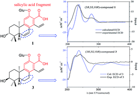
Time-dependent density functional theory (TDDFT) calculated electronic circular dichroism (ECD) and Mosher’s method were applied to establish the absolute configurations of six new highly oxygenated cleistanthane diterpenoid glucosides, phyllanembloids A–F (1–6), isolated from the roots of Phyllanthus emblica. Compounds 1–5 featured a carboxyl group at C-13 adjacent to the hydroxyl group at C-12, and are the first examples of cleistanthane diterpenoids with a salicylic acid fragment. The carboxyl group at C-13 can significantly affect the CD spectrum of cleistanthane diterpenoids, and make the excitations of phenylethylene dominate the Cotton effects (240 and 280 nm), rather than the benzene ring excitations dominating the Cotton effects at 220 and 240 nm as in the 13-methyl analogues. The characteristic Cotton effects of cleistanthane diterpenoids were summarized according to the experimental and calculated ECD curves, which can be applied as a common method to determine the absolute configurations of this type of diterpenoid.


Introduction

Cleistanthane diterpenoids featuring a benzene C ring in the skeleton are rare in nature.1 The first example was cleistanthol isolated from Cleistanthus schlechteri.2 Determination of the absolute configurations of cleistanthane diterpenoids usually depends on the sign of CD Cotton effects near 350 nm, generated by the n–π* excitation of the conjugated system comprising the benzene ring and the carbonyl group at C-7.1,3 However, most naturally occurring cleistanthane diterpenoids have no carbonyl group at C-7. Therefore, the application of this method is limited, and in most recently published work on cleistanthane diterpenoids the absolute configurations of these compounds was not determined.4–7

Phyllanthus emblica L, is an important traditional Chinese plant.8 Previous studies have revealed that its roots present tannins9 and bisabolane sesquiterpenoid glycosides.10–13 Further chemical investigation of the titled plant has led to the isolation of six new diterpenoid glucosides 1–6 (Fig. 1). These compounds possess a highly oxygenated cleistanthane skeleton, with a carboxyl group at C-13 (1–5), a 15-acetyl (5), and/or glycosylations on C-12 (1–3) and C-19 (5–6). Determination of the absolute configurations of these compounds is a challenge, because it is difficult to obtain a proper single crystal or carry out Mosher reactions, due to the presence of sugar moieties and carboxyl groups. Time-dependent density functional theory (TDDFT) based calculation of electronic circular dichroism (ECD) and optical rotations (ORs) were applied to determine the absolute configurations of 1–6. The theoretically calculated results were confirmed by Mosher’s method in the case of 1. The effects of the substitutions on the benzene ring on the ECD properties, and the characters of the Cotton effects for the cleistanthane diterpenoids were investigated and discussed. In addition, the isolated compounds were evaluated for their anti-hepatitis B virus (HBV) activities and cytotoxicities against human cancer cell lines .


image file: c4ra16624h-f1.tif
Fig. 1 Cleistanthane diterpenoids isolated from P. emblica.

Results and discussion

HRESIMS (m/z 489.2130 [M − H]) analysis returned a molecular formula of C26H34O9 for 1, corresponding to 10 degrees of unsaturation. The NMR data (Table 1) of 1 revealed the signals of one β-glucopyranosyl moiety: an anomeric proton and carbon at δH 4.81 (1H, d, J = 8.0 Hz, H-1′) and δC 104.7 (C-1′). Acidic hydrolysis of 1 afforded D-glucose [α]25D +94.3 (c = 0.63, in H2O) as the sugar residue and 1A as its aglycon. Besides, 20 carbon signals due to the aglycon part were observed in the 13C NMR and DEPT spectra, attributed to one carboxyl (δC 177.3), 10 sp2 carbons (δC 120–155) including one terminal double bond (δC 121.0, CH2), and nine aliphatic carbons, including three methyls (δC 17.5, 28.5, and 20.4) with corresponding protons as singlet signals at δH 1.00, 1.04, and 0.99 (s, each 3H) in the 1H NMR spectrum. Moreover, only one aromatic singlet proton at δH 7.01 (1H, s) was observed, indicating a pentasubstituted benzene ring in 1. Three mutually coupled olefinic protons at δH 6.91 (1H, dd, J = 11.4, 17.8 Hz), 5.44 (1H, dd, J = 2.0, 11.4 Hz), and 5.47 (1H, dd, J = 2.0, 17.8 Hz) belonged to a terminal double bond. Another two olefinic protons at δH 6.79 and 5.96 (each 1H, dd, J = 3.0, 10.0 Hz) implied a disubstituted cis-vinyl group in 1. Apart from two double bonds, one benzene ring, one carboxyl and one glucopyranosyl group accounting for eight degrees of unsaturation, it is obvious that there should be another two rings in 1. The aforementioned data indicated 1 was a cleistanthane type diterpenoid glycoside. The extensive comparison and analysis of 1D NMR (Table 1) data of 1A suggested that it had the same A, B and C rings as those of spruceanol, a cleistanthane diterpenoid reported from Croton insularis.14 However, instead of the methyl at C-17 and two aliphatic methylenes (C-6 and C-7) in spruceanol, a carboxyl carbon (δC 177.3) and a disubstituted cis-vinyl (δC 128.6 and 126.6) group were present in 1. HMBC correlations (Fig. 2) from the aromatic proton at δH 7.01 (H-11) to the carboxyl carbon (δC 177.3, C-17) and the vinyl protons at δH 5.44 and 5.47 (H2-16) to C-14 allowed the direct connectivity between the carboxyl and C-13 to be confirmed. The disubstituted cis-vinyl group was assigned as C-6 and C-7 in 1, by the HMBC correlations from H-5 (δH 1.99) to C-6/C-7, and δH 6.79 (H-7) to C-8/C-9/C-14. The HMBC correlations from the glycosyl H-1′ to C-12 (δC 153.9) confirmed the location of the glucosyl on C-12. This was further confirmed by ROESY correlation of the anomeric proton H-1′ with the aromatic proton H-11. Other 2D NMR correlations confirmed the planar structure of 1 as shown in Fig. 2. In the ROESY spectrum of 1, there were correlations of H-5 with H3-18/H-1ax (δH 1.68), H3-20 with H-1eq (δH 2.22), suggesting that the trans fused A/B ring in 1, and H-5 and Me-20 were diaxially orientated. The large coupling constant of J2b,3 (10.0 Hz) indicated an axially orientated H-3, which was further confirmed by the ROESY correlations of H-3 with H-5/H3-18.
Table 1 13C (125 MHz) and 1H (500 MHz) NMR spectroscopic data of compounds 1–3 and 1A in CD3OD (δ in ppm)
No. 1 1Aa 2 3b
δC, mult δH δC, mult δH δC, mult δH δC, mult δH
a Data were recorded in pyridine-d5.b 13C and 1H NMR data were recorded in 150 MHz and 600 MHz, respectively.
1 36.0, CH2 1.68 dt (5.0, 12.5) 35.6, CH2 1.70 dd (10.6, 12.0) 42.0, CH2 1.87 dd (2.5, 14.0) 33.2, CH2 1.85 m
2.22 dt (12.5, 3.3) 2.13 dt (13.1, 2.9) 2.56 dd (2.5, 14.0) 2.04 dt (13.1, 3.4)
2 28.6, CH2 1.77 m 29.1, CH2 2.04 m 72.5, CH 4.21 dd (2.5, 3.5) 27.2, CH2 1.73 m
1.82 dd (5.2, 10.0) 2.12 tt (3.2, 13.9)
3 79.4, CH 3.17 dd (5.5, 10.5) 78.3, CH 3.57 dd (7.5, 9.0) 79.3, CH 3.18 d (3.5) 76.4, CH 3.43 dd (3.2, 3.2)
4 39.4, C   39.3, C   38.8, C   38.9, C  
5 51.4, CH 1.99 t (3.3) 49.8, CH 2.16 t (3.0) 51.3, CH 2.10 t (2.5) 44.5, CH 1.72 m
6 128.6, CH 5.96 dd (3.0, 10.0) 126.7, CH 6.00 dd (3.0, 10.1) 128.8, CH 6.02 dd (2.5, 10.0) 19.9, CH2 1.71 m
1.85 m
7 126.6, CH 6.79 dd (3.0, 10.0) 127.3, CH 7.10 dd (3.0, 10.1) 126.4, CH 6.79 dd (2.5, 10.0) 29.6, CH2 2.57 m
2.82 dd (6.2, 17.7)
8 131.6, C   123.9, C   130.0, C   128.8, C  
9 149.8, C   154.9, C   151.2, C   153.1, C  
10 39.6, C   39.7, C   39.3, C   39.2, C  
11 111.2, CH 7.01 s 111.7, CH 7.10 s 111.5, CH 7.05 s 113.4, CH 7.15 s
12 153.9, C   162.8, C   153.9, C   151.6, C  
13 126.6, C   114.9, C   133.3, C   130.5, C  
14 133.4, C   139.7, C   133.9, C   135.9, C  
15 135.2, CH 6.91 dd (11.4, 17.8) 138.2, CH 7.71 dd (11.5, 16.9) 135.1, CH 6.91 dd (11.5, 17.5) 136.4, C 6.77 dd (11.5, 17.7)
16 121.0, CH2 5.44 dd (2.0, 11.4) 118.8, CH2 5.30 d (16.9) 121.2, CH2 5.43 d (17.5) 119.2, CH2 5.32 brd (11.5)
5.47 dd (2.0, 17.8) 5.59 d (11.5) 5.47 d (11.5) 5.50 brd (17.7)
17 177.3, C   170.0, C   176.5, C   178.2, C  
18 17.5, CH3 1.00 s 17.7, CH3 1.27 s 18.5, CH3 1.06 s 28.9, CH3 1.01 s
19 28.5, CH3 1.04 s 29.1, CH3 1.31 s 29.8, CH3 1.23 s 22.9, CH3 0.94 s
20 20.4, CH3 0.99 s 20.9, CH3 1.13 s 22.3, CH3 1.23 s 25.2, CH3 1.19 s
1′ 104.7, CH 4.81 d (8.0)     104.5, CH 4.85 d (7.5) 105.1, CH 4.78 d (7.5)
2′ 75.3, CH 3.49 dd (8.0, 9.0)     75.4, CH 3.48 m 75.2, CH 3.46 m
3′ 77.8, CH 3.47 dd (9.0, 9.0)     77.9, CH 3.46 m 77.7, CH 3.44 m
4′ 71.8, CH 3.35 dd (9.0, 9.0)     71.6, CH 3.36 dd (8.6, 8.6) 71.6, CH 3.33 m
5′ 78.7, CH 3.43 ddd (2.0, 7.0, 9.0)     78.6, CH 3.44 ddd (2.0, 7.0, 8.6) 78.7, CH 3.42 ddd (2.5, 7.0, 9.8)
6′ 62.9, CH2 3.69 dd (7.0, 12.0)     63.0, CH2 3.72 dd (7.0, 12.0) 62.9, CH2 3.68 dd (7.0, 12.1)
3.91 dd (2.0, 12.0) 3.93 dd (2.0, 12.0) 3.90 dd (2.5, 12.1)


image file: c4ra16624h-f2.tif
Fig. 2 Key 1H–1H COSY ([thick line, graph caption]) HMBC (→) correlations of 1.

The absolute configuration of 1 was established by comparison of its experimental ECD spectrum with the theoretically calculated results. The conformational searches were performed using either the Monte Carlo search applied MMFF method15 or a potential energy surface (PES) scan. The conformers were optimized using DFT-B3LYP/6-311G(d,p), and the optimized conformers with relative energy <2 kcal mol−1 were selected for ECD calculations using TD-DFT-B3LYP/6-311G(d,p). The final calculated ECD spectrum was obtained by averaging the calculated ECD curves using the Boltzmann distribution, respectively.16

The aglycon 1A displayed similar Cotton effects to its glycoside (1) in the experimental ECD curves (Fig. 3), suggesting the glycosyl moiety in 1 had little contribution to the ECD spectrum. Both the calculated and experimental ECD spectra displayed a strong positive Cotton effect at 240 nm and a weak Cotton effect at 280 nm. On the basis of the above evidence, the absolute configuration of phyllanembloid A (1) was determined to be 3R,5S,10R.


image file: c4ra16624h-f3.tif
Fig. 3 ECD and UV curves of compounds 1 and 1A.

Mosher’s method17,18 was applied to further confirm the absolute configuration of C-3 in 1. Enzymatic hydrolysis using cellulase for compound 1 was carried out to afford the aglycon 1A. Then, the carboxyl (C-13) and phenol (C-12) groups were protected by methylation reaction using CH3I with the acid scavenger K2CO3 to give 1B. Two portions of 1B were treated with (S)-(+)- and (R)-(−)-α-methoxy-α-(trifluoromethyl)phenylacetyl (MTPA) chloride in pyridine separately, to afford (R)- and (S)-MTPA ester derivatives 1r and 1s of 1B, respectively. In the 1H NMR spectra of 1r and 1s, the undisturbed signals (H-1, H-2, H-3, H-6, CH3-18, CH3-19 and CH3-20) were clearly different (see S1 in ESI) and the observed chemical shift differences (ΔδSR, Fig. 4) determined the absolute configuration of C-3 in 1B to be R, corresponding to the 3R configuration of 1. This agreed with the results obtained from theoretical ECD calculations.


image file: c4ra16624h-f4.tif
Fig. 4 The δSδR (CDCl3) values of the MTPA esters of 1B.

Phyllanembloid B (2) had a molecular formula of C26H34O10, as deduced using HREIMS (m/z 506.2133 [M]+). The NMR spectra of 2 (Table 1) were comparable to those of 1, except that the C-2 methylene signal (δC 28.6) in 1 was replaced by an oxymethine signal (δC 72.5) in 2, suggesting that 2 is the C-2 hydroxylated derivative of 1. The oxymethine carbon at δC 72.5 was assigned to be C-2, according to the 1H–1H COSY correlations of its corresponding proton at δH 4.21 (H-2) with both H-1 and H-3, and the HMBC correlation from H-2 to C-4 (δC 38.8) (see S2 in ESI). The small coupling constants of J2,3 (3.5 Hz) and J2,1 (2.5 Hz) suggested that H-2 in 2 was equatorially and α-orientated, which was confirmed by the ROESY correlations of H-1a (δH 1.87) with both H-2 and H-5 (δH 2.10), and H-5 with H-18 (δH 1.06). The ROESY correlation of H-5 with H-3 allowed the assignment of H-3 as axial and α-orientation. Compared with compound 1, the only difference in 2 was the hydroxylation at C-2. Thus, the absolute configuration of 2 could be assigned by comparing its CD spectrum with that of 1. The Cotton effects of 2 are comparable to those of 1 (Fig. 5). Therefore, the absolute configuration of phyllanembloid B (2) was determined to be 2R,3S,5S,10R.


image file: c4ra16624h-f5.tif
Fig. 5 ECD spectrum of compound 2.

Phyllanembloid C (3) displayed a quasimolecular ion peak at m/z 491.2292 [M − H] using HREIMS, corresponding to the molecular formula C26H36O9. The key difference between the NMR spectra of 3 (Table 1) and 1 was the appearance of two aliphatic methylenes at δC 19.9 and δC 29.6, instead of the Δ6,7 vinyl group in 1, suggesting that compound 3 was the C-6/C-7 hydrogenated derivative of 1. This was confirmed by the HMBC correlations (see S2 in ESI) from H-6 (δH 1.71 and 1.85) to C-5, C-10, C-18, and from H-7 (δH 2.57 and 2.82) to C-5, C-8, and C-9, and as well the 1H–1H COSY correlations between H-6 and H-7. In the ROESY spectrum of 3, correlations of H-20 (δH 1.19, 3H, s) with H-19 (δH 0.94, 3H, s), and H-5 (δH 1.72, 1H, m) with H-18 (δH 1.01, 3H, s) indicated the trans fused A/B ring, and H-5 and Me-20 were diaxially oriented. The small J2,3 (3.2 Hz) indicated an equatorial orientation of H-3, which is on the same face as Me-20 and β-orientated. This was confirmed by the ROESY correlations of H-3 with H3-18 and H3-19 (Fig. 6). The ECD spectrum of 3 (Fig. 6) demonstrated two weak positive Cotton effects at 220 and 240 nm, which were different from those of compounds 1 and 2, and the reasons are discussed below. The calculated ECD results of compound 3 with 3S,5S,10R absolute configuration agreed well with the experimental spectrum.


image file: c4ra16624h-f6.tif
Fig. 6 Key ROESY correlations and ECD spectra of 3.

Phyllanembloid D (4) had a molecular formula C26H32O10, as established using HREIMS (m/z 504.1998 [M]+). The 1H and 13C NMR spectra of 4 (Table 2) were closely related to those of 1. However, instead of the disubstituted Δ6,7 and the aliphatic methine at δC 51.4 (C-5, CH) in 1, signals for one ketone (δC 185.0, C) and one trisubstituted double bond [δC 172.8 (C), 127.1 (CH)] appeared in the 13C NMR and DEPT spectra of 4. The upfield shifted ketone signal at δC 185.0, representing an α,β-unsaturated carbonyl, was assigned as C-7 based on the key HMBC correlation (see S2 in ESI) from H-11 (δH 7.36) to the ketone carbon. The olefinic carbon signals at δC 172.8 and 127.6 were assigned to be C-5 and C-6, respectively, according to the HMBC correlations from the olefinic proton H-6 to C-8 and C-10, and from H3-18, H3-19, H3-20 and H-1 to the olefinic quaternary carbon at δC 172.8 (C-5). Based on the proton coupling constants and ROESY correlations, the relative configurations of 4 were established as shown. The calculated ECD spectra of 4 agreed well with the experimental ECD curve of 4. This determined the absolute configuration of phyllanembloid D (4) as 3R,10R (Fig. 7).

Table 2 13C (150 MHz) and 1H (600 MHz) NMR spectroscopic data of compounds 4–6 in CD3OD (δ in ppm)
No. 4 5 6
δC, mult δH δC, mult δH δC, mult δH
1 36.2, CH2 1.66 dt (13.9, 3.8) 35.8, CH2 1.73 dt (4.9, 13.5) 37.9, CH2 1.19 m
2.62 dt (13.9, 3.6) 2.22 dd (13.5, 3.5) 1.62 m
2 28.0, CH2 1.97 dq (13.8, 3.8) 28.5, CH2 1.93 m 27.5, CH2 1.63 m
2.13 dddd (3.6, 12.3, 13.8, 13.8) 1.97 dt (3.3, 13.7)
3 77.1, CH 3.38 dd (3.8, 12.3) 80.0, CH 3.37 dd (4.9, 11.6) 73.1, CH 3.76 t (7.4)
4 44.1, C   43.4, C   43.9, C  
5 172.8, C   51.0, CH 2.12 t (3.2) 46.9, CH 1.77 dd (2.3, 13.0)
6 127.1, CH 6.42 s 129.4, CH 6.16 dd (3.2, 10.0) 23.1, CH2 1.44 m
1.65 dq (4.9, 13.0)
7 185.0, C   124.1, CH 6.29 dd (3.2, 10.0) 36.2, CH2 2.24 dt (4.9, 14.0)
2.34 ddd (1.7, 4.5, 14.0)
8 123.5, C   119.9, C   141.8, C  
9 157.1, C   155.0, C   48.1, CH 1.99 dd (6.6, 10.2)
10 43.0, C   39.2, C   39.0, C  
11 112.2, CH 7.36 s 112.1, CH 6.73 s 27.3, CH2 1.57 ddd (2.2, 10.9, 13.1)
1.65 m
12 157.7, C   162.0, C   71.7, CH 3.90 t (3.9)
13 133.0, C   114.2, C   49.7, C  
14 138.4, C   142.9, C   120.9, CH 5.11 s
15 138.5, CH 7.35 dd (11.5, 17.7) 211.5, C   144.0, CH 5.85 dd (10.2, 17.6)
16 116.6, CH2 5.27 dd (1.5, 11.5) 33.6, CH3 2.42 s 117.3, CH2 5.09 dd (2.2, 17.6)
5.53 dd (1.5, 17.7) 5.14 dd (2.2, 10.2)
17 176.2, C   174.1, C   68.6, C 3.47 d (10.9)
3.60 d (10.9)
18 27.8, CH3 1.35 s 23.1, CH3 1.25 s 12.9, CH3 0.74 s
19 23.4, CH3 1.31 s 72.3, CH2 3.68 d (11.9) 73.8, CH2 3.28 d (10.1)
4.34 d (11.9) 3.82 d (10.1)
20 32.9, CH3 1.58 s 21.1, CH3 1.01 s 15.9, CH3 0.78 s
1′ 102.8, CH 5.00 d (7.7) 104.9, CH 4.29 d (7.8) 104.8, CH 4.22 d (7.9)
2′ 74.8, CH 3.58 dd (7.7, 9.3) 74.9, CH 3.26 dd (7.8, 9.3) 75.0, CH 3.20 dd (7.9, 9.0)
3′ 77.6, CH 3.54 dd (9.3, 9.3) 77.7, CH 3.41 dd (9.3, 9.3) 78.2, CH 3.34 dd (9.0, 9.0)
4′ 71.7, CH 3.35 dd (9.3, 9.3) 71.4, CH 3.35 dd (9.3, 9.3) 71.9, CH 3.26 dd (9.0, 9.0)
5′ 78.7, CH 3.52 ddd (2.2, 7.1, 9.3) 77.7, CH 3.34 m 77.7, CH 3.27 m
6′ 62.8, CH2 3.67 dd (7.1, 12.1) 62.4, CH2 3.70 dd (5.2, 12.1) 63.0, CH2 3.64 dd (5.7, 11.6)
3.96 dd (2.2, 12.1) 3.88 dd (1.5, 12.1) 3.88 dd (2.1, 11.6)


image file: c4ra16624h-f7.tif
Fig. 7 Experimental and calculated ECD curves of 4.

Phyllanembloid E (5) had a molecular formula C26H34O11, as deduced via HREIMS (m/z 521.2032). The 1H and 13C but for the presence of one acetyl (δC 211.5, 33.6, and δH 2.42) and NMR (DEPT) spectra (Table 2) were close to those of 1, except that one oxymethylene (δC 72.3) appeared in 5, instead of the Δ15,16 vinyl (δC 121.0, 135.2) and one methyl (δC 17.5) group in 1. The acetyl peaks at δC 211.5 and 33.6 were assigned respectively to be C-15 and C-16, due to the HMBC correlations (see S2 in ESI) from H-16, H-11 and H-7 to C-14, and from H-16 to δC 211.5 (C-15). The oxymethylene at δC 72.3 was determined to be C-19 due to the HMBC correlations of H-19 with C-3, C-4, C-19, and C-5. Moreover, the glucosyl moiety was located on the C-19 position, taking into account the HMBC correlation from H-19 to C-1′. Other 1H–1H COSY and HMBC correlations confirmed the planar structure of 5. The large coupling constants of J3,2 (11.6 Hz) suggested that H-3 was axially orientated and at the opposite face to the 20-methyl. The ROESY correlations of H-3 with H-5 and H-18 indicated that all the three protons were located on the same face. Together with the ROESY correlations of H-20 with H-19, the relative configurations of 5 were established.

The Monte Carlo conformer search of 5 produced two major lower energy conformers, 5a (47.4%) and 5b (39.6%) (Fig. 8), after optimization. Both 5a and 5b were 16-methyl α-oriented conformers. PES scans using 5a and 5b as the starting structures generated another two 16-methyl β-oriented conformers with the same energy level as 5a and 5b, respectively (Fig. 9 right). The distribution ratio of 16-methyl β- and α-oriented conformers was 67[thin space (1/6-em)]:[thin space (1/6-em)]32, which can explain the split signal of the 16-methyl in the 1H NMR spectrum of 5 (Fig. 9 left).


image file: c4ra16624h-f8.tif
Fig. 8 Structures of conformers 5a (left) and 5b (right).

image file: c4ra16624h-f9.tif
Fig. 9 ROESY spectrum and PES scans of 5.

ECD calculation using the optimized lower energy conformers (Fig. 10 top) could predict the experimental ECD spectrum of 5 (Fig. 10 bottom), which was comparable to those of compounds 1 and 2, and the absolute configuration of 5 was therefore assigned as 3R,4R,5S,10R.


image file: c4ra16624h-f10.tif
Fig. 10 The low energy conformers (top) and ECD spectra of 5 (bottom).

Phyllanembloid F (6), a white amorphous powder, had a molecular formula C26H42O9, deduced via HRESIMS (m/z 543.2812 [M + HCOO]). The 13C NMR and DEPT data (Table 2) of 6 showed the presence of one glucosyl moiety, in addition to 20 carbon resonances attributable to the aglycon part of 6, which were similar to those of ent-isopimara-8(14),15-dien-3β-ol.3 However, two methyls in ent-isopimara-8(14),15-dien-3β-ol were replaced by two oxymethylenes (δC 68.6 and 73.8) in 6. The oxymethylene signals at δC 68.6 and 73.8 were assigned to be C-17 and C-19, respectively, according to the HMBC correlations (see S2 in ESI) from H-16 and H-15 to C-17, from H-17 to C-12, C-13, and C-14, and from H-19 to C-3 and C-4. The location of the glucosyl moiety was determined to be at C-19 based on the HMBC correlation from the anomeric proton at δH 4.22 (1H, d, J = 7.9 Hz, H-1′) to C-19. Thus, the planar structure of 6 was determined. In the ROESY spectrum of 6, correlations of H-3 with H-5 and H-18, H-5 with H-9 revealed the β orientation for H-3, H-5 and H-9, while correlations of the 20-methyl with H-15 and H-12 indicated the α-orientated C-20, H-12, and Δ15,16 vinyl group. There is no Cotton effect in the CD spectrum used to determine the absolute configuration of 6, due to the lack of chromophore in the range of 200–400 nm.

The OR of compound 6 was calculated using TDDFT/GIAO methods.19 The calculated OR (−76.0) agreed well with the experimental value (−47.3), by which the absolute configuration of phyllanembloid F (6) was determined to be 3R,4R,5S,9R,10S,12R,13R.

Rules of the absolute configurations and Cotton effects of cleistanthane type diterpenoids

The above mentioned cleistanthane diterpenoids 1–5 feature a benzene C ring in the skeleton, and this chromophore is adjacent to the stereocenters at C-10 and C-5; the Cotton effects are sensitive to the stereo-properties of this type of diterpenoid. When a carbonyl group existed in the C-7 position, the absolute configuration of this type of diterpenoid could be determined on the basis of the sign of the CD Cotton effects near 350 nm, generated by the n–π* excitation of the benzoyl moiety.1,3 However, most naturally occurring cleistanthane diterpenoids have no carbonyl group at C-7. Herein, we establish a method to determine the absolute configuration directly using the CD spectrum without any chemical reactions.

Compounds 1, 2 and 5 displayed a similar strong Cotton effect at 240 nm and weak Cotton effect at 280 nm (Fig. 3, 5 and 10). The common characteristics of these compounds are the conjugated system comprising of a benzene C ring with a Δ6,7 vinyl group, which is adjacent to the C-5 stereocenter. The Cotton effects at 240 and 280 nm were generated by the charge transfer of 1Lb excitations occurring at the phenylethylene system, respectively. For compound 3 with an sp3 C6–C7 bond, the Cotton effects at 220 and 240 nm were generated by the benzene ring system (Fig. 6). Although phyllanflexoid (phy-) B,20 a cleistanthane diterpenoid isolated from Phyllanthus flexuosus, has a Δ6,7 vinyl group in the structure, its CD spectrum is similar to those of 3 and phy-A, whose Cotton effects were generated by the benzene ring rather than the phenylethylene system (Fig. 11). It is noted that there is a 13-carboxyl substitution at the meta-position of the Δ6,7 vinyl group in 1, 2 and 5. The induction effect of this electron-withdrawing group enhanced the conjugation of the phenylethylene system and made it display strong Cotton effects at 240 and 280 nm. It can be concluded that the carboxyl group at C-13 allowed excitations arising from Δ6,7 phenylethylene to dominate the Cotton effects of cleistanthane diterpenoids. In other cases, the cleistanthane diterpenoids with alkyl groups at C-13 (phy-A and -B), whether it has a Δ6,7 vinyl group, the CD spectra were dominated by the excitations (220 and 240 nm) arising from the benzene ring (Fig. 11). Together with the absolute configurations determined from experimental and calculated ECD of different structures of compounds 1–5 and the previously reported phy-A, -B and -C, the CD rules can become a common method to determine the absolute configurations of cleistanthane diterpenoids (Table 3).


image file: c4ra16624h-f11.tif
Fig. 11 Structures and ECD spectra of phyllanflexoids A and B.
Table 3 CD rules of 5S,10R cleistanthane diterpenoidsa
Compounds Functional group at C-13 Functional group at C6–C7 Functional group at A ring Cotton effects (nm)
a ++: strong positive; +: relatively weak positive; −−: strong negative.
1, 2, 5 –COOH CH[double bond, length as m-dash]CH   240 ++
280 +
phy-B –CH3 CH[double bond, length as m-dash]CH   220 +
250 +
3 –COOH CH2–CH2   220 +
240 +
phy-A –CH3 CH2–CH2   220 +
250 +
4 –COOH O[double bond, length as m-dash]C–C[double bond, length as m-dash]C (5)   230 ++
260 −−
phy-C –CH2–OH CH2–CH2 O[double bond, length as m-dash]C–C[double bond, length as m-dash]C (4) 220 +
240 +
320 ++


Anti-virus and cytotoxicities of cleistanthane diterpenoids

Compounds 1 and 5 were evaluated for their anti-hepatitis B virus (HBV) activities and cytotoxicities against five human cancer cell lines [lung cancer (A-549), human myeloid leukemia (HL-60), breast cancer (MCF-7), hepatocellular carcinoma (SMMC-7721), and colon cancer (SW480)], as in the previously reported procedures.21,22 Unfortunately, none of them displayed cytotoxicity at a concentration of 40 μM. Compound 1 was effective against the HBV surface antigen (HBsAg), with an IC50 value of 0.29 mg mL−1.

Conclusions

The absolute configurations of six new highly oxygenated cleistanthane diterpenoids, phyllanembloids A–F (1–6) from P. emblica, were established unambiguously by means of TDDFT calculated ECD, ORs, and Mosher’s method in the case of 1. Using the Monte Carlo search MMFF method, together with PES scans and TDDFT optimization, the preferential conformations of this type of compounds were calculated. On the basis of the relationship of the CD spectra and the structures of cleistanthane diterpenoids, the rules using CD spectra to determine the absolute configurations of cleistanthane diterpenoids were summarized. Compounds 1 and 5 were evaluated for their anti-HBV and cytotoxicities against five human cancer cell lines. Only compound 1 exhibited weak HBsAg inhibitory activity.

Experimental section

General procedures

Optical rotations were measured on a P-1020 polarimeter (JASCO, Tokyo, Japan). IR and UV spectra were measured on a Bruker Tensor 27 spectrometer with KBr pellets and Shimadzu UV 2401 PC, respectively. 1D and 2D NMR spectra were run on Bruker DRX-500 and AVANCE III-600 NMR spectrometers operating at 500 and 600 MHz for 1H, and 125 and 150 MHz for 13C, respectively. Coupling constants are expressed in hertz and chemical shifts are given on a ppm scale with solvents as internal standard. ESI-MS and HRESIMS were measured at Bruker HCT/Esquire and Agilent G6230. ECD and OR were detected at Applied Photophysics and Jasco P-1020. The apparatus of high performance liquid chromatography was a Agilent 1260 with a DAD detector. Column chromatography was performed with Sephadex LH-20 (Pharmacia Fine Chemical Co., Ltd. Uppsala, Sweden), Diaion HP20SS (Mitsubishi Chemical Co., Tokyo, Japan), Rp-8 gel (40–60 μm, Merck, Darmstadt, Germany), Toyopearl HW-40C (TOSOH, Japan), MCI CHP-20P (75–150 μm, Mitsubishi Chemical Co., Tokyo, Japan), silica gel (200–300 mesh, Qingdao Hailiang Group Co., Ltd. Qingdao, People’s Republic of China) and a 250 × 9.4 mm, i.d., 5 μm Sunfire C18 column (Waters). TLC was carried out on precoated silica gel GF254 plates, which were visualized in ultraviolet and spraying with 10% sulfuric ethanol solution. The quantum chemical calculations were carried out at the HPC Center, Kunming Institute of Botany, Chinese Academy of Sciences, People’s Republic of China.

Plant materials

The roots of P. emblica were collected in Baoshan City, Yunnan Province, People’s Republic of China, and identified by Prof. Chong-Ren Yang from the Kunming Institute of Botany, Chinese Academy of Sciences. A voucher specimen (KIB-ZL-0100020) has been deposited in the State Key Laboratory of Phytochemistry and Plant Resource in West China of Kunming Institute of Botany, Chinese Academy of Sciences.

Extraction and isolation

The extraction and pretreatment of the roots of P. emblica were carried out as in the previously reported procedure.13 Fr.A was subjected to Sephadex LH-20 (CH3OH 0–100%) to afford Fr.A1–Fr.A2. Fr.A1 (3.2 g) was passaged over a MCI CHP-20P column (CH3OH/H2O 30–90%) to give four fractions (Fr.A1.1–Fr.A1.4). Fr.A1.3 was chromatographed on Toyopearl HW-40C (CH3OH/H2O 0–30%) to give three fractions (Fr.A1.3.1–1.3.3), and Fr.A1.3.2 was separated by PHPLC (CH3CN/H2O, 10–20%) to afford 2 (8 mg). Fr.A1.4 was purified by Toyopearl HW-40C (CH3OH 0–30%) to give 1 (220 mg). Fr.B was passaged over Sephadex LH-20 (CH3OH/H2O 30–100%) to afford Fr.B1–Fr.B2. Fr.B1 was fractioned on a MCI CHP-20P column (CH3OH/H2O 30–80%) to afford Fr.B1.1–Fr.B1.6. Fr.B1.4 was purified by Toyopearl HW-40C (CH3OH/H2O 0–30%) and Rp-8 (CH3OH/H2O 30–80%) to afford 5 (25 mg). Fr.B1.6 was chromatographed on Toyopearl HW-40C (CH3OH/H2O 0–30%) to give Fr.B1.6.1–1.6.4, and Fr.B1.6.1 was purified by PHPLC (CH3CN/H2O, 10–20%) to afford 3 (2 mg). Fr.F (24.4 g) was passaged over Sephadex LH-20 (CH3OH/H2O 0–100%) to give Fr.F1–Fr.F3. Fr.F1 was fractioned on Rp-8 (CH3OH/H2O 30–80%) to give Fr.F1.1–1.9. Fr.F1.2 was chromatographed on silica gel (CHCl3/CH3OH/H2O, 8[thin space (1/6-em)]:[thin space (1/6-em)]2[thin space (1/6-em)]:[thin space (1/6-em)]0.2) to afford Fr.F1.2.1–1.2.4, and Fr.F1.2.2 was purified by PHPLC (CH3CN/H2O, 10–20%) to afford 6 (2 mg). Fr.F1.3 was passaged over Toyopearl HW-40C (CH3OH 0–30%) to give Fr.F1.3.1–1.3.5. Fr.F1.3.4 was separated by PHPLC (CH3CN/H2O, 10–20%) to afford 4 (2 mg).
Phyllanembloid A (1). White amorphous powder; [α]25D = +65.4 (c = 1.6 in methanol); 1H NMR (CD3OD, 500 MHz) and 13C NMR (CD3OD, 125 MHz) data, see Table 1; IR (KBr) νmax 3429, 2926, 1578, 1384, 1073 cm−1; UV (MeOH) λmax (log[thin space (1/6-em)]ε) 206 (1.23), 235 (1.25), 278 (0.96) nm; ECD (CH3OH, [nm], ϕ [mdeg]): 233 (25.1), 279 (5.2), 310 (−1.5); MS (ESI): m/z: 489 [M − H]; HRMS (EI): m/z calcd for: C26H33O9, 489.2130 [M − H]; found: 489.2130.
Phyllanembloid B (2). White amorphous powder; [α]25D = +17.9 (c = 1.1 in methanol); 1H NMR (CD3OD, 500 MHz) and 13C NMR (CD3OD, 125 MHz) data, see Table 1; IR (KBr) νmax 3422, 2925, 1580, 1385, 1282, 1073 cm−1; UV (MeOH) λmax (log[thin space (1/6-em)]ε) 202 (1.07), 238 (1.14), 278 (0.75) nm; ECD (CH3OH, [nm], ϕ [mdeg]): 234 (16.5), 2767 (3.5), 310 (−1.2); MS (ESI): m/z: 506 [M]; HRMS (EI): m/z calcd for: C26H34O10, 506.2147 [M]+; found: 506.2133.
Phyllanembloid C (3). White amorphous powder; [α]25D = −18.8 (c = 1.0 in methanol); 1H NMR (CD3OD, 600 MHz) and 13C NMR (CD3OD, 150 MHz) data, see Table 1; IR (KBr) νmax 3425, 2924, 1633, 1416, 1073 cm−1; UV (MeOH) λmax (log[thin space (1/6-em)]ε) 203.2 (1.22) nm; ECD (CH3OH, [nm], ϕ [mdeg]): 201 (−6.3), 225 (2.6), 242 (2.9), 285 (−1.2); MS (ESI): m/z 491 [M − H]; HRMS (ESI): m/z calcd for C26H35O9, 491.2287 [M − H]; found: 491.2292.
Phyllanembloid D (4). White amorphous powder; [α]25D = −42.6 (c = 0.45 in methanol); 1H NMR (CD3OD, 600 MHz) and 13C NMR (CD3OD, 150 MHz) data, see Table 2; IR (KBr) νmax 3431, 2924, 2854, 1640, 1583, 1463, 1382, 1066 cm−1; UV (MeOH) λmax (log[thin space (1/6-em)]ε) 207 (1.40), 246 (1.20), 298 (0.98) nm; ECD (CH3OH, [nm], ϕ [mdeg]): 218 (5.6), 256 (−9.4), 294 (2.2); MS (EI): m/z 622 [M − 1]+, 606, 379; HRMS (EI): m/z calcd for C26H32O10, 504.1990 [M]+; found: 504.1998.
Phyllanembloid E (5). White amorphous powder; [α]25D = +108.1 (c = 1.3 in methanol); 1H NMR (CD3OD, 600 MHz) and 13C NMR (CD3OD, 150 MHz) data, Table 2; IR (KBr) νmax 3429, 2926, 1622, 1447, 1387, 1358, 1076 cm−1; UV (MeOH) λmax (log[thin space (1/6-em)]ε) 230.2 (1.44), 279 (1.07), 327 (0.64) nm; ECD (CH3OH, [nm], ϕ [mdeg]): 231 (23.4), 282 (5.7); MS (ESI): m/z 521 [M − H]; HRMS (ESI): m/z calcd for C26H33O11, 521.2028 [M − H]; found: 521.2032.
Phyllanembloid F (6). White amorphous powder; [α]25D = −47.3 (c = 0.7 in methanol); 1H NMR (CD3OD, 600 MHz) and 13C NMR (CD3OD, 150 MHz) data, see Table 2; IR (KBr) νmax 3424, 2950, 1631, 1433, 1385, 1077, 1041 cm−1; UV (MeOH) λmax (log[thin space (1/6-em)]ε) 202.8 (0.97) nm; ECD (CH3OH, [nm], ϕ [mdeg]): 198 (−21.4), 289 (−0.8); MS (ESI): m/z 521 [M + Na]+; HRMS (ESI): m/z calcd for C27H43O11, 543.2811 [M + HCOO]; found: 543.2812.
Acid hydrolysis of compound 1. Compound 1 (23 mg) in 5% H2SO4–EtOH solution (2 mL) was heated at 65 °C for 4 h. After cooling down to room temperature, the mixture was extracted with CHCl3 (30 mL × 3). The organic layer was dried over Na2SO4, and evaporated in vacuum to afford a solid (10 mg), which was subjected to p-HPLC to generate compound 1A (3 mg). The water layer was neutralized with Amberlite AG IRA-401 and evaporated in vacuum to afford D-glucose (6 mg): [α]25D = +94.3 (c = 0.63 in H2O)].
Enzymatic hydrolysis of compound 1. Compound 1 (20 mg) and cellulase in H2O (2 mL) were incubated at 37 °C for 24 h. After cooling down to room temperature, the mixture was passaged over a MCI-gel CHP20P column (CH3OH/H2O 0–90%) to afford 1A (5 mg).
Compound 1A. White amorphous powder; [α]25D = +88.6 (c = 3.0 in methanol); 1H NMR (CD3OD, 600 MHz) and 13C NMR (CD3OD, 150 MHz) data, Table 1; IR (KBr) νmax 3431, 2926, 2855, 1686, 1602, 1456, 1209, 1146 cm−1; UV (MeOH) λmax (log[thin space (1/6-em)]ε) 200 (1.03), 240 (1.24), 281 (0.79), 334 (0.33) nm; ECD (CH3OH, [nm], ϕ [mdeg]): 241 (14.2), 285 (1.1), 342 (1.0); MS (ESI): m/z 327 [M − H]; HRMS (ESI): m/z calcd for C20H24O4, 328.1669 [M]+; found 328.1675.
Preparation of compound 1B. 1A (5 mg) was dissolved in 1.5 mL of anhydrous DMF/CH3CN (2[thin space (1/6-em)]:[thin space (1/6-em)]1) solution, to which K2CO3 (5 mg) and CH3I (500 μL) were added. The reaction was carried out in the dark at 40 °C for 2 h. The mixture was poured into H2O (6 mL) and extracted with EtOAc (6 mL × 3). The organic layers were combined and washed with H2O (18 mL × 3). After drying with anhydrous Na2SO4, the organic layer was evaporated to dryness. The solid was purified using a silica gel column to afford 1B (3.7 mg): MS (ESI): m/z 357 [M + H]+.
Preparation of (R)- and (S)-MTPA esters (1r and 1s). 1B (1.5 mg) was transferred to a dried tube, to which anhydrous pyridine (300 μL) and (R)-(−)-α-methoxy-α-(trifluoromethyl)phenylacetyl (MTPA) chloride (10 μL) were added. The mixture was shaken to make 1B and MTPA chloride mix evenly, and kept overnight under an N2 gas stream. The mixture was poured into H2O (1 mL) and extracted with EtOAc (1 mL × 3). The organic layers were combined and washed with H2O (1 mL × 3). After drying with anhydrous Na2SO4, the organic layer was evaporated to dryness and the solid was monitored by NMR spectrometer. 1H NMR data of (S)-MTPA ester derivative 1s of 1B (800 MHz, CDCl3; signals were assigned by 1H–1H COSY and ROESY, see S52–S57 in the ESI): δH 1.806 (1H, dt, J = 2.4, 13.0 Hz, H-1a), 2.169 (1H, dt, J = 13.0, 3.0 Hz, H-1b), 1.887 (1H, m, H-2a), 2.041 (1H, m, H-2b), 4.759 (1H, dd, J = 4.1, 11.8 Hz, H-3), 2.112 (1H, t, J = 2.7 Hz, H-5), 5.802 (1H, dd, J = 2.8, 9.9 Hz, H-6), 6.635 (1H, dd, J = 2.8, 9.9 Hz, H-7), 6.613 (1H, s, H-11), 6.701 (1H, dd, J = 11.4, 17.8 Hz, H-15), 5.296 (1H, dd, J = 1.0, 17.6 Hz, H-16a), 5.404 (1H, dd, J = 1.0, 11.4 Hz, H-16b), 0.955 (3H, s, CH3-18), 0.856 (3H, s, CH3-19), 0.976 (3H, s, CH3-20), 3.771 (3H, s, 12-CH3O), 3.765 (3H, s, 17-CH3O). In the manner described for 1s, another portion of 1B (1.5 mg) was reacted with (S)-(+)-MTPA chloride (10 μL) to give (R)-MTPA ester derivative 1r: 1H NMR (800 MHz, CDCl3; signals were assigned by 1H–1H COSY ROESY): δH 1.789 (1H, dt, J = 2.4, 13.0 Hz, H-1a), 2.137 (1H, dt, J = 13.0 Hz, H-1b), 1.769 (1H, m, H-2a and 2b), 4.725 (1H, dd, J = 4.1, 10.9 Hz, H-3), 2.111 (1H, t, J = 2.7 Hz, H-5), 5.811 (1H, dd, J = 2.8, 9.9 Hz, H-6), 6.634 (1H, dd, J = 2.8, 9.9 Hz, H-7), 6.607 (1H, s, H-11), 6.698 (1H, dd, J = 11.3, 12.0 Hz, H-15), 5.291 (1H, brd, J = 12.0 Hz, H-16a), 5.399 (1H, brd, J = 12.0 Hz, H-16b), 0.958 (3H, s, CH3-18), 0.934 (3H, s, CH3-19), 0.946 (3H, s, CH3-20), 3.821 (3H, s, 17-CH3O), 3.758 (3H, s, 12-CH3O).

Quantum chemical calculations

The structure models of the compounds were constructed based on NOE analysis. The conformation analysis was carried out using Monte Carlo searching with molecular mechanics MMFF in Spartan’06 (Wavefunction Inc. Irvine, CA). The resulting conformers were re-optimized using DFT at the B3LYP-SCRF/6-311G(d,p) level using the integral equation formalism variant of the polarizable continuum model (IEF-PCM). The free energies and vibrational frequencies were calculated at the same level to confirm their stability, and no imaginary frequencies were found. The optimized low energy conformers with energy <2 kcal mol−1 were considered for ECD calculation. The TD-DFT/B3LYP-SCRF/6-311G(d,p) method was applied to calculate the excited energies, oscillator strength and rotational strength for 50 states. All the calculations were run with Gaussian ’09.23 The excited energies and rotational strength were used to simulate ECD spectra of each conformer by introducing the Gaussian function. The final ECD spectra of each compound were obtained by averaging all the simulated ECD spectra of all conformers according to their excited energies and Boltzmann distribution. The band shape of the calculated ECD curves were all 0.5 eV if there is no illustration.

The optical rotation (OR) of compound 6 was calculated using the TDDFT/GIAO method with the B3LYP/6-311++G (2d,p) basis set with the same optimized conformers as for the ECD calculations. The final optical rotation was obtained by averaging the OR values of each conformer with the Boltzmann distribution.

Acknowledgements

The authors are grateful to the members of the analytical group and Prof. Yan Li’s group at the State Key Laboratory of Phytochemistry and Plant Resources in West China, Kunming Institute of Botany, Chinese Academy of Sciences, for measuring the spectroscopic data and the cytotoxicity screening. We also thank Fudan University for the antiviral assay. This work was supported by the NSFC 81473121, the 973 Program of Ministry of Science and Technology of P. R. China (2011CB915503), the National Science & Technology Support Program of China (2013BAI11B02), the 12th 5 Year National Science & Technology Supporting Program (2012BAI29B06), the Fourteenth Candidates of the Young Academic Leaders of Yunnan Province (Min Xu, 2011CI044) and by the West Light Foundation of the Chinese Academy of Sciences.

Notes and references

  1. A. C. Pinto, M. L. Patitucci, R. S. Dasilva, P. P. S. Queiroz and A. Kelecom, Tetrahedron, 1983, 39, 3351 CrossRef CAS.
  2. E. J. McGarry, K. H. Pegel, L. Phillips and E. S. Waight, J. Chem. Soc. D, 1969, 1074 RSC.
  3. H. A. Candy, J. M. Pakshong and K. H. Pegel, J. Chem. Soc. C, 1970, 2536 RSC.
  4. S. Liu, H. Zhu, S. Zhang, X. Zhang, Q. Yu and L. Xuan, J. Nat. Prod., 2008, 71, 755 CrossRef CAS PubMed.
  5. X. H. Xu, W. Zhang, X. P. Cao and S. Xue, Phytochem. Lett., 2011, 4, 52 CrossRef CAS PubMed.
  6. M. Takasaki, I. Yamauchi, M. Haruna and T. Konoshima, J. Nat. Prod., 1998, 61, 1105 CrossRef CAS PubMed.
  7. F. Niu, Z. Cui, Q. Li, H. Chang, Y. Jiang, L. Qiao and P.-F. Tu, Magn. Reson. Chem., 2006, 44, 724 CrossRef CAS PubMed.
  8. Z. W. Xie, China’s Medicinal Herbs, ed. Z. W. Xie and A. H. Liang, People’s Health Publishing House, Beijing, 1996, vol. 2, p. 328 Search PubMed.
  9. Y. J. Zhang, T. Abe, T. Tanaka, C. R. Yang and I. Kouno, J. Nat. Prod., 2001, 64, 1527 CrossRef CAS PubMed.
  10. Y. J. Zhang, T. Tanaka, Y. Iwamoto, C. R. Yang and I. Kouno, J. Nat. Prod., 2000, 63, 1507 CrossRef CAS PubMed.
  11. Y. J. Zhang, T. Tanaka, Y. Iwamoto, C. R. Yang and I. Kouno, J. Nat. Prod., 2001, 64, 870 CrossRef CAS PubMed.
  12. Q. Liu, Y. F. Wang, R. J. Chen, M. Y. Zhang, Y. F. Wang, C. R. Yang and Y. J. Zhang, J. Nat. Prod., 2009, 72, 969 CrossRef CAS PubMed.
  13. J. J. Lv, Y. F. Wang, J. M. Zhang, S. Yu, D. Wang, H. T. Zhu, R. R. Cheng, C. R. Yang, M. Xu and Y. J. Zhang, Org. Biomol. Chem., 2014, 12, 8764 CAS.
  14. L. A. Maslovskaya, A. I. Savchenko, V. A. Gordon, P. W. Reddell, C. J. Pierce, P. G. Parsons and C. M. Williams, Org. Lett., 2011, 13, 1032 CrossRef CAS PubMed.
  15. T. Kurtán, R. Jia, Y. Li, G. Pescitelli and Y. W. Guo, Eur. J. Org. Chem., 2012, 6722 CrossRef.
  16. P. J. Stephens, J. J. Pan, F. J. Devlin, K. Krohn and T. Kurtán, J. Org. Chem., 2007, 72, 3521 CrossRef CAS PubMed.
  17. J. A. Dale and H. S. Mosher, J. Am. Chem. Soc., 1973, 95, 512 CrossRef CAS.
  18. J. M. Seco, E. Quiñoá and R. Riguera, Chem. Rev., 2004, 104, 17 CrossRef CAS.
  19. M. Kwit, M. D. Rozwadowska, J. Gawroński and A. Grajewska, J. Org. Chem., 2009, 74, 8051–8063 CrossRef CAS PubMed.
  20. J. Q. Zhao, J. J. Lv, Y. M. Wang, M. Xu, H. T. Zhu, D. Wang, C. R. Yang, Y. F. Wang and Y. J. Zhang, Tetrahedron Lett., 2013, 54, 4670 CrossRef CAS PubMed.
  21. J. J. Lv, S. Yu, Y. F. Wang, D. Wang, H. T. Zhu, R. R. Cheng, C. R. Yang, M. Xu and Y. J. Zhang, J. Org. Chem., 2014, 79, 5432 CrossRef CAS PubMed.
  22. J. J. Lv, M. Xu, D. Wang, H. T. Zhu, C. R. Yang, Y. F. Wang, Y. Li and Y. J. Zhang, J. Nat. Prod., 2013, 76, 926 CrossRef CAS PubMed.
  23. M. J. Frisch, G. W. Trucks, H. B. Schlegel, G. E. Scuseria, M. A. Robb, J. R. Cheeseman, G. Scalmani, V. Barone, B. Mennucci, G. A. Petersson, H. Nakatsuji, M. Caricato, X. Li, H. P. Hratchian, A. F. Izmaylov, J. Bloino, G. Zheng, J. L. Sonnenberg, M. Hada, M. Ehara, K. Toyota, R. Fukuda, J. Hasegawa, M. Ishida, T. Nakajima, Y. Honda, O. Kitao, H. Nakai, T. Vreven, J. A. Montgomery, Jr., J. E. Peralta, F. Ogliaro, M. Bearpark, J. J. Heyd, E. Brothers, K. N. Kudin, V. N. Staroverov, R. Kobayashi, J. Normand, K. Raghavachari, A. Rendell, J. C. Burant, S. S. Iyengar, J. Tomasi, M. Cossi, N. Rega, J. M. Millam, M. Klene, J. E. Knox, J. B. Cross, V. Bakken, C. Adamo, J. Jaramillo, R. Gomperts, R. E. Stratmann, O. Yazyev, A. J. Austin, R. Cammi, C. Pomelli, J. Ochterski, R. L. Martin, K. Morokuma, V. G. Zakrzewski, G. A. Voth, P. Salvador, J. J. Dannenberg, S. Dapprich, A. D. Daniels, O. Farkas, J. B. Foresman, J. V. Ortiz, J. Cioslowski and D. J. Fox, GAUSSIAN 09 (Revision A.2), Gaussian, Inc., Wallingford, CT, 2009 Search PubMed.

Footnote

Electronic supplementary information (ESI) available: Copies of the 1H NMR, 13C NMR, MS, HSQC, HMBC, COSY, and ROESY spectra of 1–6, 1A, 1B, 1r, and 1s, and the low energy optimized conformers, calculated ECD curves and ORs of conformers of 1, 1A, 3, 4 and 5. See DOI: 10.1039/c4ra16624h

This journal is © The Royal Society of Chemistry 2015