Molecularly imprinted polymer nanospheres based on Mn-doped ZnS QDs via precipitation polymerization for room-temperature phosphorescence probing of 2,6-dichlorophenol

Xiao Weia, Zhiping Zhoua, Tongfan Haoa, Hongji Lib and Yongsheng Yan*b
aSchool of Material Science and Engineering, Jiangsu University, Zhenjiang 212013, China
bSchool of Chemistry and Chemical Engineering, Jiangsu University, Zhenjiang 212013, China. E-mail: jdwxtx@126.com; Fax: +86 0511 88791800; Tel: +86 0511 88790683

Received 17th December 2014 , Accepted 4th February 2015

First published on 4th February 2015


Abstract

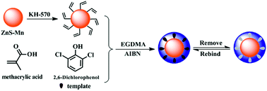
In this paper, novel molecularly imprinted polymers (MIPs) based on Mn doped ZnS quantum dots (QDs) with molecular recognition ability were successfully synthesized by precipitation polymerization using 2,6-dichlorophenol (2,6-DCP) as template, methacrylic acid (MAA) as the functional monomer and ethylene glycol dimethacrylate (EGDMA) as the cross-linker. The obtained materials (MIPs-ZnS:Mn QDs), which were composed of Mn doped ZnS QDs as phosphorescence signal and MIPs as molecular selective recognition sites, could sensitively and selectively recognize the template molecules by using the spectrofluorometer. After the experimental conditions were optimized, a linear relationship was obtained covering the range of 1.0–56 μmol L−1 with a correlation coefficient of 0.9994. The developed method was applicable to routine trace determination of 2,6-DCP in real examples. This study also provides a general strategy to fabricate MIPs-coated QDs with excellent performance and is desirable for chemical sensor application.


1 Introduction

In recent years, quantum dots (QDs) have attracted extensive attention and applications in the scientific community due to their unique optical and electronic properties, such as narrow and symmetric emission, broad excitation and excellent photostability.1,2 In particular, QDs have been widely used as probes for detecting different kinds of analytes including ions,3 small molecules,4 and biological macromolecules.5,6 To our knowledge, most of the detection methods are based on the fluorescence properties, and there are only a few studies on the use of the phosphorescence properties. Room temperature phosphorescence (RTP) as an effective detection mode has become a hotspot due to its fascinating advantages over fluorescence.7,8 The QDs with phosphorescence have longer lifetimes, which can allow an appropriate delay time and avoid the interference from autofluorescence and scattering light.9,10 It is for this reason that RTP QDs detection is very reliable. Therefore, the RTP detections based on QDs is a topic of considerable interest.

Molecular imprinting is an attractive technique for synthesizing polymer materials with specific recognition sites. The synthetic polymer materials, known as molecularly imprinted polymers (MIPs), are synthesized by the copolymerization of function monomers and a cross-linker in the presence of template molecules. After the template molecules are removed from the polymer matrixes, the binding sites with complementary size and shape of functionalities are obtained.11 Due to their high specific selectivity and stability, MIPs have been widely used in various significant applications, such as sensors,12 separation,13 drug delivery,14 and catalysis.15 In recent years, surface molecular imprinting technique has become a hot research topic at home and abroad. Compared with the traditional methods, this technique can provide a practical way to improve mass transfer, rebinding percentage and selectivity.16 Recently, a novel kind of MIPs-based RTP materials (QDs) as efficient sensors have been reported, which integrate the merits of the excellent RTP property of QDs and high selectivity of surface molecular imprinting technology.7,9,17,18 For now, most of MIPs-based RTP QDs composites were using silica as the matrix, such as MIPs (silica)/ZnS QDs for domoic acid and MIPs (silica)/ZnS QDs for pentachlorophenol.7,9 Meanwhile, a report used polymers as the matrices using a surface graft copolymerization in aqueous media for proteins.18 More novel strategies and methods for fabricating MIPs-based RTP materials (QDs) are required.

Chlorophenols (CPs), as a group of phenolic compounds, have been widely used in pharmacy, pesticides and herbicides.19 The presence of CPs in the environment is of particular concern due to their high toxicities, particularly in aquatic organisms.20 Due to their bad impact on the environment, CPs have been listed by the U.S. Environmental Protection Agency (U.S.EPA) as priority environmental pollutants.21 Thus, the detection of CPs from complex matrix is of great importance. To date, there have been a lot of methods reported, such as Gas Chromatography Mass Spectrometry (GC-MS),22 high performance liquid chromatography (HPLC),23 capillary electrophoresis,24 capillary electrochromatography25 and thin-layer chromatography26 for the determination of CPs. However, the shortcomings of these methods are that they are time consuming, use expensive reagents and require tedious sample pretreatment. Therefore, the development of simple, rapid and selective CPs detecting methods presents a challenge.

The objective of this work is to develop a new and eco-friendly kind of optical composite material combining the merits of molecular technology and phosphorescence property of Mn-doped ZnS QDs for specific recognition of 2,6-dichlorophenol (2,6-DCP, one of CPs). The vinyl modified Mn-doped ZnS QDs were used as the optical material and the core substrate. The uniform imprinted polymers was grafted onto the surface of vinyl modified Mn-doped ZnS QDs by precipitation polymerization. This imprinting layer was obtained using 2,6-DCP as the template, methacrylic acid (MAA) as the functional monomer, 2,2′-azobisisobutyronitrile (AIBN) as the initiator and ethylene glycol dimethacrylate (EGDMA) as the cross-linker. The MIPs coated ZnS:Mn QDs were characterized by transmission electron microscopy (TEM), scanning electron microscopy (SEM), X-ray diffraction (XRD) and Fourier transform infrared spectroscopy (FTIR). The RTP quenching relationship between 2,6-DCP and MIPs-ZnS:Mn QDs was investigated. This proposed RTP artificial sensor (MIPs-ZnS:Mn QDs) aims to offer a simple, rapid and selective sensing system for detecting 2,6-DCP in real samples.

2 Experimental

2.1 Reagents and chemicals

All chemicals were of analytical grade. ZnSO4·7H2O, MnCl2·4H2O, Na2S·9H2O, 3-(methacryloyloxy)propyl trimethoxysilane (KH-570), methacrylic acid (MAA), ethylene glycol dimethacrylate (EGDMA), 2,2′-azobis (2-methylpropionitrile) (AIBN), 2,6-dichlorophenol (2,6-DCP), 2,4-dichlorophenol (2,4-DCP), 2,4,5-trichlorophenol (2,4,5-TCP) and 2,4,6-trichlorophenol (2,4,6-TCP) were all purchased from Aladdin reagent Co. Ltd (Shanghai, China). Dry toluene, methanol, ethanol and acetonitrile were obtained from Sinopharm Chemical Reagent Co. Ltd (Shanghai, China). Double distilled water (DDW) was used for all the experimental procedures.

2.2 Instrument

The morphology and structure of prepared samples were observed by transmission electron microscopy (TEM, JEOL, JEM-2100) and scanning electron microscopy (SEM, JEOL, JSM-7001F). Infrared spectra (4000–400 cm−1) in KBr were recorded using Nicolet NEXUS-470 FTIR apparatus (USA). The X-ray diffraction (XRD) data were collected on a XRD-6100Lab X-ray diffractometer (Shimadzu, Japan) with Cu Kα radiation over the 2θ range of 10–80°. The phosphorescence measurements were performed on a Cary Eclipse spectrofluorometer (USA) equipped with a plotter unit and a quartz cell (1.0 cm × 1.0 cm).

2.3 Synthesis and functionalization of Mn-doped ZnS QDs

Mn-doped ZnS QDs in aqueous solution were synthesized according to previous reports with minor modifications.9,17 In a typical synthesis, ZnSO4 (6.25 mmol), MnCl2 (0.5 mmol), and 20 mL of water were mixed in a three-necked flask. After stirring at room temperature for 20 min, 5.0 mL of a 6.25 mmol Na2S solution was injected and the mixture was kept stirring overnight. Finally, the resultant Mn-doped ZnS QDs were obtained following centrifugation, washing with absolute ethanol and DDW three times and drying in vacuum oven at 60 °C overnight.

The surface of Mn-doped ZnS QDs was endowed with reactive vinyl groups through modification with KH-570. Briefly, 0.5 g of the obtained Mn-doped ZnS QDs and 2.0 mL of KH-570 were dispersed into 100 mL of dry toluene and then vigorously stirred under N2 at 90 °C for 12 h. The products were collected and washed with toluene and ethanol several times. Finally, the vinyl-modified Mn-doped ZnS QDs (KH-570–ZnS:Mn QDs) were dried under vacuum for further use.

2.4 Molecular imprinting of 2,6-DCP at the surface of KH-570–ZnS:Mn QDs

The 2,6-DCP-imprinted polymer layers on the surface of KH-570–ZnS:Mn QDs were prepared via in situ precipitation polymerization. MAA and EGDMA were used as a functional monomer and cross-linking agent, respectively. Typically, 2,6-DCP (0.1 mmol), methacrylic acid (0.4 mmol) and EGDMA (1.6 mmol) were dissolved in acetonitrile (60 mL) to self-assemble at room temperature. 50 mg of KH-570–ZnS:Mn QDs were dispersed into the above solution by ultrasonication. Then, AIBN (10 mg) as the initiator was added and the mixture was purged with nitrogen for 30 min. Finally, a two-step polymerization was carried out in a constant temperature bath oscillator with the rotation rate of 200 rpm. The slow pre-polymerization was first undertaken at 50 °C for 6 h, and the normal polymerization was completed at 60 °C for 24 h. After the polymerization reaction, the resulting MIPs-ZnS:Mn QDs were collected by centrifugation and washed with acetonitrile and ethanol several times to remove the unreacted monomers. The size and morphology of the MIPs-ZnS:Mn QDs were controlled by changing the amount of ZnS:Mn QDs and varying the total amount of polymeric monomers (MAA and EGDMA). The template 2,6-DCP in the MIPs was extracted with solvent mixture of methanol and acetic acid (9[thin space (1/6-em)]:[thin space (1/6-em)]1, v/v). The non-imprinted polymer (NIPs-ZnS:Mn QDs) were also prepared under the same conditions but without the addition of 2,6-DCP.

2.5 Measurement procedure

All the RTP detections were performed under the same conditions: the excitation wavelength was set at 320 nm with a recording emission range of 500–700 nm, and the slit widths of the excitation and emission were both 10 nm. The photomultiplier tube voltage was set at 670 V. MIPs-ZnS:Mn QDs and NIPs-ZnS:Mn QDs were dispersed in DDW to get the freshly-made stock solution (200 mg L−1). 2,6-DCP stock solution (1.0 mmol L−1, in DDW) was stocked at 4.0 °C. An appropriate quantity of MIPs-ZnS:Mn QDs and NIPs-ZnS:Mn QDs were added to a 5.0 mL colorimetric tube and a given concentration of analyte standard solution was added sequentially.

3 Results and discussion

3.1 Preparation and characterization of MIPs-ZnS:Mn QDs

Fig. 1 illustrates the principle to synthesize MIPs-ZnS:Mn QDs. First, Mn doped ZnS QDs were synthesized via a simple and practicable method at room temperature. Then, the Mn doped ZnS QDs were functionalized with KH-570 via a simple silanization reaction to form polymerizable vinyl groups. Subsequently, the precipitation polymerization was chosen as a beneficial and appropriate route to form the structural polymers on the KH-570–ZnS:Mn QDs. As shown in Fig. 1, 2,6-DCP was used as the template molecule in the formation of the imprinted polymer on the surface of ZnS QDs. In the presence of functional monomer (MAA), initiator (AIBN) and cross-linking agents (EGDMA), the 2,6-DCP@MIPs-ZnS:Mn QDs were formed by a two-step polymerization via the slow pre-polymerization at 50 °C and the normal polymerization at 60 °C. After the templates were removed from the imprinted polymers with solvent extraction, the MIPs-ZnS:Mn QDs were obtained and the generated recognition sites could selectively rebind the template molecules owing to an excellent compatibility of size, shape and chemical interactions.
image file: c4ra16542j-f1.tif
Fig. 1 Schematic procedure for the preparation of MIPs-ZnS:Mn QDs.

The thickness of the imprinted polymer plays a very important role because it influences RTP intensity and removal of the templates. In this study, a common mole ratio of template/monomer/cross-linker = 1[thin space (1/6-em)]:[thin space (1/6-em)]4[thin space (1/6-em)]:[thin space (1/6-em)]16 was used. On this basis, the size of MIPs-ZnS:Mn QDs could be controlled by two factors: the quantity of the ZnS QDs and the total amount of monomers. When the total amount of monomers was fixed, three kinds of RTP imprinted materials (P1–P3) differing in the quantity of the KH-570–ZnS:Mn QDs were prepared. The detailed compositions of the studied materials (P1–P3) are summarized in Table 1. To study their properties, the structures of the imprinted polymers were evaluated with TEM and SEM, and the images of the different imprinted polymers (P1–P3) are shown in Fig. 2. It could be seen from the figure that both the imprinted polymers consisted of spheroidal particles. When 50 mg KH-570–ZnS:Mn QDs and 2.0 mmol polymerization precursors were used, imprinted microsphere polymers with a diameter of 200 nm (P1) were obtained (Fig. 2a and b). When the amount of the KH-570–ZnS:Mn QDs was increased to 150 mg, we could see from Fig. 2c and d that the diameter of the imprinted polymers was about 100 nm (P2). Upon increasing the amount of the KH-570–ZnS:Mn QDs to 250 mg, as shown in Fig. 2e and f, the diameter of the imprinted polymers reduced to about 60 nm (P3). The sizes are listed in Table S1 in detail. As far as we know, if the imprinted polymer was too thick, the removal of templates would become difficult.27 Therefore, P3 was selected as the optimized imprinted polymer for further experiments. In addition, the NIPs-ZnS:Mn QDs were the same as the MIPs-ZnS:Mn QDs in size and morphology.

Table 1 Chemical composition of the studied MIPs and NIPs
Polymers KH-570–ZnS:Mn QDs (mg) Template (mmol) MAA (mmol) EGDMA (mmol) AIBN (mg)
P1 50 0.1 0.4 1.6 10
P2 150 0.1 0.4 1.6 10
P3 250 0.1 0.4 1.6 10


image file: c4ra16542j-f2.tif
Fig. 2 TEM images of different MIPs-ZnS:Mn QDs (P1: (a), P2: (c), P3: (e)) and SEM images of different MIPs-ZnS:Mn QDs (P1: (b), P2: (d), P3: (f)).

The formation of Mn-doped ZnS QDs could be confirmed by wide angle X-ray diffraction characterization. Fig. 3a shows the X-ray diffraction patterns of the Mn-doped ZnS QDs (curve 1), KH-570–ZnS:Mn QDs (curve 2) and MIPs-ZnS:Mn QDs (curve 3). The crystal structures of the three samples were cubic zinc blende, with peaks indexed as (111), (220), and (311) planes. The XRD patterns of KH-570–ZnS:Mn QDs and MIPs-ZnS:Mn QDs were similar to that of Mn-doped ZnS QDs, indicating that the surface modification and coating of polymer layer did not change the crystalline structure of Mn-doped ZnS QDs. The intensities of the (111), (220), and (311) diffraction peaks of MIPs-ZnS:Mn QDs were weaker than that of KH-570–ZnS:Mn QDs and Mn-doped ZnS QDs due to the imprinted polymer shell coating the quantum dots.


image file: c4ra16542j-f3.tif
Fig. 3 X-ray diffraction patterns (a) of ZnS:Mn QDs (1), KH-570–ZnS:Mn QDs (2) and MIPs-ZnS:Mn QDs (3), and FT-IR spectra (b) of ZnS:Mn QDs (1), KH-570–ZnS:Mn QDs (2), MIPs-ZnS:Mn QDs (3) and NIPs-ZnS:Mn QDs (4).

The FT-IR spectra of Mn-doped ZnS QDs (curve 1), KH-570–ZnS:Mn QDs (curve 2), MIPs-ZnS:Mn QDs (curve 3) and NIPs-ZnS:Mn QDs (curve 4) are shown in Fig. 3b. To our knowledge, the peak at 620 cm−1 belongs to the ZnS QDs corresponding to sulfuret and the wide peak between 3000 and 3700 cm−1 and the characteristic peak at 1614 cm−1 are observed due to O–H vibration of water.9,28 After the modification of KH-570, as shown in Fig. 5 (curve 2), there arose new absorptions at 1617 cm−1, 1718 cm−1 and 550–850 cm−1, which could be attributed to the C[double bond, length as m-dash]C stretching mode, C[double bond, length as m-dash]O stretching vibration and C–Cl vibration of KH-570 respectively.29 This phenomenon indicated that the surface of the Mn-doped ZnS QDs was successfully endowed with reactive vinyl groups through modification with KH-570. As shown in Fig. 5 (curve 3 and curve 4), the MIPs-ZnS:Mn QDs and NIPs-ZnS:Mn QDs showed similar locations and appearance of the major bands. The peak around 2968 cm−1 was the C–H band, and the absorption band at 3440 cm−1 (O–H stretching) of the polymers could be attributed to MAA molecules. The two characteristic peaks at 1730 cm−1 (C[double bond, length as m-dash]O stretching) and 1100–1200 cm−1 (C–O–C stretching) confirmed the presence of EGDMA in the polymers.30 All these bands showed that the MIPs were grafted on the surface of the ZnS QDs.

3.2 Addition of MIPs-ZnS:Mn QDs

The addition of MIPs-ZnS:Mn QDs in the range from 6.0 mg L−1 to 18 mg L−1 was used to investigate the effects on the 2,6-DCP detection system. Both linear range and detection sensitivity are important for the analysis of the RTP detection system. The change of RTP intensity [(P0P)/P0] (P0 and P represent the RTP intensity before and after adding 2,6-DCP into the system) determined the sensitivity of the analysis. High RTP intensity of MIPs-ZnS:Mn QDs could obtain wide linear range.31 As shown in Fig. 4, when the concentration of MIPs-ZnS:Mn QDs was low, a little amount of 2,6-DCP could lead to obvious phosphorescence quenching, and high sensitivity could be obtained with a narrow linear range. When the MIPs-ZnS:Mn QDs were at higher levels, RTP intensity was increased, whereas the phosphorescence quenching rate was relatively small. As a result, a concentration of 12 mg L−1 was chosen for the detection of 2,6-DCP.
image file: c4ra16542j-f4.tif
Fig. 4 Effects of the addition of MIPs-ZnS:Mn QDs on RTP intensity. Curve (a): the function of variation rate of RTP intensity of detection system (containing MIPs-ZnS:Mn QDs + 16 μmol L−1 2,6-DCP) vs. addition of MIPs-ZnS:Mn QDs; curve (b): the function of relative RTP intensity vs. addition of MIPs-ZnS:Mn QDs.

3.3 Stabilities

The RTP stability of MIPs-ZnS:Mn QDs was estimated by RTP intensity as a function of time at room temperature. As shown in Fig. 5a, the RTP of MIPs-ZnS:Mn QDs was stable for 7 measurements within 1.0 h. The maintained RTP intensity within 1.0 h may well be because the ZnS:Mn QDs were well protected by the polymer shell of the MIPs and the composite had excellent stability.
image file: c4ra16542j-f5.tif
Fig. 5 Stabilities of MIPs-ZnS:Mn QDs (a) and effect of time on RTP intensity (experimental conditions: MIPs-ZnS:Mn QDs, 12 mg L−1; 2,6-DCP, 16 μmol L−1) (b).

3.4 Detection time

In order to determine the optimal detection time of the RTP quenching procedure, a certain amount of 2,6-DCP (16 μmol L−1) was mixed with MIPs-ZnS:Mn QDs (12 mg L−1). The RTP intensities were recorded at different time intervals and the results are shown in Fig. 5b. It was found that the RTP intensity decreased at the beginning, and then remained constant after 50 min. As a result, the MIPs-ZnS:Mn QDs had a good and steady response rate for 2,6-DCP, and 50 min was selected as the optimal detection time for the following experiments.

3.5 MIPs-ZnS:Mn QDs and NIPs-ZnS:Mn QDs with template 2,6-DCP of different concentrations

Under the optimal conditions, MIPs-ZnS:Mn QDs were used as the optical material to detect 2,6-DCP based on the RTP quenching between ZnS:Mn QDs and the target molecule. The test was carried out in a colorimetric tube with water, the incubation target 2,6-DCP and MIPs-ZnS:Mn QDs for 50 min at room temperature, and NIPs-ZnS:Mn QDs were used as a contrast. Fig. 6a and b show the spectral response of MIPs-ZnS:Mn QDs and NIPs-ZnS:Mn QDs with the template 2,6-DCP at different concentrations, respectively. The RTP quenching followed the Stern–Volmer equation:32,33
 
P0/P = 1 + KSV[c] (1)
where P and P0 are the RTP intensities of the MIPs-ZnS:Mn QDs and NIPs-ZnS:Mn QDs in the absence and presence of the template 2,6-DCP, respectively, KSV is the Stern–Volmer constant, and [c] is the concentration of 2,6-DCP. The equation was applied to quantify the different quenching constants in this study, and the ratio of KSV,MIP to KSV,NIP was defined as the imprinting factor (IF). It was found (Fig. 6c and d) that the RTP intensities of the MIPs-ZnS:Mn QDs and NIPs-ZnS:Mn QDs decreased linearly with the increase of the concentrations of 2,6-DCP. As shown in Fig. 6c, the KSV,MIP was found to be 46[thin space (1/6-em)]560 M−1 and the linear range of the calibration curve was 1.0–56 μmol L−1 with a correlation coefficient of 0.9994. On the other hand, it could be found from Fig. 6d that the KSV,NIP was 7380 M−1, the linear range of 2,6-DCP was also 1.0–56 μmol L−1 but with a correlation coefficient of 0.9983. Under this condition, the imprinting factor was 6.309, indicating that the MIPs-ZnS:Mn QDs had a better selectivity than the NIPs-ZnS:Mn QDs. In addition, the detection limit (3σ/k) was 0.23 μmol L−1, in which k is the slope of the calibration line and σ is the standard deviation of blank measurements (n = 10).

image file: c4ra16542j-f6.tif
Fig. 6 RTP emission spectra of MIPs-ZnS:Mn QDs (a) and NIPs-ZnS:Mn QDs (b) (12 mg L−1) with the addition of the indicated concentrations of 2,6-DCP in water solution and the Stern–Volmer plots for MIPs-ZnS:Mn QDs (c) and NIPs-ZnS:Mn QDs (d).

Much excellent work about MIPs-ZnS:Mn QDs has been reported, some of which have been summarized and compared with the present work, with the characteristics and conclusions listed in Table 2. It is found that most common route for MIPs-ZnS:Mn QDs is a sol–gel reaction that employs 3-aminopropyltriethoxysilane (APTES) as the functional monomer and tetraethoxysilane (TEOS) as the cross-linker. The step-growth polymerization of TEOS with APTES could anchor the QDs to the silica monoliths and interact with template molecules to form the imprinted cavities. The surface graft copolymerization was also used to prepare MIPs-ZnS:Mn QDs for proteins in aqueous media. In contrast to these reported studies, a two-step precipitation polymerization method was used to prepare MIPs-ZnS:Mn QDs in this work. The proposed methodology possesses the same superior detection limit and linear range. In addition, the obtained MIPs-ZnS:Mn QDs integrate the advantages of the excellent RTP property of the ZnS QDs and high selectivity of the molecular imprinted polymers.

Table 2 Comparison between present method and literature
Synthetic methods Target object Response mode Linear range LOD Reference
Sol–gel reaction Domoic acid RTP enhancement 0.25–3.5 μM 67 nM 7
Sol–gel reaction Pentachlorophenol RTP quenching 0.2–19.1 μM 86 nM 9
Sol–gel reaction 2,4,5-Trichlorophenol RTP quenching 5.0–50 μM 17
Surface graft copolymerization Bovine hemoglobin RTP quenching 0.1–5.0 μM 38 nM 18
Precipitation polymerization 2,6-Dichlorophenol RTP quenching 1.0–56 μM 230 nM This work


3.6 Selectivity

Several kinds of phenols structurally related to 2,6-DCP (namely, 2,4-DCP, 2,4,5-TCP and 2,4,6-TCP) were utilized to evaluate the selectivity of the MIPs-ZnS:Mn QDs. As shown in Fig. 7, MIPs-ZnS:Mn QDs had a strong response to 2,6-DCP, which caused a significant change of RTP intensity with a high quenching amount. The changes in RTP intensity of the MIPs based on ZnS QDs for 2,6-DCP were more obvious than other phenols. By calculation, the difference in the quench amount (P0/P − 1) of MIPs-ZnS:Mn QDs and NIPs-ZnS:Mn QDs were 2.2079, 0.3885, 0.15127, 0.069 at 56 μmol L−1 for 2,6-DCP, 2,4-DCP, 2,4,5-TCP and 2,4,6-TCP, respectively. The results suggested that MIPs-ZnS:Mn QDs were specific to 2,6-DCP but nonspecific to other phenols and there was no selective recognition sites in the NIPs-ZnS:Mn QDs. This result can be reasonably explained as follows: in the process of synthesis of the MIPs-ZnS:Mn QDs, many specific imprinted cavities with the memory of the template 2,6-DCP were generated and the template could be bound strongly to the imprinted particles and cause changes in the RTP intensity.
image file: c4ra16542j-f7.tif
Fig. 7 Quenching amount of MIPs-ZnS:Mn QDs and NIPs-ZnS:Mn QDs by different kinds of 56 μmol L−1 chlorophenols (2,6-DCP, 2,4-DCP, 2,4,5-TCP, 2,4,6-TCP).

3.7 Analytical applications in Yangtze River sample

To demonstrate the applicability of this proposed method, MIPs-ZnS:Mn QDs were utilized for 2,6-DCP detection in water samples which were collected from Yangtze River. The samples were filtered through 0.45 μm Supor filters and stored in pre-cleaned glass bottles. As no phenols were detectable in the Yangtze River samples, a recovery study was carried out by using two methods: one was the RTP analysis and the other the ultraviolet analysis, which was used as a reference method. The experimental results are shown in Table 3. It was found that the results of RTP quenching method were more sensitive and accurate than those of the UV method, but the stability was less. The results showed a good agreement between the two methods. The values determined by the MIPs-ZnS:Mn QDs show the optical and selective recognition ability to provide accurate measures of 2,6-DCP concentrations on unknown environmental samples. Therefore, the MIPs-ZnS:Mn QDs could be regarded as an optional scheme for the direct analysis of relevant real samples.
Table 3 Recovery of 2,6-DCP in Yangtze River sample with 2,6-DCP solution at different concentration levels (n = 5)
Sample Concentration taken (μmol L−1) Found (μmol L−1) Recovery (%) RSD (%)
FL UV FL UV FL UV
1 10 10.35 10.43 103.5 104.3 5.2 3.6
2 20 20.32 20.48 101.6 102.4 3.8 2.7
3 40 40.2 40.52 100.5 101.3 3.4 2.3


4 Conclusion

In summary, a facile and versatile strategy was studied for the preparation of the MIPs-ZnS:Mn QDs that possessed Mn doped ZnS QDs as the phosphorescence species for selective recognition of a target phenol (2,6-DCP). The MIPs-ZnS:Mn QDs integrated the advantages of the excellent RTP property of the ZnS QDs and high selectivity of the molecular imprinted polymers. Under optimal conditions, the MIPs-ZnS:Mn QDs had a strong response to 2,6-DCP, which caused a significant change of RTP intensity with a high quenching amount. The results indicated that the MIPs-ZnS:Mn QDs had good selectivity for the template 2,6-DCP over other phenols. Furthermore, the proposed RTP analysis method with MIPs-ZnS:Mn QDs can successfully provide a simple, direct and selective detection of 2,6-DCP in real samples.

Acknowledgements

This work was financially supported by the National Natural Science Foundation of China (no. 21107037, no. 21176107, no. 21174057, no. 21277063, no. 21407057 and no. 21407064), National Basic Research Program of China (973 Program, 2012CB821500), Natural Science Foundation of Jiangsu Province (no. BK20140535), Ph.D. Innovation Programs Foundation of Jiangsu Province (no. KYLX_1032), National Postdoctoral Science Foundation (no. 2014M561595), Postdoctoral Science Foundation funded Project of Jiangsu Province (no. 1401108C).

References

  1. Z. Y. Tang, N. A. Kotov and M. Giersig, Spontaneous Organization of Single CdTe Nanoparticles into Luminescent Nanowires, Science, 2002, 297, 237 CrossRef CAS PubMed .
  2. M. Bruchez, M. Moronne, P. Gin Jr, S. Weiss and A. P. Alivisatos, Semiconductor Nanocrystals as Fluorescent Biological Labels, Science, 1998, 281, 2013 CrossRef CAS .
  3. W. J. Jin, M. T. Fernández-Argüelles, J. M. Costa-Fernández, R. Pereiro and A. Sanz-Medel, Photoactivated luminescent CdSe quantum dots as sensitive cyanide probes in aqueous solutions, Chem. Commun., 2005, 883 RSC .
  4. D. B. Cordes, S. Gamsey and B. Singaram, Fluorescent Quantum Dots with Boronic Acid Substituted Viologens To Sense Glucose in Aqueous Solution, Angew. Chem., Int. Ed., 2006, 45, 3829 CrossRef CAS PubMed .
  5. L. Y. Wang, L. Wang, F. Gao, Z. Y. Yu and Z. M. Wu, Application of functionalized CdS nanoparticles as fluorescence probe in the determination of nucleic acids, Analyst, 2002, 127, 977 RSC .
  6. H. Q. Yao, Y. Zhang, F. Xiao, Z. Y. Xia and J. H. Rao, Quantum Dot/Bioluminescence Resonance Energy Transfer Based Highly Sensitive Detection of Proteases, Angew. Chem., Int. Ed., 2007, 46, 4346 CrossRef CAS PubMed .
  7. L. Dan and H. F. Wang, Mn-Doped ZnS Quantum Dot Imbedded Two-Fragment Imprinting Silica for Enhanced Room Temperature Phosphorescence Probing of Domoic Acid, Anal. Chem., 2013, 85, 4844 CrossRef CAS PubMed .
  8. H. Yan and H. F. Wang, Turn-on room temperature phosphorescence assay of heparin with tunable sensitivity and detection window based on target-induced self-assembly of polyethyleneimine capped Mn-doped ZnS quantum dots, Anal. Chem., 2011, 83, 8589 CrossRef CAS PubMed .
  9. H. F. Wang, Y. He, T. R. Ji and X. P. Yan, Surface molecular imprinting on Mn-doped ZnS quantum dots for room-temperature phosphorescence optosensing of pentachlorophenol in water, Anal. Chem., 2009, 81, 1615 CrossRef CAS PubMed .
  10. Y. Q. Wang and W. S. Zou, 3-Aminopropyltriethoxysilane-functionalized manganese doped ZnS quantum dots for room-temperature phosphorescence sensing ultratrace 2,4,6-trinitrotoluene in aqueous solution, Talanta, 2011, 85, 469 CrossRef CAS PubMed .
  11. W. Lian, S. Liu, J. Yu, X. Xing, J. Li, M. Cui and J. Huang, Electrochemical sensor based on gold nanoparticles fabricated molecularly imprinted polymer film at chitosan–platinum nanoparticles/graphene–gold nanoparticles double nanocomposites modified electrode for detection of erythromycin, Biosens. Bioelectron., 2012, 38, 163 CrossRef CAS PubMed .
  12. R. N. Liang, D. A. Song, R. M. Zhang and W. Qin, Potentiometric Sensing of Neutral Species Based on a Uniform-Sized Molecularly Imprinted Polymer as a Receptor, Angew. Chem., Int. Ed., 2010, 49, 2556 CrossRef CAS PubMed .
  13. Y. Li, C. K. Dong, J. Chu, J. Y. Qi and X. Li, Surface molecular imprinting onto fluorescein-coated magnetic nanoparticles via reversible addition fragmentation chain transferpolymerization: A facile three-in-one system for recognition and separation of endocrine disrupting chemicals, Nanoscale, 2011, 3, 280 RSC .
  14. J. F. Yin, Y. Cui, G. L. Yang and H. L. Wang, Molecularly imprinted nanotubes for enantioselective drug delivery and controlled release, Chem. Commun., 2010, 46, 7688 RSC .
  15. J. Orozco, A. Cortes, G. Z. Cheng, S. Sattayasamitsathit, W. Gao, X. M. Feng, Y. F. Shen and J. Wang, Molecularly imprinted polymer-based catalytic micromotors for selective protein transport, J. Am. Chem. Soc., 2013, 135, 5336 CrossRef CAS PubMed .
  16. B. J. Gao, J. Wang, F. Q. An and Q. Liu, Molecular imprinted material prepared by novel surface imprinting technique for selective adsorption of pirimicarb, Polymer, 2008, 49, 1230 CrossRef CAS PubMed .
  17. X. Wei, Z. P. Zhou, J. D. Dai, T. F. Hao, H. J. Li, Y. Q. Xu, L. Gao, J. M. Pan, C. X. Li and Y. S. Yan, Composites of surface imprinting polymer capped Mn-doped ZnS quantum dots for room-temperature phosphorescence probing of 2,4,5-trichlorophenol, J. Lumin., 2014, 155, 298 CrossRef CAS PubMed .
  18. L. Tan, C. C. Kang, S. Y. Xu and Y. W. Tang, Selective room temperature phosphorescence sensing of target protein using Mn-doped ZnS QDs-embedded molecularly imprinted polymer, Biosens. Bioelectron., 2013, 48, 216 CrossRef CAS PubMed .
  19. M. Czaplicka, Sources and transformations of chlorophenols in the natural Environment, Sci. Total Environ., 2004, 322, 21 CrossRef CAS PubMed .
  20. I. Rodríguez, M. P. Llompart and R. Cela, Solid-phase extraction of phenols, Solid-phase extraction of phenols, J. Chromatogr. A, 2000, 885, 291 CrossRef .
  21. L. H. Kieth and W. A. Telliard, Priority pollutants: I-a perspective view, Environ. Sci. Technol., 1979, 13, 416 CrossRef .
  22. E. Pitarch, C. Medina, T. Portolés, F. J. López and F. Hernández, Determination of priority organic micro-pollutants in water by gas chromatography coupled to triple quadrupole mass spectrometry, Anal. Chim. Acta, 2007, 583, 246 CrossRef CAS PubMed .
  23. M. Rizzo, D. Ventrice, M. A. Varone, R. Sidari and A. Caridi, HPLC determination of phenolics adsorbed on yeasts, J. Pharm. Biomed. Anal., 2006, 42, 46 CrossRef CAS PubMed .
  24. Y. Iinuma and H. Herrmann, Method development for the analysis of particle phase substituted methoxy phenols and aromatic acids from biomass burning using capillary electrophoresis/electrospray ionization mass spectrometry (CE/ESI-MS), J. Chromatogr. A, 2003, 1018, 105 CrossRef CAS PubMed .
  25. Y. S. Fung and Y. H. Long, Determination of phenols in soil by supercritical fluid extraction-capillary electrochromatography, J. Chromatogr. A, 2001, 907, 301 CrossRef CAS .
  26. O. P. Sharma, T. K. Bhat and B. Singh, Thin-layer chromatography of gallic acid, methyl gallate, pyrogallol, phloroglucinol, catechol, resorcinol, hydroquinone, catechin, epicatechin, cinnamic acid, p-coumaric acid, ferulic acid and tannic acid, J. Chromatogr. A, 1998, 822, 167 CrossRef CAS .
  27. J. D. Dai, X. Wei, Z. J. Cao, Z. P. Zhou, P. Yu, J. M. Pan, T. B. Zou, C. X. Li and Y. S. Yan, Highly-controllable imprinted polymer nanoshell at the surface of magnetic halloysite nanotubes for selective recognition and rapid adsorption of tetracycline, RSC Adv., 2014, 4, 7967 RSC .
  28. M. J. Iqbal and S. Iqbal, Synthesis of stable and highly luminescent beryllium and magnesium doped ZnS quantum dots suitable for design of photonic and sensor material, J. Lumin., 2013, 134, 739 CrossRef CAS PubMed .
  29. C. B. Liu, Z. L. Song, J. M. Pan, Y. S. Yan, Z. J. Cao, X. Wei, L. Gao, J. Wang, J. D. Dai, M. J. Meng and P. Yu, A simple and sensitive surface molecularly imprinted polymers based fluorescence sensor for detection of λ-cyhalothrin, Talanta, 2014, 125, 14 CrossRef CAS PubMed .
  30. K. Yoshimatsu, K. Reimhult, A. Krozer, K. Mosbach, K. Sode and L. Ye, Uniform molecularly imprinted microspheres and nanoparticles prepared by precipitation polymerization: The control of particle size suitable for different analytical applications, Anal. Chim. Acta, 2007, 584, 112 CrossRef CAS PubMed .
  31. H. Wei, Y. Wang and E. Song, New Method for Detection of Norfloxacin Nicotinate Based on the CdTe Quantum Dots, Acta Chim. Sin., 2011, 69, 2039 CAS .
  32. H. B. Li, Y. L. Li and J. Cheng, Molecularly Imprinted Silica Nanospheres Embedded CdSe Quantum Dots for Highly Selective and Sensitive Optosensing of Pyrethroids, Chem. Mater., 2010, 22, 2451 CrossRef CAS .
  33. X. Wei, M. J. Meng, Z. L. Song, L. Gao, H. J. Li, J. D. Dai, Z. P. Zhou, C. X. Li, J. M. Pan, P. Yu and Y. S. Yan, Synthesis of molecularly imprinted silica nanospheres embedded mercaptosuccinic acid-coated CdTe quantum dots for selective recognition of λ-cyhalothrin, J. Lumin., 2014, 153, 326 CrossRef CAS PubMed .

Footnote

Electronic supplementary information (ESI) available. See DOI: 10.1039/c4ra16542j

This journal is © The Royal Society of Chemistry 2015
Click here to see how this site uses Cookies. View our privacy policy here.