Antineoplastic agents 596. Isolation and structure of chromomycin A5 from a Beaufort Sea microorganism

George R. Pettit*a, Rui Tana, Robin K. Pettitab, Dennis L. Doubeka, Jean-Charles Chapuisa and Christine A. Webera
aDepartment of Chemistry and Biochemistry, Arizona State University, P.O. Box 871604, Tempe, Arizona 85287-1604, USA. E-mail: bpettit@asu.edu
bA. T. Still University, School of Osteopathic Medicine in Arizona, 5850 E. Still Circle, Mesa, AZ 85206, USA

Received 30th October 2014 , Accepted 19th December 2014

First published on 19th December 2014


Abstract

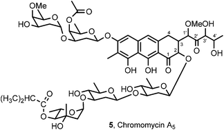
A microorganism identified as Streptomyces sp. was isolated from a sedimentary specimen collected on the Beaufort Sea coast of Alaska. Fermentation scale-up and cancer cell line-guided separation of the constituents led to isolation and structural elucidation (high resolution MALDI-TOF mass and 2D NMR spectral analyses) of a diastereomer of chromomycin A2 (2) designated chromomycin A5 (5). Chromomycin A5 displayed very potent cancer cell growth inhibitory activity (reaching with MCF-7 breast cancer GI50 0.00073 μg mL−1) against a mini-panel of murine and human cancer cell lines.


Introduction

Marine microorganisms represent a vast resource for discovery of new and greatly improved drugs for improving human cancer treatments, and a broad spectrum of other very serious to lethal medical problems. Fortunately, this approach to new drug discovery has been accelerating. Not surprisingly, marine Streptomyces sp. continue to be well represented with respect to cancer cell growth inhibitory constituents,2a–d and a variety of other marine microorganisms including fungi continue to be productive sources of anticancer2e,f and other types of potentially useful biologically activities.2g–l

In 1998, as part of an expedition along the Beaufort Sea coast of north Alaska, we collected specimens of marine sediments that led us to isolate a Streptomyces sp. that contained a very powerful cancer cell growth inhibitor. We soon determined it was related to one of the early anticancer and antibiotic drugs mithramycin (1),3 and even closer to other members of this anticancer “aureolic acid” family namely chromomycin A2 (2), A3 (3)4 and olivomycin A (4).5 Interestingly, chromomycins A2 and A3 have recently been reported as anticancer constituents of a marine Streptomyces sp.4a

The mithramycin group of anticancer drugs has stimulated extensive chemical, biological,6 and clinical investigation4a,7 with a focus on discovering structural modifications with improved therapeutic windows, greatly reduced toxicity, and increased human anticancer treatment results. With those eventual objectives in mind, we elected to scale-up the availability of our marine Beaufort Sea Streptomyces constituents, isolate the potent cancer cell growth inhibitor, determine its structure, and evaluate its anticancer potential (Fig. 1).


image file: c4ra16517a-f1.tif
Fig. 1 Selected aureolic acids.

Results and discussion

Toward those objectives, a 360 L scale-up fermentation of the Beaufort Sea Streptomyces sp. was divided into four batches and each was extracted (4×) with DCM. After removal of solvent in vacuo, the residue was partitioned (4×) between DCM and water (1[thin space (1/6-em)]:[thin space (1/6-em)]1). The organic fractions were dried immediately under vacuum and the residue was successively partitioned between hexane and 9[thin space (1/6-em)]:[thin space (1/6-em)]1 methanol–water. The methanol–water layer was then diluted with water to 3[thin space (1/6-em)]:[thin space (1/6-em)]2 and extracted (4×) with an equal volume of DCM. Upon solvent removal (in vacuo) a brown gum was obtained from the four batches (1.7 g, total) and showed murine P388 leukemia activity at ED50 0.27–1.4 μg mL−1.

The brown gum (1.7 g) was subjected to two sequences of gel permeation (methanol) and partition chromatography (hexane–toluene–methanol 3[thin space (1/6-em)]:[thin space (1/6-em)]1[thin space (1/6-em)]:[thin space (1/6-em)]1) on Sephadex LH-20; the most P388 active fraction exhibited ED50 0.059 μg mL−1. Further separation of this fraction by normal phase HPLC using a silica gel C8 column and a gradient of MeOH–DCM provided chromomycin A5 (5, 2.5 mg) as a yellow amorphous powder. The structure of chromomycin A5, except for the chirality at C-3′ (and possibly C-4′), was solved using a combination of high resolution mass and 1D/2D NMR spectral analyses as follows.

A mass spectral evaluation of chromomycin A5 revealed m/z at 1233.5002 for [M + Na]+ (calcd for C59H86O26Na 1233.5300) using a MALDI-TOF technique. The coordinated analyses of 1H, 13C NMR, APT and HMQC initially located the following carbon groups: two methoxyls, eleven methyls, six methylenes, twenty-seven methines, nine quaternary carbons, and three carbonyl groups that accounted for fifty-eight carbons and seventy-eight protons. In concert with the MS result, and assuming only carbon, hydrogen and oxygen comprised the molecule; the molecular formula would be C59H86O26. In turn this result suggested that chromomycin A5 might contain eight hydroxyl groups, which could not be readily observed in 1H NMR or related 2D NMR. Seventeen degrees of unsaturation were therefore derived based on the above analysis. That result implied a highly unsaturated molecule which was further supported by its UV spectrum with maximum UV absorptions at λmax 230, 279, 317.5, 332 and 412 nm suggesting an extended chromophore system.

Assignments for the carbon and proton groups were accomplished by interpretation of cross-signals in the 2D NMR spectra (COSY, TOCSY and HMBC). From a starting point with the five CH moieties from HSQC at downfield, δC/δH 98.2/5.20, 96.4/5.12, 102.2/5.08, 100.0/4.58 and 95.1/5.03, five carbohydrate units featured with 2,6-dideoxypyranose rings were further defined by analysis of H, H-COSY, TOCSY and HMBC cross signals and a series of 1H, 13C NMR data evaluations (see NMR data of pyranose units A to F in Table 1). The downfield behavior of the 13C signals of these anomeric CH units implied their linkages with adjacent units involved C-1 oxyls.8

Table 1 NMR data (500 MHz, CD3OD) for chromomycin A5 (5)
Position δC δH (Multi. J) HMBCb
a Signals overlapped or missing due to limited material.b HMBC correlations are from protons stated to the indicated carbon.
Aglycone
1 CO 204.0a    
2 CH 78.2 3.40 (1H, overlap)  
3 CH 43.6 2.78 (1H, m) 1′
4 CH2 28.1 2.80 (1H, m)a 1′
2.57 (1H, m)b  
5 CH 102.0 6.48 (1H, s)  
6 C 160.4    
7 C 112.1    
8 C 165.1a    
9 C 156.5    
10 CH 118.2 6.59 (1H, s)  
4a C 136.5    
8a C 109.3a    
9a C 108.6    
10a C 139.7    
7 C-CH3 8.5 2.15 (3H, s)  
1′ CH 83.2 4.83 (1H, overlap) OCH3-1′
2′ CO 213.6    
3′ CH 80.1 4.23 (1H, s, br) 5′
4′ CH 69.4 4.27 (1H, q, 6.5) 5′
5′ CH3 19.8 1.28 (3H, d, 6.5)  
1′C-OCH3 59.6 3.44 (3H, s) 1′
[thin space (1/6-em)]
Sugar A
A1 CH 98.2 5.23 (1H, m) 2a, 2b, 5
A2 CH2 34.1 2.05 (1H, m)a 4
2.09 (1H, dd, 9.0, 2.5)b  
A3 CH 71.4 4.16 (1H, overlap) 2a, 2b, 4, 1(α-D-sugar B)
A4 CH 69.1 5.18 (1H, d, 2.5) 5, 6
A5 CH 70.8 3.89 (1H, q, 6.5) 6
A6 CH3 17.1 1.21 (3H, d, 6.5) 5
A4 acetyl-CO 172.5   4, acetyl-CH3
A4 acetyl-CH3 20.7 2.15 (3H, s)  
[thin space (1/6-em)]
Sugar B
B1 CH 96.4 5.12 (1H, s, br) 2a, H-5
B2 CH2 33.8 1.62 (1H, dd, 12.5, 5.0)a 4
1.86 (1H, ddd, 12, 12, 3.5)b  
B3 CH 67.4 3.98 (1H, m) 1, 2a, 2b, 4
B4 CH 82.9 3.23 (1H, d, 2) 2a, 3, 5, 6, CH3-4
B5 CH 68.5 3.95 (1H, q, 7.0) 1, 6
B6 CH3 17.4 1.26 (3H, d, 5.5) 5
B4 OCH3 62.3 3.57 (3H, s) 4
[thin space (1/6-em)]
Sugar C
C1 CH 102.0 5.08 (1H, overlap)  
C2 CH2 8.3 1.62 (1H, dd, 12.5, 5.0)a  
2.59 (1H, m)b  
C3 CH 81.6a 3.70 (1H, m)  
C4 CH 76.0 3.05 (1H, d, 2.5) 6
C5 CH 73.4 3.32 (1H) 6
C6 CH3 18.6 1.33 (3H, d, 6.0)  
[thin space (1/6-em)]
Sugar D
D1 CH 100.2 4.58 (1H, overlap)  
D2 CH2 37.4 1.43 (1H, m)a  
2.30 (1H, m)b  
D3 CH 76.0 3.62 (1H, m)  
D4 CH 76.6 3.02 (1H, m) 6
D5 CH 73.8 3.35 (1H) 6
D6 CH3 18.6 1.34 (3H, d, 6.0)  
[thin space (1/6-em)]
Sugar E
E1 CH 95.1 5.03 (1H, s, br) 2
E2 CH2 45.0 1.94 (2H, m)a,b CH3-3
E3 C 71.4   2, 4, CH3-3
E3 CH3 23.1 1.44 (3H, s) 2, 4
E4 CH 80.4 4.68 (1H, d, 10) 2, 5, 6, CH3-3
E5 CH 66.8 4.13 (1H, dq, 10, 6.5) 4, 6
E6 CH3 18.3 1.12 (3H, d, 6.5) 4, 5
[thin space (1/6-em)]
E4-isobutanoyl
1′′ CO 178.1   4, 2′′, 3′′
2′′ CH 35.5 2.62 (1H, hept, 7)  
3′′ CH3 19.3 1.19 (6H, d, 7) 2′′
3′′ CH3 19.4 1.19 (6H, d, 7)  


An acetyl carbonyl was assigned to C-4 of pyranose A owing to the downfield shift of 4-H compared to that of a methine-proton with a hydroxyl group only and (was) confirmed by its correlation with the CO (δC 172.5/δH 5.18) in the HMBC. The smaller coupling constant (2.5 Hz) implied an equatorial orientation for H-4 in a chair-conformation for the pyranose unit. In pyranose B a methoxyl group linked to C-4 was deduced by observation of a HMBC cross peak of the methyl carbon with 4-H (δC 62.3/δH 3.23). The COSY and HMBC spectra demonstrated the presence of an isobutanoyl segment that was connected to the C-4 in pyranose E as confirmed by the HMBC cross signal of CO and 4-H (δC 178.1/δH 4.68). The 4-H in pyranose ring E appeared upfield (δ 4.68) and with a wider coupling of 10 Hz, which indicated an axial orientation. In pyranose rings B and E the 1-H appeared both as a broad single peak that implied their smaller couplings with the 2-Heq and 2-Hax. In turn that indicated an equatorial orientation for 1-H, namely α-glycosidic linkages for the B and E units.4b Further analysis of the COSY relationship of the C-1 H with the corresponding C-2 H in the pyranose segments revealed more evidence for the anomeric carbon being an α- or β-bond linkage. The equatorial oriented 1-H in α-bond linkage units B and E was observed cross peaks with both axial- and equatorial-oriented protons of C-2 (δH/δH 5.12/1.62, 1.86 for B; and 5.03/1.94, 1.94 for E); whereas the axial 1-H evidenced a cross peak with only the axial-oriented 2-H (δH/δH 5.23/2.05; 5.08/1.62; and 4.58/1.43), β-bond linkage was then assigned to the anomeric carbons of pyranoses A, C and D.

Additional 2D NMR analyses of COSY, TOCSY, and HMBC data revealed polyoxygenated segment –CH(O)–CH(CH2)–CH(OCH3)–CO–CH(OH)–CH(OH)–CH3. The remaining ten sp2-carbons downfield in the 13C NMR resembled those found in a hexa-substituted naphthalene system analogous to the aglycone found in the family of aureolic acids.5b,9 That important fact was ascertained by NMR comparison which showed limited cross peaks in the HMBC spectrum of A5, with matching two aromatic protons as singlet (δH 6.48 and 6.59), three C–O bonds at C-6, 8, and 9 (δC 160.4, 165.1 and 156.5), and one methyl (δC 8.5) at C-7. By considering the evidence for five 2,6-dideoxypyranoses, the aglycone frame was readily ascertained by connecting the polyoxygenated segment –CH(O)–CH(CH2)–CH(OCH3)–CO–CH(OH)–CH(OH)–CH3 with an additional carbonyl group introduced at position 4a and cycling at 9a.4

A disaccharide anomeric α-bond connection from pyranose B to A was confirmed by a key HMBC signal from 1-H of pyranose B to the C-3 of pyranose A (δH 5.12/δC 71.4). The connection of the remaining three pyranose rings was not immediately proved by correlations in the HMBC results, but rather by analyses of 1D and 2D NMR data to provide enough information (Fig. 2 and Table 1) to conclude pyranose E was bonded through 1α-linkage to C-3 of pyranose D. In turn unit D was joined through a β-bond to C-3 of pyranose C. Those facts indicated the trisaccharide assembly was related to the chromomycins4b series rather than mithramycins.3a


image file: c4ra16517a-f2.tif
Fig. 2 COSY correlations of chromomycin A5.

The disaccharide in the chromomycins was defined as β-A(3 → 1)-α-B and the trisaccharide as β-C(3 → 1)-β-D-(3 → 1)-α-E based on 1H, 13C NMR research4b and extensive circular dichroism (CD) studies,10 whereas β-A(3 → 1)-β-B and β-C(3 → 1)-β-D-(3 → 1)-β-E were reported for mithramycin following reinvestigation of its 1D and 2D NMR evidence.3a As is typical of antibiotic class, disaccharide and trisaccharide were assigned bonded to C-6 and C-2, respectively.

Measurement of the specific rotation of the compound chromomycin A5 (5) showed [α]24D + 56.7° (EtOH), which was opposite to that of chromomycin A2 (2) [α]24D − 61° (EtOH), reported by Miyamoto.4c Most compounds identified in this series, such as chromomycins A3 (3), A4 (6), mithramycin (1), and olivomycins A (4), B (7), C, and D, gave their [α]20–24D all in negative values.3b,4c,5d Two chromomycin derivatives, 02-3D (8) and 02-3G (9),11 exhibited positive [α]20D values (Table 2). However, there was no discussion on their stereostructures, although the compounds were reported as deacetyl and dideacetyl derivatives of chromomycin A3.11

Table 2 Comparison of optical rotations (EtOH) for chromomycins A2–A4 (2, 3 and 6),4c A5 (5), mithramycin (1),3b olivomycins A (4), B (7),5d 02-3D (8), and 02-3G (9)11
Compound 2 (ref. 4c) 3 (ref. 4c) 6 (ref. 4c) 5 1 (ref. 3b) 4 (ref. 5d) 7 (ref. 5d) 8 (ref. 11) 9 (ref. 11)
[α]20–24D −61.4° −57° −47° +57.6° −51° −36° −28° +55.6° +21.1°


Given the ketone groups at C-1 and C-2′ bonded to potentially labile units at C-2, C-1′ and C-3′ one or more of those chiral positions might have been epimerized during the extensive fermentation scale-up operations and/or during isolation procedures. Also, the 4′-CH might have epimerized by biosynthesis. When using the same kinds of NMR solvents, very similar resonance values were observed for the aglycone units of aureolic acids derivatives. The most obvious distinction of [α]24D − 61° for A2 and [α]24D + 56.7° for A5 raises some question about the 1H NMR at 2–4 H for A5 (5) being different from that of the 1H NMR (Table 3) for chromomycin A3 (3),3a mithramycin (1), and olivomycin A (4).5b At this point, we concluded our new anticancer antibiotic was a stereoisomer of chromomycin A2 (ref. 4c) at C2 and/or C3 and was named chromomycin A5 (5). The stereochemistry of A5 was not further assigned owing to a lack of definitive evidence and exhaustion of sample. That may have to be of augmented by a future resupply of chromomycin A5 and X-ray crystal structure determination.

Table 3 Comparison of partial NMR data for chromomycins A2 (2), A3 (3),9a A5 (5), mithramycin (1),5b olivomycin A (4),5b,9a olivomycin B (7),9a 02-3D (8), and 02-3G (9)11
Compound 2 (ref. 9a) 3 (ref. 9a) 3 (ref. 9a) 5 1 (ref. 5b) 4 (ref. 5b) 4 (ref. 9a) 7 (ref. 9a) 8 (ref. 11) 9 (ref. 11)
a Data was measured at a temperature of 60 °C.b This reference reported assignment of C-4 at 45.2 (1) and 43.9 (4), however this carbon is commonly found to resonate at 27–28 ppm.9ac 1H NMR data not reported.
13C NMR solvent CDCl3–CD3OHa CDCl3–CD3OHa CDCl3 CD3OD CD3OD CDCl3 CDCl3–CD3OHa CDCl3CD3OHa CDCl3 CD3CO2D
C 2 77.0 77.0 75.9 78.2 78.4 76.6 76.8 76.9 77.2 77.8
C 3 43.5 43.4 43.8 43.6 43.4 43.5 43.1 43.1 43.1 43.7
C 4 27.3 27.3 27.0 28.1 45.2b 43.9b 27.4 27.4 27.2 27.9
C 1′ 82.1 82.4 82.0 83.2 83.2 79.6 82.1 82.1 81.8 83.1
C 2′ 211.9 211.9 211.2 213.6 213.3 210.9 211.8 211.8 211.8 212.3
C 3′ 79.1 79.0 78.4 80.1 80.0 78.3 79.1 79.1 78.8 79.3
C 4′ 68.5 68.4 67.9 69.4 69.4 67.9 68.5 68.5 68.3 69.1
[thin space (1/6-em)]
1H NMR solvent   CDCl3 CD3OD CD3COCD3 CDCl3  
H 2 c c 4.73 3.40 4.78 4.74 c c c c
H 3     2.60 2.78 2.82 2.62        
H 4     3.10 2.80 2.97 3.09        
H 4     2.67 2.57 2.65 2.69        
H 1′     4.72 4.83 4.84 4.71        
H 3′     4.23 4.23 4.26 4.23        
H 4′     4.37 4.27 4.26 4.36        


The acetylation of compound A5 (5) with acetic anhydride in pyridine yielded a heptaacetate product that was evaluated by mass spectral analysis giving of m/z 1257.6 for [M + Na]+ which confirmed seven of the eight hydroxyls were converted, and matched previous work by Koenuma.9b However, the obvious decomposition loss of a major portion of the chromomycin A5 specimen during acetylation was a setback and prevented further characterization. Clearly, further evaluation of chromomycin A5 chemistry will require a future large scale production. As found for other aureolic acid analogues,7a chromomycin A5 revealed strong cancer cell growth inhibition against murine lymphocytic leukemia P388 (ED50 0.031 μg mL−1) and a mini-panel of human cancer cell lines (GI50 at 0.0024 μg mL−1 for pancreatic BXPC-3; 0.00073 μg mL−1 for breast cancer MCF-7; 0.0021 μg mL−1 for central nerve system cancer SF-268; 0.0014 μg mL−1 for lung cancer NCI-H460; 0.0043 μg mL−1 for colon cancer KM20L2 and 0.0019 μg mL−1 for prostate cancer DU-145). That remarkably strong level of cancer cell growth inhibition suggests chromomycin A5 would be a prime candidate for linkage to monoclonal antibodies capable of targeting cancer cells.

Experimental

General procedures

All solvents used in the extraction and isolation processes were redistilled prior to use. HPLC solvents were obtained from EMD Chemicals Inc., Gibbstown, NJ. Gel permeation column chromatography was performed with Sephadex LH-20 from Pharmacia. Optical rotations were measured using an Autopol IV Polarimeter from Rudolph Research Analytical. UV spectra were acquired using a Cary-14 UV/VIS spectrophotometer by Olis Inc. MALDI-TOF-MS were recorded with a Laser Desorption Time-of-Flight Mass Spectrometer from Vestec corporation. The 1H, 13C NMR and 2D NMR data were obtained employing Varian Unity 400 and Varian VXR 500S spectrometers. Semi-preparative HPLC separations were performed using a Waters 600E multi-solvent delivery system with a Waters 2487 dual λ Absorbance Detector. A Phenomenex Luna C8 silica gel column (250 × 10 mm, 5μ micron) and a Zorbax SB-C18 (5 μm, 4.6 × 250 mm) were used, respectively.

Specimen collection and fermentation

In 1998, low tide sedimentary specimens along the Beaufort Sea near Prudhoe Bay were collected by one of us (G. R. P.). The samples were shipped by air in clean plastic bags to our laboratories.

Scale-up fermentation to 360 L was performed in 1/3 strength YpSs broth (yeast extract and starch media) at rt with aeration. Optimization experiments performed in advance of scale-up fermentation indicated that activity peaked at 5 days. The actinomycete was identified by 16S rRNA gene sequence similarity (Accugenix, Newark, DE). Results from the MicroSeq database indicated that the isolate is Streptomyces polychromogenes (% difference = 1.8; confidence level to genus).

Solvent extraction and solvent partitioning of the microbial broth

By 2005 we completed a 360 L scale-up fermentation of Streptomyces sp. Fermentation broth was next subjected to solvent extraction and partitioning in four, 9 L batches employing the following procedure. Each portion was extracted with DCM four times (60 L of DCM per pass). The DCM extract was concentrated to dryness in vacuo and re-dissolved in 2 L of DCM. Two liters of water were added, and partitioning against water repeated four times to remove water soluble material from the DCM fraction. The dried residue from removal (in vacuo) of the DCM phase was next partitioned between hexane and methanol–water (9[thin space (1/6-em)]:[thin space (1/6-em)]1) four times. The methanol–water phase was adjusted to 3[thin space (1/6-em)]:[thin space (1/6-em)]2 by the addition of water, and then partitioned with DCM four times; after evaporation of the DCM a brown gum was obtained amounting to a total of 1.7 g from all four batches. The DCM residue showed ED50 at 0.27–1.4 μg mL−1 against the P388 murine lymphocytic leukemia cells and GI50 0.35–0.17 μg mL−1 against six human cancer cell lines (pancreatic BxPC-3, breast cancer MCF-7, central nerve system cancer SF-268, lung cancer NCI-H460, colon cancer KM20L2, and prostate cancer DU-145).

Isolation of chromomycin A5 (5)

The 1.7 g dried material from the DCM fraction was dissolved in methanol and subjected to a gel permeation column chromatography on Sephadex LH-20. The methanol fractions were each evaluated against the P388 and human cancer cell lines. The first few fractions were found to display anticancer activities. These fractions were combined and separated on another Sephadex LH-20 chromatographic column with hexane–toluene–methanol (3[thin space (1/6-em)]:[thin space (1/6-em)]1[thin space (1/6-em)]:[thin space (1/6-em)]1) as eluting solvent. One of the fractions, which showed ED50 0.0059 μg mL−1 against the P388 cell line, was further separated using normal phase HPLC with a Luna C8 silica gel column in a gradient of 5–8% MeOH–CH2Cl2 solvent system. Chromomycin A5 (5, 2.5 mg) was obtained from the peak fraction of rt 10 minutes as an amorphous yellow powder. [α]24D + 56.7 (EtOH, c 0.035); UV (EtOH) λmax nm (log[thin space (1/6-em)]ε) 230 (4.34), 271 (4.64, sh), 279 (4.72), 317.5 (3.92), 332 (3.82) and 412 (3.91); the observed MALDI-TOF-MS mass peak was at m/z 1233.5002 for (M + Na)+ (calcd for C59H86O26Na 1233.5300). For the 1H NMR 13C NMR, APT, HMQC, and HMBC data see Table 1. The COSY spectrum results appear in Fig. 2.

Acetylation of chromomycin A5 (5)

Octadiol 5 (2.2 mg) was added to a solution of anhydrous pyridine (300 μL) and acetic anhydride (170 μL) at room temperature. The reaction was allowed to proceed for four days. The resulting solution was diluted with ice and extracted four times with DCM. The organic layer was dried in vacuo, and the product mixture was separated on reverse-phase HPLC using a Zorbax SB-C18 column. Elution with 70% of acetonitrile–water at a flow rate of 1 mL min−1 yielded impure chromomycin A5 heptaacetate. MALDI-TOF-MS peak at m/z 1527.6 (M + Na)+ for C73H100O33Na. Further attempts at purification did not yield enough specimen for reliable collection of optical rotation, NMR and other structural data.

Conclusions

A Beaufort Sea Streptomyces sp. led to our isolation of a very potent cancer cell growth inhibitor designated chromomycin A5 (5). The structure was determined by results of a series of high resolution mass and 2D NMR spectral analyses. Chromomycin A5 exhibited nano- to subnano molar inhibition against a mini-panel of human cancer cell lines.

Acknowledgements

We are pleased to acknowledge financial support provided by Outstanding Investigator Grant CA44344-03-12 and R01 CA90441-01-05 awarded by the Division of Cancer Treatment and Diagnosis, National Cancer Institute, DHHS; the Arizona Biomedical Research Commission; the Robert B. Dalton Endowment Fund; Dr Alec D. Keith, and Dr John C. Budzinski. Other helpful assistance was provided by Drs Noeleen Melody; Fiona Hogan; John C. Knight; J. M. Schmidt; L. Williams; and Linda K. Butts, United States Department of the Interior, Bureau of Land Management, Northern Division Office, Dalton Management Unit, 1150 University Avenue, Fairbanks, Alaska 99709-3899.

Notes and references

  1. For Antineoplastic Agents Part AA595 refer: G. R. Pettit, N. Melody, F. Hempenstall, J.-C. Chapuis, T. L. Groy and L. Williams, J. Nat. Prod., 2014, 77(4), 863–872 CrossRef CAS PubMed.
  2. (a) X. Alvarez-Mico, P. R. Jensen, W. Fenical and C. C. Hughes, Org. Lett., 2013, 15, 988–991 CrossRef CAS PubMed; (b) Y. Igarashi, D. Asano, K. Furihata, N. Oku, S. Miyanaga, H. Sakurai and I. Saiki, Tetrahedron Lett., 2012, 53, 654–656 CrossRef CAS PubMed; (c) H.-B. Huang, T.-T. Yang, X.-M. Ren, J. Liu, Y.-X. Song, A.-J. Sun, J.-Y. Ma, B. Wang, C.-G. Huang, C.-S. Zhang and J.-H. Ju, J. Nat. Prod., 2012, 75, 202–208 CrossRef CAS PubMed; (d) T. Hosoya, T. Hirokawa, M. Takagi and K. Shin-ya, J. Nat. Prod., 2012, 75, 285–289 CrossRef CAS PubMed; (e) Y. Sun, K. Takada, Y. Takemoto, M. Yoshida, Y. Nogi, S. Okada and S. Matsunaga, J. Nat. Prod., 2012, 75, 111–114 CrossRef CAS PubMed; (f) A. A. Sy-Cordero, T. N. Graf, A. F. Adcock, D. J. Kroll, Q. Shen, S. M. Swanson, M. C. Wani, C. J. Pearce and N. H. Oberlies, J. Nat. Prod., 2011, 74, 2137–2142 CrossRef CAS PubMed; (g) E. Julianti, H. Oh, K. H. Jang, J. K. Lee, S. K. Lee, D.-C. Oh, K.-B. Oh and J. Shin, J. Nat. Prod., 2011, 74, 2592–2594 CrossRef CAS PubMed; (h) P.-Y. Zhang, X.-F. Sun, B. Xu, K. Bijian, S.-B. Wan, G.-G. Li, M. Alaoui-Jamali and T. Jiang, Eur. J. Med. Chem., 2011, 46, 6089–6097 CrossRef CAS PubMed; (i) M. A. M. Mondol, F. S. Tareq, J. H. Kim, M. A. Lee, H.-S. Lee, Y.-J. Lee, J.-S. Lee and H. J. Shin, J. Nat. Prod., 2011, 74, 2582–2587 CrossRef CAS PubMed; (j) L. Chen, T.-J. Zhu, Y.-Q. Ding, I. A. Khan, Q.-Q. Gu and D.-H. Li, Tetrahedron Lett., 2012, 53, 325–328 CrossRef CAS PubMed; (k) G. Pandy and S. Madhuri, Drug Invent. Today, 2009, 1, 7–9 Search PubMed; (l) G. R. Pettit, R. Tan, R. K. Pettit, T. H. Smith, S. Feng, D. L. Doubek, L. Richert, J. Hamblin, C. Weber and J.-C. Chapuis, J. Nat. Prod., 2007, 70, 1069–1072 CrossRef CAS PubMed.
  3. (a) S. E. Wohlert, E. Künzel, R. Machinek, C. Méndez, J. A. Salas and J. Rohr, J. Nat. Prod., 1999, 62, 119–121 CrossRef CAS PubMed; (b) G. P. Bakhasva, Y. A. Berlin, E. F. Boldyreva, O. A. Chunrunova, M. N. Kolosov, V. S. Soifer, T. E. Vasiljeva and I. V. Yartseva, Tetrahedron Lett., 1968, 32, 3595–3598 CrossRef; (c) J. E. Philip and J. R. Schenck, Antibiot. Chemother., 1953, 3, 1218–1220 CAS.
  4. (a) J.-S. Lu, Y.-H. Ma, J.-J. Liang, Y.-Y. Xing, T. Xi and Y.-Y. Lu, Microbiol. Res., 2012, 167, 590–595 CrossRef CAS PubMed; (b) J. Thiem and B. Meyer, J. Chem. Soc., Perkin Trans. 2, 1979,(10), 1331–1336 RSC; (c) M. Miyamoto, Y. Kawamatsu, K. Kawashima, M. Shinohara, K. Tanaka, S. Tatsuoka and K. Nakanish, Tetrahedron, 1967, 23, 421–437 CrossRef CAS; (d) M. Shibata, K. Tanabe, Y. Hamada, X. Nakazawa, A. Miyake, M. Hitomi, M. Miyamoto and K. Mizuno, J. Antibiot., Ser. B, 1960, 13, 1–4 CAS.
  5. (a) J. H. Dodd, R. S. Garigipaati and S. M. Weinreb, J. Org. Chem., 1982, 47, 4045–4049 CrossRef CAS; (b) J. Thiem and B. Meyer, Tetrahedron, 1981, 37, 551–558 CrossRef CAS; (c) G. P. Bakhaeva, Y. A. Berlin, O. A. Chuprunova, M. N. Kolosov, G. Y. Peck, L. A. Piotrovich, M. M. Shemyakin and I. V. Vasina, Chem. Commun., 1967, 10–11 RSC; (d) Y. A. Berlin, S. E. Esipov, M. N. Kolosov and M. M. Shemyakin, Tetrahedron Lett., 1966, 15, 1643–1647 CrossRef CAS; (e) G. F. Gauze, R. S. Ukholina and M. A. Sveshnikova, Antibiotiki, 1962, 7, 34–38 CAS.
  6. J. D. Skarbek and M. K. Speedie, ChemBioChem, 1981, 1, 191–235 CAS.
  7. (a) Y.-C. Hu, A. P. D. M. Espindola, N. A. Stewart, S.-G. Wei and B. A. Posner, Bioorg. Med. Chem., 2011, 19, 5183–5189 CrossRef CAS PubMed; (b) F. Lombó, N. Menéndez and J. A. Salas, Appl. Microbiol. Biotechnol., 2006, 73, 1–14 CrossRef PubMed; (c) N. Menéndez, N.-A. Mohammad, A. F. Braña, J. Rohr, J. A. Salas and C. Méndez, Chem. Biol., 2004, 11, 21–32 Search PubMed.
  8. E. Breimaier and W. Voelter, 13C NMR Spectroscopy, 3rd edn, 1987, pp. 379–402 Search PubMed.
  9. (a) Y. Yoshimura, M. Koenuma, K. Matsumoto, K. Tori and Y. Terui, J. Antibiot., 1988, 41, 53–67 CrossRef CAS; (b) M. Koenuma, Y. Yoshimura, K. Matsumoto and Y. Terui, J. Antibiot., 1988, 41, 68–72 CrossRef CAS.
  10. R. Riccio and K. Nakanishi, J. Org. Chem., 1982, 47, 4589–4592 CrossRef CAS.
  11. T. Kawano, T. Hidaka, K. Furihata, J. Mochizuki, H. Nakayama, Y. Hayakawa and H. Seto, J. Antibiot., 1990, 110–113 CrossRef CAS.

Footnotes

See ref. 1.
Electronic supplementary information (ESI) available. See DOI: 10.1039/c4ra16517a

This journal is © The Royal Society of Chemistry 2015
Click here to see how this site uses Cookies. View our privacy policy here.