
Stable manganese carbonyl radicals as a rapid colorimetric thiol and hydrazine sensor†
Hwa Tiong Poha,
Tsz Sian Chweeb and
Wai Yip Fan*a
aDepartment of Chemistry, National University of Singapore, 3 Science Drive 3, Singapore 117543. E-mail: chmfanwy@nus.edu.sg; Fax: +65-6779-1691
bInstitute of High Performance Computing, Agency for Science, Research and Technology (A*STAR), 1 Fusionopolis Way, Singapore 138632, Singapore
First published on 27th January 2015
Abstract
Blue cyclopentadienyl manganese dicarbonyl anilinyl radical complexes have been developed as a rapid and sensitive colorimetric thiol and hydrazine sensor with sensitivity limits approaching 3 ppm.
Transition metal carbonyl radicals have found many important uses in polymerisation, light-driven processes and heterogeneous catalysis.1–3 Previously a class of stable cyclopentadienyl manganese dicarbonyl radicals, CpMn(CO)2X (X is –NHR (R = aryl) or –SR (R = aryl and alkyl)) has been synthesized.4,5 Notably, the colours of the radical complexes are highly dependent on the type of ligand. Radical complexes with alkylthio ligands are red while a deep blue-green colouration is observed with those carrying anilinyl ligands. As these radical complexes have very intense absorptions in the visible region, they can be exploited as colorimetric sensors.
Fluorescence and colorimetric techniques are some of the most viable and attractive options for the detection of thiols.6,7 Such optical probes have the advantage of sensitivity, simplicity and most importantly, potential for in vivo bioimaging, a field of increasing importance in biomedical science.8,9 In particular, the sensing of biological thiols such as glutathione, cysteine and homocysteine is essential as these they regulate a variety of biological processes. These optical probes function via a variety of reaction mechanisms ranging from Michael additions,10 cyclization with aldehydes,11 cleavages of sulfonamides and sulfonate esters,12,13 cleavage of selenium–nitrogen bonds,14 redox processes in metal complexes,15,16 and cleavage of disulfide bonds.17–20
Hydrazines are a source of global pollution and can seriously affect human health. It is an explosive fuel widely used in missile propulsion systems21,22 and is also a key reactant in the syntheses of dyes, pesticides, chemicals, blowing agents and pharmaceuticals.23,24 The widespread use of hydrazines can degrade the environment if its transportation, production and use are not carefully regulated and monitored. Current hydrazine detection methods rely on instrumentation such as HPLC,25 GC26 and capillary electrophoresis27 which are generally cumbersome to be employed in the field and silver nanoparticles, which too requires instrumentation for analysing the sample.28–30 Hence it is desirable that a simple and rapid colorimetric sensor be developed for hydrazines.
In this work, we have prepared two stable blue CpMn(CO)2X radical complexes [where X = –NHPh (anilinyl) (1) and –NHC6H2(OMe)3 (3,4,5-trimethoxyanilinyl) (2)] as colorimetric thiol and hydrazine sensors. We have found that these radical complexes change colour rapidly upon reaction with thiol and hydrazine hence enabling their monitoring and quantitation to be carried out using a simple UV-visible absorption spectrometer.
The syntheses of 1 and 2 are easily accomplished. Briefly, a diglyme solution of cyclopentadienyl manganese tricarbonyl CpMn(CO)3 is irradiated with an equimolar volume of aniline or 3,4,5-trimethoxyaniline (Scheme 1).31 The resulting orange carbonyl complex CpMn(CO)2(NH2R) formed upon photolysis is rapidly oxidized when exposed to air, and generates a blue solution containing 1 or 2. Complex 1 is intense blue while complex 2 shows a much lighter blue colouration.
 | ||
| Scheme 1 Reaction scheme for the syntheses of 1 and 2. | ||
The sensitivity of 1 towards aliphatic thiols is first evaluated. The UV-visible spectrum (Fig. 1) shows a broad absorption in the visible region with λmax centered at 672 nm. This absorption has previously been described as a ligand-to-metal charge transfer (LMCT) transition from the anilinyl ring onto the Mn center.4 The addition of an aliphatic thiol rapidly turns the solution dark red and produces a new absorption peak at 510 nm due to the formation of the CpMn(CO)2(–SR) radical complex.5 As the reaction progresses, an isosbestic point (≈555 nm) is observed indicating the clean conversion of 1 to the red complex. The visual colour changes are displayed in Fig. 2 (from a to b).
 | ||
| Fig. 1 Changes in UV-visible spectrum upon addition of 0.50 mM of 1-octanethiol to 0.80 mM of 1 in diglyme monitored over 20 minutes at 1 minute interval. The black line represents the initial absorbance of 1. | ||
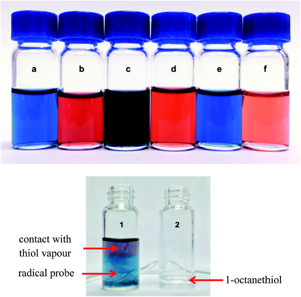 | ||
| Fig. 2 (Top) colour changes to the CpMn(CO)2(anilinyl) radical 1 (0.50 mM) observed upon addition of 0.5 mM of the molecule to be detected. (a) 1 only (b) 1-octanethiol (c) p-thiocresol (d) cysteine (e) glutathione (f) phenylhydrazine. (Bottom) colour change of probe 1 (vial 1) when exposed to vapours diffusing out of vial 2 containing 1-octanethiol placed 1 m away. The purple colouration observed is the result of a colour combination of the red CpMn(CO)2(–SR) radical and the blue complex 1. | ||
The detection of biological thiols such as cysteine, homocysteine and glutathione is also attempted by adding a drop of the blue diglyme solution containing 1 into an aqueous solution containing the thiol. As diglyme and water are miscible, a resultant homogeneous red solution is observed for both cysteine and homocysteine (Fig. 2a to d). However, there is no colour change associated with glutathione, which could be a result of steric bulk hindering the formation of the thiol complex and subsequently, the radical (Fig. 2a to e). Nevertheless the negative result may be useful to distinguish between glutathione and either cysteine or homocysteine.
The addition of an aromatic thiol, p-thiocresol, to a solution containing 1 produces a darker blue coloration (see Fig. 2a to c). If the thiol concentration is very low, it becomes difficult to ascertain whether a reaction has indeed taken place due to the same shade of blue being produced. In this case, we have used the lighter blue radical complex 2 as the sensor so that the difference in the colours is more pronounced. Compared to 1, the λmax of the absorption of complex 2 is red-shifted to 714 nm. Upon the addition of p-thiocresol, the UV-visible spectrum rapidly changes and shows the appearance of two peaks at 614 nm and 480 nm which are assigned to CpMn(CO)2(–SC6H4CH3), in agreement with previously published data.5 An isosbestic point is also observed close to 600 nm indicating the clean conversion of 2 to CpMn(CO)2(–SC6H4CH3) (Fig. 3).
 | ||
| Fig. 3 Changes in UV-visible spectrum upon addition of 0.20 mM of p-thiocresol to 0.50 mM of 2 in diglyme monitored over 10 minutes at 1 minute interval. The black line represents the initial absorbance of 2. | ||
Both 1 and 2 do not respond to the presence of aliphatic amines. However a test with hydrazine or phenylhydrazine gives a colour change from blue to orange-red, suggesting that 1 or 2 is selective towards hydrazines and not amines (Fig. 2a to f). Similar to the thiol case, an isosbestic point is observed near 550 nm in the UV-visible absorption spectrum showing a clean conversion of 1 to the CpMn(CO)2(–NHNHPh) radical complex which absorbs at λmax = 498 nm (Fig. 4).
 | ||
| Fig. 4 Changes in UV-visible spectrum upon addition of 0.35 mM of phenylhydrazine to 0.30 mM of 1 in diglyme monitored over 2 hours at 5 minute interval. The black line represents the initial absorbance of 1. | ||
The detection limit of 1 or 2 towards thiol detection is then assessed via visual inspection and UV-visible absorption spectroscopy. We have found that it is possible to observe the colour change from blue to red with the addition of only 0.1 mM (∼15 ppm) of 1-octanethiol, with 1 in a slight excess. Furthermore, a solution of 1 changes colour when exposed to the vapour emitted by a solution of 1-octanethiol (Fig. 2 bottom). Thus 1 can even be exploited as a visual thiol vapour detector. However, a more quantitative determination of the detection limit has been carried out where the spectral absorbances of the generated radical complexes are measured (Fig. 5). The detection limits for 1-octanethiol, p-thiocresol (an aromatic thiol) and phenylhydrazine, have been determined to be 3.0 ppm, 2.9 ppm and 3.4 ppm respectively (Fig. 5). As the absorption band of the complex is broad, the value chosen for estimation of the detection limits cannot be too low as it will introduce a small background contributed by the tailing end of the absorption band. Hence, a value of 0.05 absorbance units was chosen to maintain a good signal to noise ratio as well as to minimize signal contribution from the radical probe.
 | ||
| Fig. 5 Absorbance vs. concentration plot for 1-octanethiol (green), p-thiocresol (blue) and phenylhydrazine (orange). 1 is used for 1-octanethiol and phenylhydrazine sensing while 2 is used for p-thiocresol sensing. UV measurements were made under room temperature conditions using varying concentrations of the analyte. | ||
Although related radical complexes of 1 and 2 have been prepared and structurally characterised, their reactivities towards organic molecules have not been investigated before.4,5 Using the reaction of 1 with 1-octanethiol as an example (Scheme 2), we believe that the first step of the reaction is a hydrogen atom (of the S–H bond) abstraction by 1. This regenerates the N–H bond of the aniline ligand which is subsequently displaced by the incoming thio radical. For the reaction to proceed spontaneously, the S–H bond has to be weaker than the aniline N–H bond. Similarly, the differences in the reactivity of 1 with other molecules can be traced mainly to the difference in the strength of the N–H bond of aniline and the corresponding bond of the other molecule.
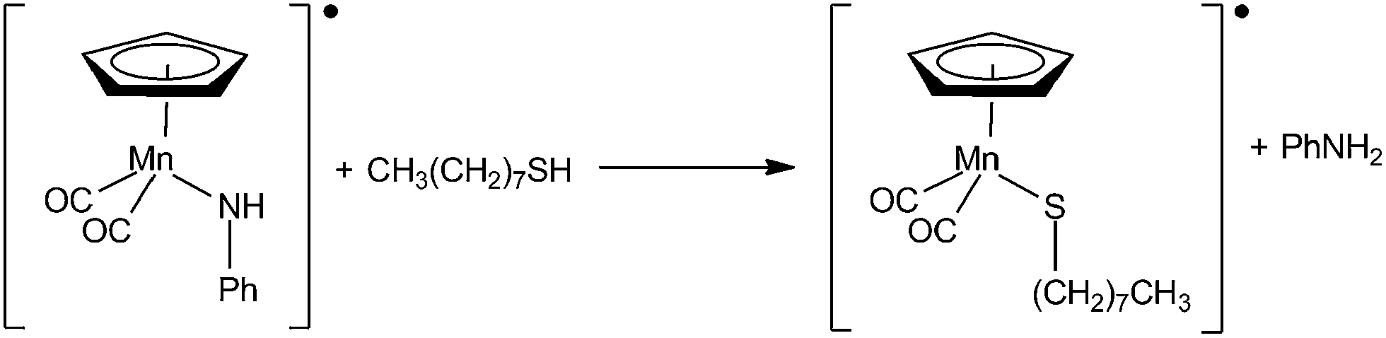 | ||
| Scheme 2 The displacement of the anilinyl moiety of 1 with 1-octanethiol. | ||
In order to better understand how 1 and 2 are able to sense thiols and hydrazines through reactions, the enthalpy and free energy changes of these reactions have been computed using density functional method in Table 1. To reduce computational time, the aliphatic thiol, aromatic thiol and hydrazine molecules are represented by their simplest members which are methanethiol, benzenethiol and hydrazine. Complex 1 is used to represent the sensor. The molecular structures are first optimized at b3lyp/lanl2dz level before single-point energy calculations are carried out using a larger basis set 6-311 + g(d,p) in the Gaussian 09 suite of programs (Cartesian coordinates of the optimised molecular structures are found in the ESI† file).
| ΔH kJ mol−1 | ΔG kJ mol−1 | |
|---|---|---|
| [Mn]–NHPh + CH3SH → [Mn]–SCH3 + NH2Ph | −73.2 | −78.2 |
| [Mn]–NHPh + N2H4 → [Mn]–NHNH2 + NH2Ph | −21.7 | −30.6 |
| [Mn]–NHPh + PhSH → [Mn]–SPh + NH2Ph | −73.1 | −72.2 |
Table 1 shows that the three substitution reactions have been calculated to be exothermic and exergonic as well, with the thiol reactions more spontaneous than the hydrazine counterpart. The aliphatic and aromatic S–H and hydrazine N–H bond energies are indeed low enough for 1 to act as a thiol and hydrazine sensor. The calculations are also in agreement with typical experimental dissociation energies (BDE) of the X–H bond (where X = S or N) which are in the order; aromatic N–H 380 kJ mol−1 > aliphatic S–H 350 kJ mol−1 > aromatic S–H 345 kJ mol−1 > hydrazine N–H 303 kJ mol−1.23 However, attempts at locating the transition state of the reaction are unsuccessful, hence we are unable to obtain values for the activation barriers.
Experimental
Materials
Cyclopentadienyl manganese tricarbonyl (CpMn(CO)3, 98%) was purchased from Alfa Aesar. 1-Octanethiol, p-thiocresol, glycine, phenylhydrazine, aniline, 3,4,5-trimethoxyaniline, homocysteine, cysteine, reduced glutathione were purchased from Sigma-Aldrich. Diethylene glycol dimethyl ether (diglyme) was purchased from Merck. All solvents and reagents were used without further purification.Instrumentation
Photochemical experiments were conducted with Nd-YAG pulsed laser system (Quantel Brilliant B, 10 ns pulse, 355 nm). All infrared (IR) absorption spectra were obtained on a Shimadzu IR Prestige-21 spectrometer, using a 0.1 mm path length CaF2 cell for liquid samples. UV-visible absorption spectroscopy was carried out using a Shimadzu UV-1600 Series spectrometer with a 1 cm path length quartz cell.Preparation of CpMn(CO)2(NHR) radicals (R = C6H5 (1) or C6H2(OMe)3 (2))
All reactions were carried out using standard vacuum line and Schlenk techniques unless otherwise stated. CpMn(CO)3 (10 mg, 0.049 mmol) was dissolved in 4 ml of diglyme. One molar equivalent of PhNH2 (aniline) was then added before the flask was evacuated. The reaction mixture was then subjected to laser photolysis at 355 nm for 90 minutes upon which an orange solution of CpMn(CO)2(PhNH2) was obtained. IR (in diglyme): 1916 cm−1, 1842 cm−1. Oxidation of the complex to the CpMn(CO)2(NHPh) was performed by breaking the vacuum and allowing passage of air into the flask. A colour change from orange to an intense dark blue solution was observed. IR (in diglyme): 1959 cm−1, 1904 cm−1. The same method was applied to the preparation of the CpMn(CO)2(trimethoxyanilinyl) radical. IR (diglyme): 1949 cm−1, 1894 cm−1. The stability of the radical was determined by monitoring the UV-visible spectrum over 7 days under atmospheric conditions (Fig. S1 and S2†). Briefly, a 0.1 mM solution of both sensors was prepared in diglyme and placed in an isolated fume cabinet not used for the handling of thiols and hydrazines. Their respective UV-spectra were then obtained at the same time of the day over a period of 7 days. No significant changes to the colour or their UV-spectra were observed.UV-visible absorption measurements
An initial UV-visible absorption spectrum was recorded for a solution containing 0.80 mM of 1 in 4 ml of diglyme. Following that, 0.50 mM of 1-octanethiol was added and the solution was thoroughly mixed before recording the change in the spectrum. Subsequent measurements were taken at 1 minute interval for 20 minutes. The time period was chosen such that the final spectrum showed very little or no change in the band intensities. A similar procedure was repeated to acquire the UV-visible spectrum in the case of p-thiocresol with complex 2 and phenylhydrazine with complex 1 over the appropriate time interval and period. For the detection of water soluble thiols and hydrazines such as cysteine, glutathione and hydrazine hydrate, a 0.1 mM solution of the respective analyte was prepared and subjected to detection by the sensor. In all cases, the detection was performed under atmospheric conditions using the same concentration of complex 1 or 2. Even though the kinetics of detection varied among the different analytes, the detection limit did not depend on the reaction time because the final product was stable for more than 24 hours to allow detection via UV-visible spectrometry.Detection limit calibration
Differing concentrations (0.1 mM to 0.5 mM) of 1-octanethiol in 5 cm3 of diglyme were prepared in separate flasks. A 0.5 cm3 diglyme solution containing complex 1 was then added to each flask such that the thiol was reacted completely. Only a slight excess of 1 was used in each case in order to minimize background contribution from the absorbance of 1 itself. The UV-visible spectrum of the product was then recorded to detect and measure the CpMn(CO)2(–SR) peak absorbance at 510 nm. A graph showing the absorbance vs. thiol concentration was plotted, as shown in Fig. 5. The same procedure was carried out to obtain the calibration plots for phenylhydrazine and p-thiocresol. Calculations of the detection limit were performed as follow: first, the equation for the calibration curve was obtained via linear regression. The equation is then equated to a y-value of 0.05 as the detection limit. From there, solving the equation for x gives the minimum concentration of the analyte that can be detected. The concentration is then converted to units of ppm. For p-thiocresol, complex 2 was used as the colorimetric sensor.Real time monitoring
A vial of 0.1 ml 1-octanethiol to simulate the presence atmospheric thiols was placed at different distances (1 meter, 1.5 m and 2 m) away from another vial containing 0.1 mM of sensor 1. The time taken for the sensor to change colour is monitored and the absorbance measured with a UV-visible spectrometer.Notes and references
- M. C. Baird, Chem. Rev., 1988, 88, 1217–1227 CrossRef CAS.
- C. H. Bamford and R. Denyer, Nature, 1968, 217, 59–60 CrossRef CAS.
- M. S. Kharasch, P. S. Skell and P. Fisher, J. Am. Chem. Soc., 1948, 70, 1055–1059 CrossRef CAS.
- H. T. Poh, J. W. Kee, T. S. Chwee and W. Y. Fan, J. Organomet. Chem., 2014, 759, 11–14 CrossRef CAS PubMed.
- P. Lau, H. Braunwarth, G. Huttner, D. Guenauer, K. Evertz, W. Imhof, C. Emmerich and L. Zsolnai, Organometallics, 1991, 10, 3861–3873 CrossRef CAS.
- H. S. Jung, X. Chen, J. S. Kim and J. Yoon, Chem. Soc. Rev., 2013, 42, 6019–6031 RSC.
- C. Yin, F. Huo, J. Zhang, R. Martinez-Manez, Y. Yang, H. Lv and S. Li, Chem. Soc. Rev., 2013, 42, 6032–6059 RSC.
- X. Chen, T. Pradhan, F. Wang, J. S. Kim and J. Yoon, Chem. Rev., 2012, 112, 1910–1956 CrossRef CAS PubMed.
- Y. Yang, Q. Zhao, W. Feng and F. Li, Chem. Rev., 2013, 113, 192–270 CrossRef CAS PubMed.
- M. E. Moragues, R. Martinez-Manez and F. Sancenon, Chem. Soc. Rev., 2011, 40, 2593–2643 RSC.
- Z. Yao, X. Feng, C. Li and G. Shi, Chem. Commun., 2009, 5886–5888 RSC.
- J. Bouffard, Y. Kim, T. M. Swager, R. Weissleder and S. A. Hilderbrand, Org. Lett., 2008, 10, 37–40 CrossRef CAS PubMed.
- S. Ji, J. Yang, Q. Yang, S. Liu, M. Chen and J. Zhao, J. Org. Chem., 2009, 74, 4855–4865 CrossRef CAS PubMed.
- B. Tang, L. Yin, X. Wang, Z. Chen, L. Tong and K. Xu, Chem. Commun., 2009, 5293–5295 RSC.
- S. Wang, H. Ma, J. Li, X. Chen, Z. Bao and S. Sun, Talanta, 2006, 70, 518–521 CrossRef CAS PubMed.
- B. Rezaei and A. Mokhtari, Spectrochim. Acta, Part A, 2007, 66, 359–363 CrossRef CAS PubMed.
- B. Han, J. Yuan and E. Wang, Anal. Chem., 2009, 81, 5569–5573 CrossRef CAS PubMed.
- P. K. Pullela, T. Chiku, M. J. Carvan 3rd and D. S. Sem, Anal. Biochem., 2006, 352, 265–273 CrossRef CAS PubMed.
- A. M. Piggott and P. Karuso, Anal. Chem., 2007, 79, 8769–8773 CrossRef CAS PubMed.
- F. X. Zhang, L. Han, L. B. Israel, J. G. Daras, M. M. Maye, N. K. Ly and C.-J. Zhong, Analyst, 2002, 127, 462–465 RSC.
- J.-W. Mo, B. Ogorevc, X. Zhang and B. Pihlar, Electroanalysis, 2000, 12, 48–54 CrossRef CAS.
- S. D. Zelnick, D. R. Mattie and P. C. Stepaniak, Aviat., Space Environ. Med., 2003, 74, 1285–1291 CAS.
- U. Ragnarsson, Chem. Soc. Rev., 2001, 30, 205–213 RSC.
- S. Garrod, M. E. Bollard, A. W. Nicholls, S. C. Connor, J. Connelly, J. K. Nicholson and E. Holmes, Chem. Res. Toxicol., 2005, 18, 115–122 CrossRef CAS PubMed.
- H. Bhutani, S. Singh, S. Vir, K. K. Bhutani, R. Kumar, A. K. Chakraborti and K. C. Jindal, J. Pharm. Biomed. Anal., 2007, 43, 1213–1220 CrossRef CAS PubMed.
- M. Sun, L. Bai and D. Q. Liu, J. Pharm. Biomed. Anal., 2009, 49, 529–533 CrossRef CAS PubMed.
- J. Liu, W. Zhou, T. You, F. Li, E. Wang and S. Dong, Anal. Chem., 1996, 68, 3350–3353 CrossRef CAS PubMed.
- L. Zhang, C. Jiang and Z. Zhang, Nanoscale, 2013, 5, 3773–3779 RSC.
- Y. Han, S. Liu, B. Liu, C. Jiang and Z. Zhang, RSC Adv., 2014, 4, 2776–2782 RSC.
- C. Jiang, R. Liu, G. Han and Z. Zhang, Chem. Commun., 2013, 49, 6647–6649 RSC.
- D. Sellmann and J. Müller, J. Organomet. Chem., 1985, 281, 249–262 CrossRef CAS.
Footnote |
| † Electronic supplementary information (ESI) available: Computational data for the manganese complexes and other organic molecules. See DOI: 10.1039/c4ra16483k |
| This journal is © The Royal Society of Chemistry 2015 |