
Novel amino-cyclodextrin cross-linked oligomer as efficient carrier for anionic drugs: a spectroscopic and nanocalorimetric investigation†
Valentina Giglioa,
Carmelo Sgarlataab and
Graziella Vecchio*a
aDipartimento di Scienze Chimiche, Università degli Studi di Catania, V.le A. Doria 6, 95125 Catania, Italy. E-mail: gr.vecchio@unict.it
bConsorzio I.N.B.B., Viale delle Medaglie d'Oro 305, 00136 Roma, Italy
First published on 26th January 2015
Abstract
A large variety of cross-linked cyclodextrin polymers exhibiting interesting properties towards various guests have been characterized. Some polymeric nanoparticles based on cyclodextrins have been tested in clinical trials as nanotherapeutics for cancer. Recently, oligomers have been also synthesized and the advantages of their low molecular weight have been highlighted. In this context, we have synthesized a new cross-linked amino-β-cyclodextrin oligomer of 3A-amino-3A-deoxy-2A(S),3A(R)-β-cyclodextrin of about 11 kDa. The inclusion capability of the oligomer for the model drug diclofenac was investigated in neutral aqueous solution by spectroscopy and nano-isothermal titration calorimetry and compared to both β-cyclodextrin and a soluble cross-linked β-cyclodextrin polymer. The log![[thin space (1/6-em)]](https://www.rsc.org/images/entities/char_2009.gif) K values and the thermodynamic parameters for the interaction of the hosts with diclofenac have been determined. Results suggested that the presence of the polymer backbone does not perturb the molecular recognition ability of the cyclodextrin cavities and the amino groups play a key role in the inclusion process of diclofenac.
K values and the thermodynamic parameters for the interaction of the hosts with diclofenac have been determined. Results suggested that the presence of the polymer backbone does not perturb the molecular recognition ability of the cyclodextrin cavities and the amino groups play a key role in the inclusion process of diclofenac.
Introduction
Cyclodextrins (CyDs) are cyclic oligosaccharides α(1 → 4) of glucose which have been extremely appealing in different fields over a long time.1,2 Investigations on native cyclodextrins and their exhaustively modified products have been widely reported.3 CyDs have been frequently used as building blocks as they can be linked both non covalently and covalently to different molecules. The covalent conjugation to molecules with different properties provides a very attractive approach in the building of multifunctional systems devoted to applications in supramolecular, bioinorganic, organic, pharmaceutical, material chemistry and separation sciences.4–6 A number of CyD conjugates with amines, amino acids, peptides and aromatic systems have been reported over the years.7 The functionalization with suitable moieties has been shown to improve the metal binding ability leading to a wide range of applications.8,9 Many examples of CyD derivatives whose OH groups have been alkylated, esterified or easily randomly derivatized have been reported in the literature and mostly used for the formation of inclusion complexes.10 The total or partial modification of the OH groups affects the solubility of CyDs in water or in organic solvent and, in some cases, allows for the introduction of charged groups.11 A successful example is sugammadex, a cyclodextrin derivative with carboxylic groups designed to selectively reverse the effect of rocuronium.12 Some cyclodextrins, such as hydroxypropyl β-cyclodextrins (HP-CyD), have been approved by FDA as therapeutic agents to remove cholesterol from blood. In 2010 HP-CyD was administrated in twin girls suffering from Niemann Pick Type C, a congenital neurodegenerative disease.13 Cyclodextrin-based carriers may enhance the capability of encapsulating guest molecules, improve the stability of drug and efficiently regulate the drug release rate.14 In the last years, nanosystems have been gaining importance and also CyDs have been used to build nanoparticles. A variety of systems, from organic polymers to inorganic nanoparticles, decorated with cyclodextrins has been reported.15–17 A large variety of cross-linked CyD polymers exhibiting a dramatic enhancement of the inclusion ability towards various guests have been synthesized.18,19 CyD polymers have also been modified through the incorporation of anionic or cationic groups to increase their drug loading features.20 Choline or carboxyl moieties have been added and their effect on the upload of charged drugs was investigated.19 Cyclosert is a very promising class of linear polymers based on CyDs. Cyclosert, partnered with Cerulean Pharma, Inc., is the first nanoparticle drug carrier platform to be designed specifically to overcome some limitations in existing technologies used for the systemic transport of therapeutics. CRLX-101 is a nanoparticle that consists of the drug camptothecin (CPT) conjugated to CyD polymers based on the cyclosert delivery platform. CRLX-101 has been tested in clinical trials as nanotherapeutics for cancer.21Recently, oligomers have been also synthesized as drug carriers. They are able to include molecules and the few quantitative data reported suggest that these systems could be very promising. Furthermore the advantages of their low molecular weight have been highlighted. In fact, oligomers can be excreted by renal tubules without degradation.
In order to contribute to the knowledge regarding the properties of NPs based on CyD oligomers, we have synthesized a new amino-cyclodextrin oligomer of about 11 kDa using epichlorohydrin (EPI) as the cross linking agent. The inclusion capability of the oligomer for the model drug diclofenac (DCF) was investigated. β-CyD and a commercial water-soluble β-CyD polymer (pCyDcom) were studied for comparison. DCF is an anti-inflammatory drug widely used in inflammatory and painful conditions. Inclusion complexes of DCF into cyclodextrins have been widely investigated22 and some formulations of DCF with cyclodextrins have been patented and are available in the market.10 In this work, the oligomer of 3A-amino-3A-deoxy-2A(S),3A(R)-β-cyclodextrin (pCyDNH2, Fig. 1) was synthesized and its inclusion properties towards DCF were investigated in neutral aqueous solution by spectroscopy (CD and UV-vis) and nano-isothermal titration calorimetry (ITC). In particular, the latter technique allowed for the determination of the binding constants and the energetics of the complex formation by direct measurements of the reaction heats thus providing a complete thermodynamic characterization of the host–DCF systems. pCyDNH2, pCyDcom and β-CyD were used as hosts.
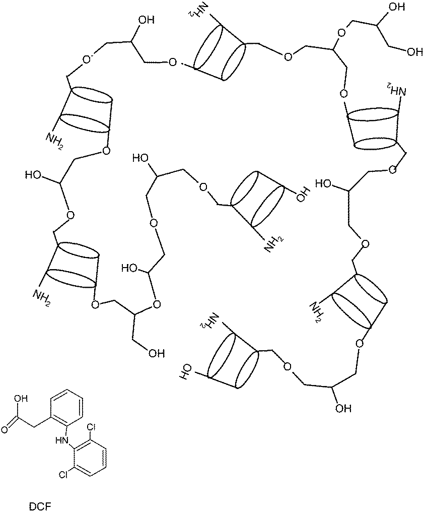 | ||
| Fig. 1 Diclofenac (DCF) and one of the possible structures of pCyDNH2. | ||
Results and discussion
Synthesis and characterization of the oligomer
pCyDNH2 was synthesized starting from 3A-tert-butoxycarbonyl-amino-3A-deoxy-2A(S),3A(R)-β-cyclodextrin (CyD3NHBoc). The polymerization reaction was carried out under basic conditions using epichlorohydrin (EPI) as a cross-linking agent. A low EPI/monomer ratio (EPI/CyD3NHBoc = 5) was used in order to obtain an oligomer.23 Boc protected 3-amino-CyD was used to avoid the reaction of the amino group with EPI. After Boc deprotection, a water-soluble product was isolated. Its solubility was more than 100 mg mL−1. pCyDNHBoc and pCyDNH2 were characterized with 13C, 1H NMR and light scattering (Fig. 2 and S1–S3†).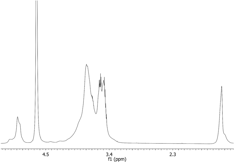 | ||
| Fig. 2 1H NMR spectrum (D2O, 500 MHz) of pCyDNHBoc. | ||
In the NMR spectra of pCyDNH2 and pCyDNHBoc peaks are broad, as typically observed for these classes of compounds.23 In the 1H NMR spectra of pCyDNHBoc the t-butyl protons resonate at 1.45 ppm. Cross-peak correlations between t-butyl protons and CyD protons at 4.2–3.2 ppm can be observed in the ROESY spectra as expected (Fig. S2†). Moreover, the Hs of the cross linker chains can be identified in the region around 3.46 and 3.50 ppm together with the broad signal of CyD protons. In the 13C and HSQC spectra is possible to identify the carbons of cyclodextrins as well as those of the cross linker chains (CH and CH2) (Fig. S1†).
The integrated intensities of the signal of Hs-1 of CyD and the broad signal at 4.2–3.2 ppm provide the ratio of the number of hydrogens of CyDs to that of the cross linker propylenic chains in the oligomer; this value was used to determine the number of protons of the linker chains in the oligomer as reported elsewhere.23 This value suggests that about 70% of CyDNHBoc was contained in the oligomer.
The spectra of pCyDNH2 are very similar to that of the Boc oligomer. The signals due to the t-butyl group disappeared in the spectra as expected (Fig. S3†). The relative amount (70%) of CyDNH2 in the oligomer was confirmed through the integration of the relevant signals in the 1H NMR spectra of pCyDNH2.
Dynamic light scattering (DLS) measurements show that pCyDNH2 and pCyDNHBoc form stable nanoparticles of about 3 nm diameter (Fig. S4†). Positive Zeta potential values were measured. The Zeta potential value for pCyDNH2 depends on the pH value of the solution of the sample, in keeping with the protonation state of the amino groups.
The weight-averaged molecular weight (Mw) of the oligomer was determined through light scattering measurements. The best set of data was obtained in the case of pCyDNHBoc (Fig. 3). The Boc deprotection does not modify the number of cavities in the oligomer and thus the Mw was determined for pCyDNHBoc. The plot of KC/RθP vs. the concentration of pCyDNHBoc is shown in Fig. 3. The intercept of the straight line was used to obtain the Mw value, which was found to be 12 ± 1 kDa.
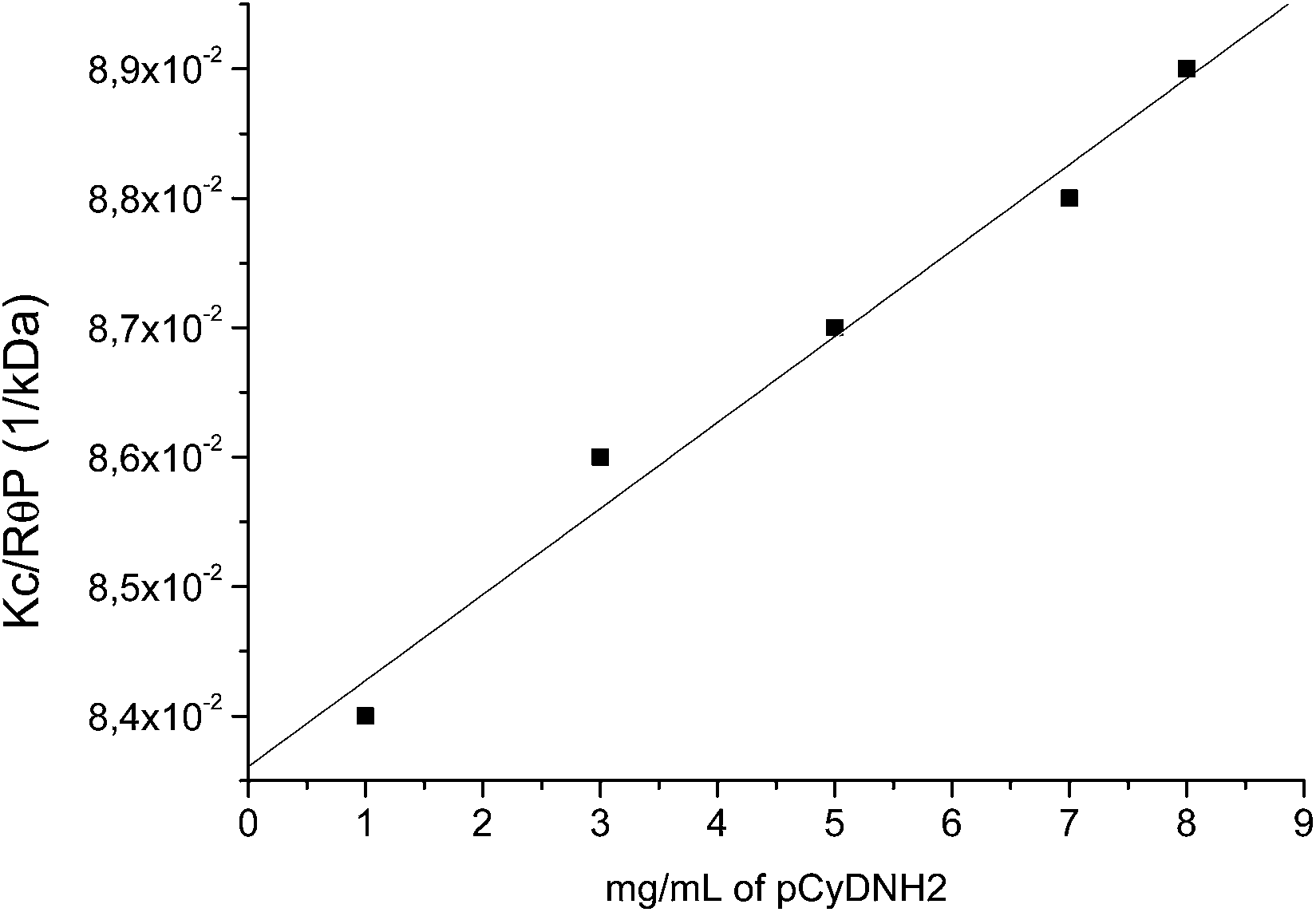 | ||
| Fig. 3 Intensity of scattered light (KC/RθP) vs. pCyDNHBoc concentration. | ||
The newly synthesized oligomers have a low molecular weight due to the use of a smaller amount of EPI in the synthetic reaction and a more diluted reagent solution if compared to the procedure reported elsewhere.19 In particular, an average number of about 7 cavities can be hypothesized on the basis of the 70% of CyDNH2 in the oligomer obtained from NMR data and the Mw of 12 kDa. The weight-averaged molecular weight of pCyDNH2 can be calculated by subtracting the molecular weight of the Boc groups and the estimated value is about 11 kDa.
CD spectroscopy
The inclusion ability of pCyDNH2 for DCF was investigated by CD spectroscopy. β-CyD, an uncharged and soluble CyD polymer (pCyDcom) and pCyDNHBoc were also tested for comparison. Fig. 4 shows the induced CD spectra obtained when DCF was added to a pCyDNH2 solution at pH 7.1. The spectra show a negative band at 285 nm and a positive band at 310 nm. The intensity of the bands increased when the guest–host molar ratio increased thus suggesting that DCF can interact with the oligomer. The ICD spectra obtained in the presence of either β-CyD, pCyDcom or pCyDNHBoc were recorded and shown in Fig. 5. For an easier comparison among hosts which have a different number of cavities, CD spectra were carried out at the same concentration of CyD cavities for all the samples. The highest intensity of the CD bands was observed in the case of pCyDNH2. This is about twice the intensity detected for the DCF–pCyDcom system. Indeed, when pCyDNHBoc was used as the host, a lower intensity in the CD bands was observed (Fig. 5). The different intensity of the CD spectra suggests that the amino groups play a key role in the inclusion process of DCF.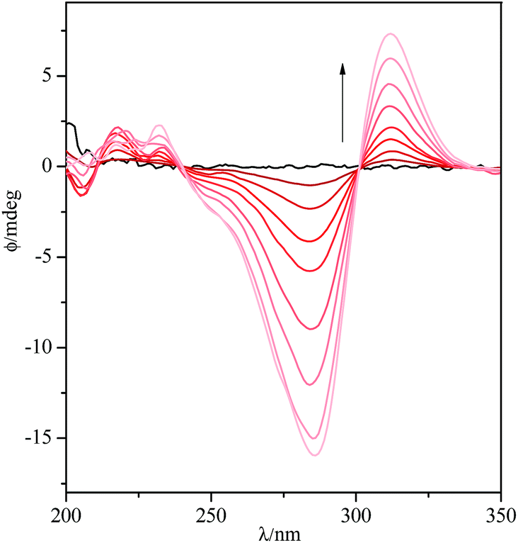 | ||
| Fig. 4 CD spectra of DCF (read from black to light red: 0, 0.5, 1.0, 2.0, 3.0, 5.0, 7.0, 10, 13 × 10−5 M) with pCyDNH2 (β-CyD cavity concentration: 1.3 × 10⁻³ M) at pH 7.4 (10 mM Tris buffer). | ||
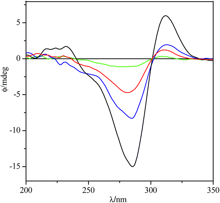 | ||
| Fig. 5 CD spectra of DCF (7.0 × 10⁻⁵ M) with different hosts (β-CyD cavity concentration: 1.3 × 10⁻³ M): pCyDNH2 (black); pCyDcom (blue); pCyDNHBoc (red); β-CyD (green). | ||
Binding features and thermodynamic parameters
Preliminary spectroscopic titrations carried out for the determination of the binding affinity of DCF with either β-CyD (used as a reference compound) or pCyDcom in neutral aqueous solution (40 mM phosphate buffer, pH 6.9) resulted in changes of the observable which were not appropriate for fitting purposes. Similar findings were reported for the investigation of the simple DCF–β-CyD system by absorption spectroscopy in water at neutral pH.24 Consequently, we resorted to nano-ITC which is a powerful tool for the evaluation of the energetics of molecular recognition processes in solution.25,26The driving forces for the formation of inclusion complexes include specific, non-specific, weak interactions27 between DCF and CyD cavities as well as contributions resulting from the desolvation of both the host and the guest.28,29 These factors may have different and often opposing enthalpic and entropic contributions30 and thus splitting the free energy term into the ΔH° and ΔS° components may help understanding the host–guest formation equilibria and unveil aspects that are not expressed in the ΔG° term.31,32
Calorimetric experiments were carried out in buffered aqueous solution (40 mM phosphate buffer, pH 6.9) to minimize any contribution resulting from the interaction of either the guest or the host with protons. Initially, ITC titrations of β-CyD into a DCF solution were run to obtain thermodynamic data to be used as reference values for the study of the binding features of the CyD polymers under homogeneous experimental conditions. Values found for the DCF–β-CyD 1:1 inclusion complex (see Table 1) are in good agreement with those previously reported,33 provided due allowance is made for the different experimental procedures and conditions employed. The binding constant value we determined is slightly smaller than that reported by Chadha et al. at different ionic strength conditions (i.e., in the absence of background electrolyte) while it nicely reproduces that obtained by Barbato et al.34 at pH 7 in phosphate buffer.
| logK |
ΔH° (kJ mol−1) | ΔS° (J deg−1 mol−1) | Number of CyD cavitiesb | |
|---|---|---|---|---|
| a Units are expressed as “per moles of host site”.b Number of cavities estimated by dividing the “overall enthalpy of complexation” of either pCyDcom or pCyDNH2 host–guest complex by the ΔH° of formation of a simple DCF–β-CyD complex (−12.69 kJ mol−1). | ||||
| β-CyD | 2.41(3) | −12.69(2) | 3.6(5) | |
| pCyDcom | 2.8(1) | −17.54(8)a | −5(1)a | 55–58 |
| pCyDNH2 | 3.47(6) | −9.58(2)a | 34(2)a | 7–8 |
Typical thermograms for the interaction of DCF with pCyDcom or pCyDNH2 at 25 °C are shown in Fig. 6 and 7, respectively.
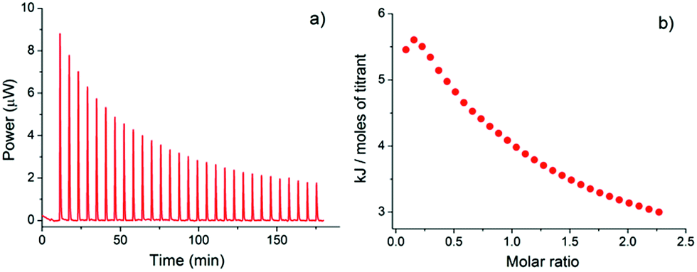 | ||
| Fig. 6 (a) Typical ITC titration of DCF into pCyDcom in buffered aqueous solution (pH 6.9) at 25 °C; (b) integrated heat data (the molar ratio is referred to the β-CyD cavity concentration of the host). | ||
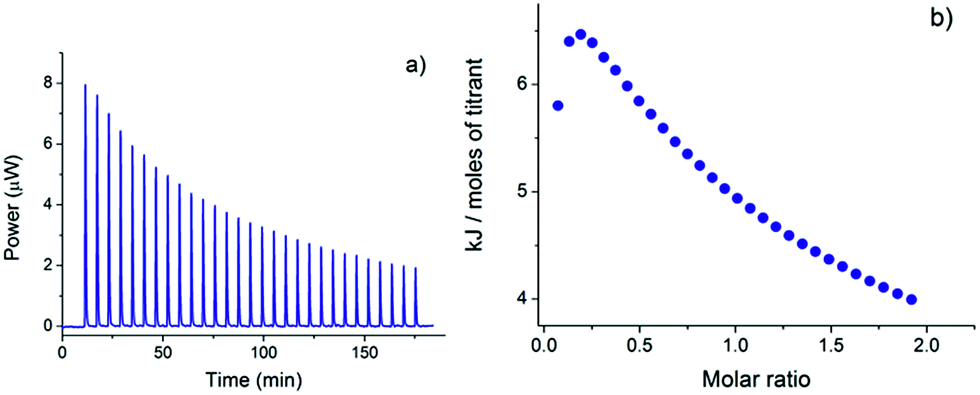 | ||
| Fig. 7 (a) Typical ITC titration of DCF into pCyDNH2 in buffered aqueous solution (pH 6.9) at 25 °C; (b) integrated heat data (the molar ratio is referred to the β-CyD cavity concentration of the host). | ||
Titrations were carried out by adding a number of equivalents of DCF (used as the titrant) to the host in the ITC cell in order to satisfy the saturation fraction criteria proposed for the formation of a 1:1 complex.35 Usually, a 5 mM DCF solution was used to titrate CyD derivative solutions having a 0.01–0.08 mM total polymer concentration. Such small concentration values emphasize the role of nano-ITC as versatile technique for the study of binding interactions in solution by using amounts of reactants which are comparable to those commonly employed for spectrophotometric measurements.
The simultaneous host–guest equilibria occurring in solution between DCF and the multiple binding sites (cavities) present on the CyD polymer backbone make the data fitting process rather cumbersome. Consequently, a simplified model which assumes the formation of a formal 1:1 complex stoichiometry was chosen. For data analysis, the host concentration was expressed as “β-CyD cavity” concentration, calculated on the base of the estimated average Mw and β-CyD% content (w/w) of the polymer.18,36 According to this 1:1 complex model, the equilibrium constant is referred to a single host–guest interaction regardless of the number of binding sites (cavities) actually accessible on the host structure. Assuming that all the binding sites for the guest are identical within the polymer, logK values are expected to be nearly comparable along the whole set of cavities, if progressive saturation of the binding sites is not considered. This approach seems reasonable when assuming that, on average, some of the CyD cavities of the polymer will not be fully available or properly exposed for guest inclusion whereas others may act in a cooperative way resulting in a more effective guest binding.37
As reported for similar multi-site polymeric hosts,38,39 when β-CyD cavity concentration is used as the input value for data analysis, convenient “ΔH° per host site” values (expressed in terms of kJ mol−1 of host site) may be obtained by the refinement process. On the other hand, the use of the total polymer concentration for data analysis does not take into account the effective number of binding sites of the host and, in the absence of any cooperative effect among the cavities, would provide an “overall enthalpy of complexation” value which should be n times that obtained for a single β-CyD unit, with n = number of cavities of the polymer.37 Intriguingly, the value of n calculated both for pCyDcom and pCyDNH2 by simply dividing the “overall enthalpy of complexation” by the ΔH° of formation of the DCF–β-CyD 1:1 complex (−12.69 kJ mol−1) nicely compares with the number of cavities (55–58 and 7–8, respectively) estimated for the two different CyD polymers by DLS measurements (see Table 1).
The logK values and the thermodynamic parameters for the interaction of DCF with pCyDcom and pCyDNH2, determined by using a software which refines data from multiple titrations, are reported in Table 1. The binding constants of the two CyD polymers, assuming a 1:1 complex stoichiometry, are comparable with that of the complex formed by β-CyD with DCF thus indicating that the polymerization process does not affect the molecular recognition ability of the CyD cavities on the polymer backbone. This result has been observed for other drug–CyD polymer systems in which the presence of a macromolecular arrangement does not weaken or negatively influence the stability and efficiency of the inclusion complexes.40–42
The larger logK value found for the DCF–pCyDNH2 complex may be ascribed to the supplementary contribution provided, in addition to hydrogen bonds and van der Waals forces, by the interactions between the negatively charged guest and the charged amino groups present on the CyD cavities of the oligomer. In fact, the pKa value reported24 for the carboxylic group of DCF is 4.84 while the amino moieties on the CyD units are expected to be nearly protonated at pH 6.9. On the whole, the favorable combination of different attractive forces makes pCyDNH2 an efficient receptor for the simultaneous complexation of several target guests.
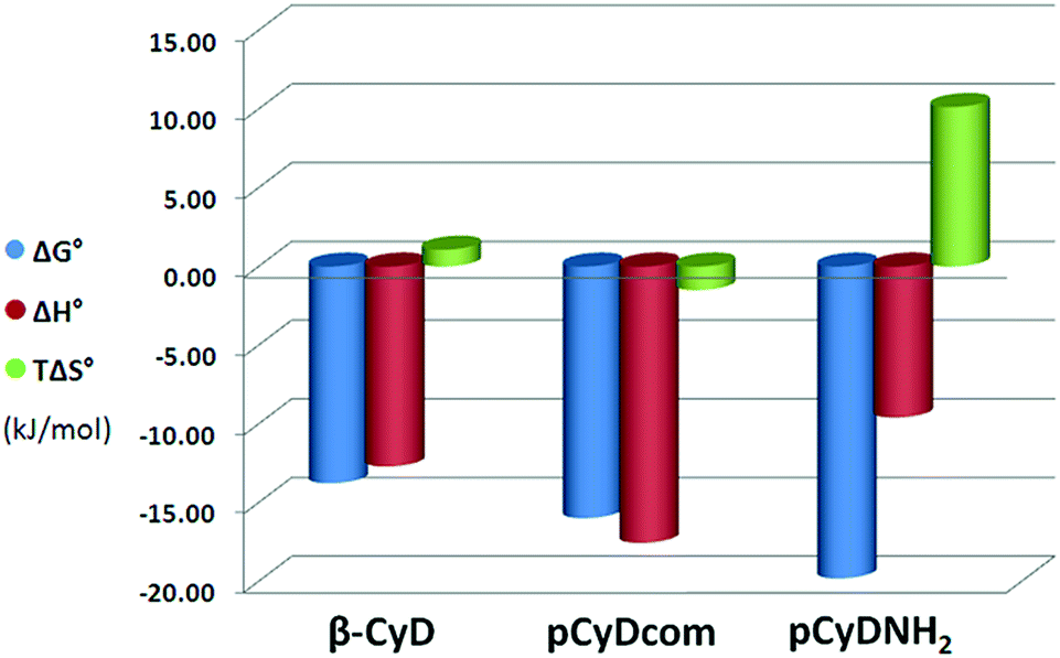
The immediate perception of the enthalpic and entropic contribution to the free energy term of each host–guest complex is shown in Fig. 8. The complexation of DCF is enthalpically favored (ΔH° < 0) for all CyD derivatives and driven by enthalpy (|ΔH°| > |TΔS°|) for both β-CyD and pCyDcom and by entropy (although by a small extent) in the case of pCyDNH2.
 | ||
| Fig. 8 Thermodynamic parameters for the different host–guest complexes at 25 °C in buffered aqueous solution (pH 6.9). | ||
Factors affecting the thermodynamic parameters due to guest inclusion into CyDs are the result of a balance of several effects: the insertion of the hydrophobic portion of the guest molecule into the CyD cavity, the formation of weak interactions (such as hydrogen bonds, van der Waals and/or electrostatic forces), the desolvation of the guest, the release of water molecules from the CyD cavity and conformational changes of the host upon guest complexation.42,43
For both CyD polymers, a combination of hydrogen bonds and van der Waals interactions between DCF and the CyD cavities triggers the host–guest complex formation. These enthalpically favorable contributions override the cost in energy needed for the desolvation of all the interacting components. However, the differences observed in both the enthalpy and entropy values suggest that the mechanism of binding is slightly different for the two CyD polymers. In particular, the larger amount of cavities of pCyDcom enhances the enthalpically favorable attractive interactions with the guest resulting in a ΔH° value larger than that found both for the shorter pCyDNH2 polymer and the simple β-CyD. On the other hand, the large and favorable ΔS° value obtained for the DCF–pCyDNH2 complex may be ascribed to a combination of (i) a reorganization of water molecules released upon guest inclusion and (ii) a significant translational and conformational freedom experienced by this flexible and less constrained polymer when guest molecules are entrapped within its cavities. Such increase of the entropic gain, along with a small decrease of the ΔH° value, has been reported in the literature in the case of similar host–guest complexes employing β-CyD oligo-polymeric structures as multi-site receptors.38,44,45
DFC solubility study
The effect of the pCyDNH2 polymer on the DCF solubility was investigated at pH 5.5 since the solubility of DCF is very low (about 2.5 × 10⁻⁵ M) at this pH value. pCyDcom and β-CyD were used for comparison. Results of the phase solubility studies are reported in Fig. 9 which shows the solubility profiles of DCF as a function of increasing concentrations of β-CyD, pCyDcom or pCyDNH2 in aqueous solution at pH 5.5. The host concentration has been reported as β-CyD cavity concentration for a more appropriate comparison among the different systems. Interestingly the solubility of the drug increased linearly with the increase of the host concentration. This linear correlation between DCF and host concentration, with a slope of less than 1, suggests the formation of a 1:1 host–guest inclusion complex with the CyD unit. The solubility of DCF in the presence of either pCyDNH2 or pCyDcom increases of about 10 times. The oligomer is a slightly more efficient solubilizing agent than pCyDcom. Interestingly, the length and size of the polymeric chain seem not to improve the solubilizing effect of the polymer in comparison with that of the oligomer.
 | ||
| Fig. 9 Solubility of DCF vs. β-CyD cavity concentration of different hosts at pH 5.5: pCyDNH2 (●); pCyDcom (▲); β-CyD (■). | ||
Experimental
Materials and reagents
Commercially available reagents were used as received unless otherwise noted. 3A-Amino-3A-deoxy-2A(S),3A(R)-β-cyclodextrin was synthesized starting from the corresponding 2-tosylate-CyD derivative as reported elsewhere.46 The water soluble polymer pCyDcom (92 kDa, 70% of CyDs) was purchased from CyClolab. Thin layer chromatography (TLC) was carried out on silica gel plates (Merck 60-F254). Carbohydrate derivatives were detected on TLC by UV and/or by the anisaldehyde test.3A-tert-Butoxycarbonyl-amino-3A-deoxy-2A(S),3A(R)-β-cyclodextrin (CyD3NHBoc)
3A-Amino-3A-deoxy-2A(S),3A(R)-β-CyD (100 mg, 88.1 μmol) and di-tert-butyldicarbonate (19.24 mg, 88.1 μmol) were dissolved in dry DMF and the reaction mixture was stirred at room temperature for 8 h. The solvent was evaporated to dryness in vacuo. The crude product was suspended in water and purified by dialysis (Float-A-LyZer G2, MWCO 1000). The product (CyD3NHBoc) was freeze dried to give a white powder. Yield: 91%.Rf = 0.56 (PrOH/AcOEt/H2O/NH3 5:2:2:4), ESI-MS m/z: 1234.4 (M + 1).
Synthesis of amino-oligomer pCyDNH2
500 mg of CyD3NHBoc (0.4 mmol) in 3 mL of a 7 M NaOH solution was stirred overnight at room temperature. EPI (0.160 mL, 2 mmol) was slowly added and the mixture was heated to 40 °C. After 6 h, acetone was added and a solid was obtained. The suspension was maintained at 50 °C for 12 h. Then acetone was evaporated and HCl 6 N was added until neutral pH and the mixture was ultra-filtrated (molecular weight cut-off 5000). The final product was precipitated by ethanol and dried under vacuum to obtain Boc protected pCyDNH2. pCyDNHBoc was deprotected by reaction with TFA for 2 h and purified through ultrafiltration. Finally, the product was freeze dried to give a white powder. Yield: 68%.NMR spectroscopy
1H and 13C NMR spectra were recorded at 25 °C with a Varian UNITY PLUS-500 spectrometer at 499.9 and 125.7 MHz respectively. NMR spectra were obtained by using standard pulse programs from Varian library. The 2D experiments (COSY, TOCSY, gHSQCAD, gHMBC, ROESY) were acquired using 1K data points, 256 increments and a relaxation delay of 1.2 s. The spectra were referred to the solvent signal.UV-visible and circular dichroism spectroscopy
UV-vis spectra were recorded using an Agilent 8452A diode array spectrophotometer. Circular dichroism measurements were performed with a JASCO J-1500 spectropolarimeter. The spectra were recorded at 25 °C on freshly prepared solutions in 10 mM Tris buffer (pH 7.1).Mass spectrometry
ESI-MS measurements were carried out by using a Finnigan LCQ DECA XP PLUS ion trap spectrometer operating in the positive or negative ion mode and equipped with an orthogonal ESI source (Thermo Electron Corporation, USA).Light scattering measurements
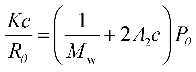
Dynamic light scattering (DLS) measurements were carried out using a Zetasizer Nano ZS (Malvern Instruments, UK) equipped for backscattering at 173° with a 633 nm He–Ne laser. Weight average molecular weight (Mw) determination was performed through static light scattering measurements as reported elsewhere.47 Samples were studied at different concentrations and at 25 °C applying the Rayleigh equation, which reports the intensity of the light scattered from the oligomer/polymer solution:wherein Rθ is the ratio of scattered light to incident light of the sample; Mw is the weight-averaged molecular weight; A2 is the second virial coefficient; c is the sample concentration; Pθ is the angular dependence of the sample scattering intensity; K is the optical constant defined as:
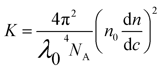
wherein NA, Avogadro's constant; λ0, laser wavelength; n0, solvent refractive index; dn/dc, differential refractive index increment. A dn/dc value of 0.137 g mL−1 was used as reported in the literature for similar systems.47 Toluene was used as the reference. KC/RθP can be plotted as a function of c. The slope (2A2) gives the second virial coefficient and the intercept (1/Mw) gives the molecular weight. Aqueous buffered solutions of oligomer (10 mM Tris buffer, pH 7.1) were filtered with nylon membrane filter (0.22 μm). The size distribution of the sample were measured during the determination of molecular weight. Triplicate measurements were carried out. Every measurement was the average of at least 12 runs.
Phase solubility studies
Phase solubility studies were performed in solutions at pH 5.5. Excess amounts of DCF (7 mg) were added to a 2 mL aqueous solutions containing different concentrations of β-CyD, pCyDNH2 or pCyDcom in acetate buffer (0.1 M, pH 5.5). Suspensions were vigorously stirred at 25 °C for 16 h. Samples were centrifuged and the DCF concentration in the supernatant was determined by UV-vis spectroscopy.ITC titrations
ITC titrations were carried out at 25 °C in water (40 mM phosphate buffer, pH 6.9) by using a nano-isothermal titration calorimeter Nano-ITC2G (TA Instruments, USA) having an active cell volume of 0.988 mL and a 250 or 100 μL stirring syringe. Measurements were run in the overfilled mode; the reaction mixture in the sample cell was continuously stirred at 250 rpm during the titration. Intervals between injections were 350–500 s to allow complete equilibration. The power curve (heat flow as a function of the time) was integrated through NanoAnalyze (TA Instruments, USA) to obtain the gross heat evolved/absorbed in the reaction. The instrument was chemically calibrated by titrating an HCl solution (1 mM) into buffered Tris (30 mM containing 10 mM HCl) by following the procedure described previously.48 The equipment was also checked by running an electrical calibration. ITC measurements were carried out by titrating aqueous solution of β-CyD (14.80–16.53 mM) into a DCF solution (0.605–1.07 mM) and aqueous solutions of DCF (4.40–5.47 mM) into either pCyDcom (0.0108–0.0526 mM as total polymer concentration) or pCyDNH2 (0.0685–0.0878 mM as total polymer concentration). Based on the average Mw and β-CyD% content of the polymers, the above total polymer concentrations correspond to “β-CyD cavity” concentrations of 0.616–2.99 mM and 0.480–0.615 mM for pCyDcom and pCyDNH2, respectively. Solutions of both DCF and CyD derivatives were prepared by dissolving weighed amounts in 40 mM phosphate buffer (pH 6.9). Prior to the titration experiments, all solutions were degassed with gentle stirring under vacuum for about 15 min. 4–5 independent experiments were typically run for each host–guest system. The heats of dilution were determined in separate blank experiments by titrating solutions of either β-CyD or DCF (in 40 mM phosphate buffer) into a solution containing 40 mM phosphate buffer only. The net heats of reaction, obtained by subtracting the heat evolved/absorbed in the blank experiments, were analyzed by HypΔH.49 This software determines the equilibrium constants and/or formation enthalpies of complex species in solution by a non-linear least-squares analysis of the function U = ∑(Qobs − Qcalc)2, where Qobs and Qcalc are the observed heats, corrected for the dilution (blank) effects, and the calculated heats, respectively. HypΔH also allows for the simultaneous refinement of data from multiple titrations.Conclusions
CyDs are very promising multifunctional tools as drug carriers as well as stabilizing and solubilizing agents. Nevertheless the functionalization can widely improve their features. We have synthesized and characterized a new water soluble amino oligomer starting from 3-amino CyD and the results showed that the presence of the macromolecular arrangement does not weaken the stability of the inclusion complex with DCF and, consequently, it may be employed as a potential drug carrier. Thermodynamic data obtained by nano-ITC for the complex formation of DCF with pCyDNH2 and the commercial polymer pCyDcom indicated that the process is always enthalpically driven and that the guest is included by concerted hydrogen bonds and van der Waals interactions. The amino group of pCyDNH2 also plays a relevant role in the recognition process. Interestingly, the length and size of the chain seem not to improve some of the features of the polymer when compared with those of the oligomer and this highlights the potential of short polymeric chains as new drug carriers with more favourable pharmacokinetic profiles.Acknowledgements
The authors acknowledge support from the Italian Ministero dell'Università e della Ricerca (PON01_01078 and FIRB RINAME RBAP114AMK) and the Consorzio Interuniversitario di Ricerca in Chimica dei Metalli nei Sistemi Biologici (CIRCMSB).Notes and references
- J. Szejtli, Chem. Rev., 1998, 98, 1743 CrossRef CAS PubMed.
- G. Crini, Chem. Rev., 2014, 114, 10940 CrossRef CAS PubMed.
- L. Jicsinszky, E. Fenyvesi, H. Hashimoto and A. Ueno, Cyclodextrin derivatives, in Comprehensive Supramolecular Chemistry, ed. J. L. Atwood, J. E. Davies, D. D. MacNicol, F. Vögtle and J. M. Lehn, Pergamon, Oxford, 1996, vol. 3 Search PubMed; Cyclodextrins, ed. J. Szejtli and T. Osa, Pergamon, Oxford, 1996 Search PubMed.
- J. R. Lakkakula and R. W. Maçedo Krause, Nanomedicine, 2014, 9, 877 CrossRef CAS PubMed.
- G. Cravotto, A. Binello, E. Baranelli, P. Carraro and F. Trotta, Curr. Nutr. Food Sci., 2006, 2, 343 CrossRef CAS.
- Z. Li, S. Chen, Z. Gu, J. Chen and J. Wu, Trends Food Sci. Technol., 2014, 35, 151 CrossRef PubMed.
- F. Bellia, D. La Mendola, C. Pedone, E. Rizzarelli, M. Saviano and G. Vecchio, Chem. Soc. Rev., 2009, 38, 2756 RSC.
- V. Oliveri, F. Attanasio, A. Puglisi, J. Spencer, C. Sgarlata and G. Vecchio, Chem.–Eur. J., 2014, 20, 8954 CrossRef CAS.
- A. Puglisi, J. Spencer, V. Oliveri, G. Vecchio, X. Kong, J. Clarke and J. Milton, Dalton Trans., 2012, 41, 2877 RSC.
- S. V. Kurkov and T. Loftsson, Int. J. Pharm., 2013, 453, 167 CrossRef CAS PubMed.
- R. Khan, P. Forgo, K. J. Stine and V. T. D'Souza, Chem. Rev., 1998, 98, 1977 CrossRef PubMed and references therein.
- R. K. Jones, J. E. Caldwell, S. J. Brull and R. G. Soto, Anesthesiology, 2008, 109, 816 CrossRef CAS PubMed.
- I. Rosenbaum, G. Zhang, J. D. Warren and F. R. Maxfield, Proc. Natl. Acad. Sci. U. S. A., 2010, 107, 5477 CrossRef PubMed.
- K. Uekama, F. Hirayama and T. Irie, Chem. Rev., 1998, 98, 2045 CrossRef CAS PubMed.
- G. U. Ruiz-Esparza, S. Wu, V. Segura-Ibarra, F. E. Cara, K. W. Evans, M. Milosevic, A. Ziemys, M. Kojic, F. Meric-Bernstam, M. Ferrari and E. Blanco, Adv. Funct. Mater., 2014, 24, 4753 CrossRef CAS.
- V. Oliveri, R. D'Agata, V. Giglio, G. Spoto and G. Vecchio, Supramol. Chem., 2013, 25, 465 CrossRef CAS.
- C. Tudisco, V. Oliveri, M. Cantarella, G. Vecchio and G. G. Condorelli, Eur. J. Inorg. Chem., 2012, 32, 5323 CrossRef.
- R. Anand, M. Malanga, I. Manet, F. Manoli, K. Tuza, A. Aykac, C. Ladaviere, E. Fenyvesi, A. Vargas-Berenguel, R. Graf and S. Monti, Photochem. Photobiol. Sci., 2013, 12, 1841 CAS.
- B. Gidwani and A. Vyas, Colloids Surf., B, 2014, 114, 130 CrossRef CAS PubMed.
- J. R. Kanwar, B. M. Long and R. K. Kanwar, Curr. Med. Chem., 2011, 18, 2079 CrossRef CAS.
- S. Avnesh, M. D. Thakor, S. Sanjiv and M. D. Gambhir, Ca-Cancer J. Clin., 2013, 63, 395 CrossRef PubMed.
- M. Zeitlinger, A. Rusca, A. Z. Oraha, B. Gugliotta, M. Muller and M. O. Ducharme, Int. J. Clin. Pharmacol. Ther., 2012, 50, 383 CrossRef CAS.
- R. Renald, A. Deratani, G. Volet and B. Sebille, Eur. Polym. J., 1997, 33, 49 CrossRef.
- J. A. Arancibia and G. M. Escandar, Analyst, 1999, 124, 1833 RSC.
- F. P. Schmidtchen, Chem. Soc. Rev., 2010, 39, 3916 RSC.
- K. Bouchemal and S. Mazzaferro, Drug Discovery Today, 2012, 17, 623 CrossRef CAS PubMed.
- A. Meyer, R. K. Castellano and F. Diederich, Angew. Chem., Int. Ed., 2003, 42, 1210 CrossRef CAS PubMed; M. Nishio, Tetrahedron, 2005, 61, 6923 CrossRef PubMed.
- F. Biedermann, W. M. Nau and H. J. Schneider, Angew. Chem., Int. Ed., 2014, 53, 11158 CrossRef CAS PubMed.
- M. Rekharsky and Y. Inoue, J. Am. Chem. Soc., 2000, 122, 10949 CrossRef CAS; M. Rekharsky and Y. Inoue, J. Am. Chem. Soc., 2000, 122, 4418 CrossRef.
- E. Grunwald and C. Steel, J. Am. Chem. Soc., 1995, 117, 5687 CrossRef CAS; K. N. Houk, A. G. Leach, S. P. Kim and X. Zhang, Angew. Chem., Int. Ed., 2003, 42, 4872 CrossRef PubMed.
- C. Bonaccorso, A. Ciadamidaro, V. Zito, C. Sgarlata, D. Sciotto and G. Arena, Thermochim. Acta, 2012, 530, 107 CrossRef CAS PubMed; C. Bonaccorso, C. Sgarlata, G. Grasso, V. Zito, D. Sciotto and G. Arena, Chem. Commun., 2011, 47, 6117 RSC.
- C. Bonaccorso, G. Brancatelli, G. Forte, G. Arena, S. Geremia, D. Sciotto and C. Sgarlata, RSC Adv., 2014, 4, 53575 RSC.
- R. Chadha, N. Kashid, A. Kumar and D. V. S. Jain, J. Pharm. Pharmacol., 2002, 54, 481 CrossRef CAS.
- F. Barbato, B. Cappello, M. I. La Rotonda, A. Miro and F. Quaglia, J. Inclusion Phenom. Macrocyclic Chem., 2003, 46, 179 CrossRef CAS.
- D. A. Derenleau, J. Am. Chem. Soc., 1969, 91, 4044 CrossRef.
- R. Anand, F. Manoli, I. Manet, S. Daoud-Mahammed, V. Agostoni, R. Gref and S. Monti, Photochem. Photobiol. Sci., 2012, 11, 1285 CAS.
- V. H. Soto Tellini, A. Jover, J. Carrazana Garcia, L. Galantini, F. Meijide and J. Vazquez Tato, J. Am. Chem. Soc., 2006, 128, 5728 CrossRef PubMed.
- S. Daoud-Mahammed, P. Couvreur, K. Bouchemal, M. Cheron, G. Lebas, C. Amiel and R. Gref, Biomacromolecules, 2009, 10, 547 CrossRef CAS PubMed.
- T. T. Nielsen, V. Wintgens, K. L. Larsen and C. Amiel, J. Inclusion Phenom. Macrocyclic Chem., 2009, 65, 341 CrossRef CAS; T. T. Nielsen, V. Wintgens, C. Amiel, R. Wimmer and K. L. Larsen, Biomacromolecules, 2010, 11, 1710 CrossRef PubMed.
- V. Wintgens and C. Amiel, Eur. Polym. J., 2010, 46, 1915 CrossRef CAS PubMed.
- M. Di Cagno, T. T. Nielsen, K. Lambertsen Larsen, J. Kuntsche and A. Bauer-Brandl, Int. J. Pharm., 2014, 468, 258 CrossRef CAS PubMed.
- C. Moers, L. Nuhn, M. Wissel, R. Stangenberg, M. Mondeshki, E. Berger-Nicoletti, A. Thomas, D. Schaeffel, K. Koynov, M. Klapper, R. Zentel and H. Frey, Macromolecules, 2013, 46, 9544 CrossRef CAS.
- M. V. Rekharsky and Y. Inoue, Chem. Rev., 1998, 98, 1875 CrossRef CAS PubMed; Y. Inoue, T. Hakushi, L. Tong, B. Shen and D. Jin, J. Am. Chem. Soc., 1993, 115, 475 CrossRef.
- M. Othman, K. Bouchemal, P. Couvreur and R. Gref, Int. J. Pharm., 2009, 379, 218 CrossRef CAS PubMed.
- M. Othman, K. Bouchemal, P. Couvreur, D. Desmaele, E. Morvan, T. Pouget and R. Gref, J. Colloid Interface Sci., 2011, 354, 517 CrossRef CAS PubMed.
- V. Cucinotta, A. Giuffrida, D. La Mendola, G. Maccarrone, A. Puglisi, E. Rizzarelli and G. Vecchio, J. Chromatogr. B: Anal. Technol. Biomed. Life Sci., 2004, 800, 127 CrossRef CAS PubMed.
- A. Puskás, A. Szemjonov, É. Fenyvesi, M. Malanga and L. Szente, Carbohydr. Polym., 2013, 94, 124 CrossRef PubMed.
- C. Sgarlata, V. Zito and G. Arena, Anal. Bioanal. Chem., 2013, 405, 1085 CrossRef CAS PubMed.
- P. Gans, A. Sabatini and A. Vacca, J. Solution Chem., 2008, 37, 467 CrossRef CAS.
Footnote |
| † Electronic supplementary information (ESI) available: NMR spectra and DLS measurements of oligomers. See DOI: 10.1039/c4ra16064a |
| This journal is © The Royal Society of Chemistry 2015 |