
Bio-inspired double-layer structure artificial microreactor with highly efficient light harvesting for photocatalysts†
Qiang Yangab,
Jian Liua,
Huizeng Liab,
Yanan Liab,
Jue Houab,
Mingzhu Li*a and
Yanlin Song*a
aKey Laboratory of Green Printing, Beijing National Laboratory for Molecular Sciences (BNLMS), Center for Molecular Sciences, Institute of Chemistry, Chinese Academy of Sciences, Beijing 100190, P. R. China. E-mail: ylsong@iccas.ac.cn; mingzhu@iccas.ac.cn
bUniversity of Chinese Academy of Sciences, Beijing, 100190, P. R. China
First published on 8th January 2015
Abstract
Submerged aquatic leaf structures with two layers of epidermis tissue for light focusing and trapping, and spongy tissue for light scattering were introduced as templates to fabricate photocatalysts. The artificial TiO2 leaf microreactors showed better photocatalytic efficiency than aquatic leaf with three layers structures, over 7 times larger than that of nanocrystalline of TiO2.
Confronting the environmental pollution and energy crisis, photocatalysis provides us a convenient and green method to keep the environment clean and meet the energy demand.1 To enhance the photocatalytic capability, many researchers employed nanostructured photocatalysts due to their advantages of high surface area.2 However, the light was not efficiently harvested and utilized due to the lack of light-harvesting structures. Recently, some researchers constructed light-harvesting structures, such as photonic crystal structures3–5 and bio-template mimicked structures,6,7 to enhance light harvesting efficiency. G. A. Ozin et al. reported that the photocatalytic performance of TiO2 with photonic crystal structures could be enhanced dramatically by the slow photon effect.8 In our group, Liu et al. fabricated Pt-loaded TiO2 photonic crystals to improve the hydrogen generation efficiency by utilizing the slow photon effect.4 Even though the slow photons could enhance the light harvesting efficiency,8 the photonic structures restricted their sizes in the range of sub-micrometer scale, which sacrificed their surface area.4 Recently, many researchers paid attention to the bio-mimicking leaflike-structures to improve light harvesting.6,9 As reported by D. Zhang's group,10 the artificial leaves with leaflike structures showed excellent light harvesting capability, which was reflected by their superior hydrogen production efficiency.
As most photocatalytic processes are performed in aqueous solution, the structure of submerged aquatic plant leaf, adapting for the underwater environment with weak light intensity and sparse oxygen, provides a superior template for the photocatalyst fabrication.10 In this communication, a submerged aquatic plant leaf with needle-like shape and double-layer tissues morphology was used to fabricate photochemical microreactor.11 The artificial leaves with channel walls took advantages of mass transfer, thermodynamics, high surface area, especially prestigious light harvesting capability derived from the leaf structure. The artificial leaves microreactors had better photocatalytic capability than common commercial TiO2 nanocrystalline (Degussa P25), and their photocatalytic efficiency was over 7 times larger than that of nanocrystalline of TiO2 (Nc-TiO2). The bio-mimicking microreactors with highly light-harvesting capability could be applied in pollutants elimination, solar cells and hydrogen evolution.
A typical submerged rootless aquatic macrophyte, Ceratophyllum demersum L., has evolved to adapt to the underwater environment where shared weak light intensity and sparse oxygen.12 Accordingly, the leaf structure must have the capability to satisfy the requirements. The leaf of Ceratophyllum (lC) showed slender needle-like shape (Fig. 1a and b) which could concentrate light and exchange substances with environment in all directions. In comparison, most leaves primarily employed the upper epidermis layer for light concentration.6,10 The lC had two layers of tissues, the outer layers of epidermis cells and the inner layers of spongy cells (Fig. 1b and c). The epidermis cells exhibited rectangle and convex lens-like shape and rough surface along the long axis (Fig. 1b). The convex and rectangle shape was beneficial for focusing the light into the leaf interior and holding the leaf to be straight, respectively. The rough surface of the leaf could play a role of antireflection layer, which could enhance the light absorption ability.13 In addition, it was worth mentioning that the convex epidermis cells played a concave structure role with respect to the spongy cells. Besides the function of focusing light, accordingly, the epidermis cells with concave shape could trap the scattered light from the spongy cells in the leaf interior to be reused, which would further enhance the light harvesting efficiency. The spongy cells connected directly to the epidermis cells without the palisade cells (Fig. 1b and c) that existed in most leaves. The spongy cells had random shape and size, which could scatter light randomly14 so as to enhance the light harvesting capability.
 | ||
| Fig. 1 Schematic illustration of Ceratophyllum leaf structure and its light harvesting simulation. (a) Optical image of the leaves underwater. (b) Schematic 3-dimensional illustration of the Ceratophyllum leaf structures. (c and d) Schematic illustration of the simulation models for the cross-sections of the Ceratophyllum (c) and Vallisneria (d) leaves. (e) The absorption spectra of the two aquatic leaves with different structures from FDTD simulation, and the absorption spectra of the chlorophyll a and b (right Y-axis). (f) The absorption spectra of TiO2 replicas of the two aquatic leaves from the FDTD simulation, and the emission spectrum of UV lamp employed in the photocatalysis (right Y-axis). | ||
In order to illustrate the superior light harvesting capability of lC, an aquatic leaf with three layers of tissues (Fig. 1d), leaf of Vallisneria (lV) reported in our previous work,6 was employed to be a control sample. The FDTD simulation method was utilized to simulate the light absorption in the two kinds of structures. The schematic structures for simulation were showed in Fig. 1c and d for lC and lV, respectively. In the simulation, the index for the cell walls was set to 1.6, while the background index was set to the index of water (1.33). The simulation results, showed in Fig. 1e, indicated that lC exhibited better light absorption ability (ca. 1.16 times in average of absorptivity) than lV. In photosynthesis, the main light absorption compounds were chlorophyll a and b whose absorption curves were illustrated in right axis of Fig. 1e. There were two absorption regions (blue and red region) for chlorophyll a and b. In the blue region, the light absorptivity of chlorophyll a for lC (0.2879 and 0.2614) was 1.23 and 1.12 times higher than that for lV (0.2340 and 0.2328) at the absorption peak of 430 and 455 nm, respectively. The results indicated that the light absorption capability of lC for blue light was better than that of lV. In the red region, the light absorption capability for lC and lV didn't show great difference. In summary, the lC had better light harvesting capability than lV, especially for short wavelength light.
With the special structure, the leaf morphology was characterized by Scanning Electronic Microscope (SEM), Confocal Laser Scanning Microscopy (CLSM) and Micro X-ray Computed Tomography (MicroXCT), and shown in Fig. 2. The morphologies and internal structures of lC were characterized anatomically and exhibited by SEM images (Fig. 2a and b). The results indicated that the leaf showed needle-like shape with cross diameters in the range of 200 to 500 μm (Fig. 2a), which could act as a light guide from leaf head to leaf bottom to enhance light utilization. Additionally, epidermis cells showed convex and rectangle lens-like shape, whose length was in the range of 100–300 μm and length–width ratio was in the range of 2 to 5 (inset of Fig. 2a). These structures were beneficial for focusing and trapping light into the leaf interior, and holding the leaf to be straight underwater. The cross section image (Fig. 2b) indicated the double layers for the leaf, which showed that the spongy layer directly connected with the epidermis layer. The direct connection shorten the path of light reaching the spongy cells and accelerated mass transfer of spongy cells with the surrounding environment, which would promote the process of photosynthesis. Furthermore, the cross section and longitudinal section anatomies of the lC were characterized by CLSM, as illustrated in Fig. 2c and d and insets, respectively. The emitted red chlorophyll fluorescence upon 488 nm excitation indicated that the individual mesophyll cell contained a number of chloroplasts. As chloroplasts were the major sites for photosynthesis, the mesophyll cells with lots of chloroplasts were in favor of facilitating the photocatalytic performance.
 | ||
| Fig. 2 Characterization of Ceratophyllum leaf. (a) SEM image of the Ceratophyllum leaf. The inset shows the epidermal cells of the leaf. (b) SEM image of the cross-section of freeze-fracture leaves, which indicates the double-layer structure of the leaf. (c and d) CLSM images for the transverse (c) and cross-section (d) of leaves, respectively, with the insets showing the chlorophyll fluorescence in the chloroplasts of leaves. | ||
Employing the leaf as a template, photocatalyst could be obtained by replication of leaf structure with photocatalytic molecules or precursors. The replicas of cells could be regarded as microreactors, which had the advantages of high mass transfer rate, raw materials utilization and reaction efficiency or yield.15 Furthermore, the cells replicas herein had more advantages of superior light harvesting capability inherited from the leaf cells. According to the simulation, the average absorptivity (0.5252) of TiO2 replicas of lC (index: 2.5) showed about 1.1 times larger than that (0.4716) of lV in the UV lamp emission wavelengths ranging from 360 to 380 nm, which could activate TiO2 (Fig. 1f). The results indicated that the light harvesting capability for lC microreactors was better than that of lV microreactors. Accordingly, the synergic effect of light harvesting structures and functions of microreactors would contribute greatly to enhance the photocatalytic performance of the bio-mimicked artificial leaves.
With the superior structural qualities of light harvesting, lC templated photocatalysts were fabricated. The microreactors of leaves replication were synthesized by soaking the as-fixed leaves into TiO2 precursor sols under vacuum for 24 hours to ensure the sol penetrating into the interior of leaves and attaching to the skeletons and tissues. After calcination at 600 °C which was the optimal temperature for photocatalysis,6 the TiO2 replicas (Ti-rC-600, the number denotes the calcination temperature) microreactors were obtained with retaining the morphologies of leaf cells (Fig. 3). The rectangle and convex lens-like structures of leaf epidermis cells (Fig. 3a and inset of Fig. 3b) were perfectly inherited, which was very important for light focusing and trapping to enhance the photocatalytic performance. In addition, the cross section SEM image (Fig. 3b) of the leaves replicas showed that the spongy cells structures were mimicked. Moreover, the TiO2 nanoparticles with sizes of about 20–50 nm were formed (Fig. 3c), which could increase the surface area and mass transfer rate. The synergic effect of light scattering of spongy cells replicas and surface area enhancement of nanoparticles would improve the photocatalytic efficiency of the material. In addition, the HRTEM image of the Ti-rC-600 exhibited that the lattice distance of the nanocrystalline was 0.35 nm (Fig. 3d), and the crystal phase of the nanocrystalline particles was anatase (101, Fig. S1†) that was the best crystalline phase for the TiO2 photocatalytic performance.16
 | ||
| Fig. 3 Morphology characterization of Ceratophyllum leaf TiO2 replicas under calcination of 600 °C. (a and b) Morphologies of epidermis cells replicas (a) and cross-section replicas (b) of Ceratophyllum leaf with TiO2 precursor. (c) The nanostructure formed in the leaf replicas. (d) HRTEM image of TiO2 replicas, which shows the lattice distance of 0.35 nm, which should be assigned to anatase TiO2 (101). | ||
In order to verify the light harvesting capability enhancement of the microreactors with leaflike structures, the photocatalytic degradation of Methylene Blue (MB) in aqueous solution was employed to characterize the photocatalytic efficiency of the as-prepared photocatalysts under UV light irradiation. Although the visible light driven photocatalysts were more promising, the TiO2 replicas herein with N self-doped showed unobvious visible light activity (Fig. S2†).17 Fig. 4a showed the photocatalytic kinetics of the Ti-rC-600 microreactors on the MB degradation under UV light irradiation. The concentration of MB was monitored by absorption peak of MB molecules at 665 nm. The degradation process was recorded by the peak intensity variation derived from the absorption spectra of MB aqueous solution at specific increasing time intervals. Herein, the TiO2 microreactors of lV (Ti-rV-600) were fabricated with the same preparation process of Ti-rC-600. The photocatalytic kinetic curves showed in Fig. 4a demonstrated that the Ti-rC-600 microreactors exhibited the best photocatalytic performance. Even though the surface area of Ti-rC-600 microreactors (38.4 m2 g−1) was 64% smaller than that of Ti-rV-600 (59.7 m2 g−1) (Table S1† and Fig. 4b), the apparent rate constant (0.0964 min−1) derived from the photocatalytic kinetics of Ti-rC-600 microreactors was 1.29 times bigger than that of Ti-rV-600 (0.0745 min−1, Fig. 4b). The result indicated that the light harvesting capability of lC microreactors was better than that of lV microreactors. The enhanced light harvesting capability was attributed to the light focusing and trapping of epidermis cells, and superior light scattering of spongy cells. Moreover, the photocatalytic capabilities of Ti-rC-600 microreactors in comparison with that of P25 and Nc-TiO2 were demonstrated in Fig. 4b. The results suggested that the Ti-rC-600 microreactors with the least surface area (38.4 m2 g−1), in comparison with P25 (54 m2 g−1)3 and Nc TiO2 (58.5 m2 g−1), showed the best photocatalytic capability reflected by its apparent rate constant of 0.0964 min−1 compared with 0.0884 min−1 for P25 and 0.0133 min−1 for Nc-TiO2 (Fig. 4b, Table S1†). The photocatalytic performance advantage of Ti-rC-600 microreactors was contributed to the light harvesting capability improvement from the leaflike structures and the reaction efficiency enhancement from the microreactors.
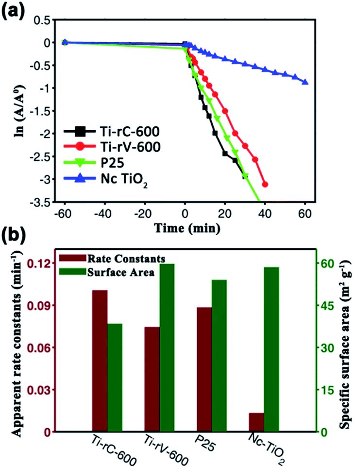 | ||
| Fig. 4 (a) Photocatalytic degradation kinetics of MB for Ti-rC-600, Ti-rV-600, P25, and Nc-TiO2, respectively. (b) The corresponding apparent rate constants and specific surface area of the above samples. Every data was achieved from the average value of at least three times. | ||
Conclusions
In summary, the aquatic leaf with two layers of tissues was employed as a template to fabricate photocatalysts. In the leaf, the epidermis cells showed rectangle and convex lens-like structure, which could focus and trap the light into the leaf interior. The spongy cells that directly connected to the epidermis cells scattered the light to make full use it. As a result, the leaf showed very good light harvesting capability. Mimicking the needle-like structure with two layers of tissues, the TiO2 replicas successfully inherited the leaf structures and functions. Moreover, the cells replicas would be regarded as microreactors which would further promote the photocatalytic efficiency. Consequently, the photocatalytic performance of the artificial leaf microreactors showed better than artificial leaves replicas with three layers of tissues and commercially used P25, which was ascribed to the superior light harvesting structure of the leaf structures and highly reactive efficiency of microreactors.Acknowledgements
This work is supported by the National Nature Science Foundation NSFC (no. 51173190, 21003132, 91127038, and 21121001), the 973 Program (no. 2013CB933004, 2011CB932303, and 2011CB808400), Beijing Nova Program (no. Z131103000413051), and the “Strategic Priority Research Program” of the Chinese Academy of Sciences (no. XDA09020000).Notes and references
- D. G. Nocera, Acc. Chem. Res., 2012, 45, 767 CrossRef CAS PubMed; W. Y. Wang, J. Chen, C. Li and W. M. Tian, Nat. Commun., 2014, 5, 4647 Search PubMed; J. Marshall, Nature, 2014, 510, 22 CrossRef PubMed; X. Pang, W. Chang, C. Chen, H. Ji, W. Ma and J. Zhao, J. Am. Chem. Soc., 2014, 136, 8714 CrossRef PubMed; J. H. Ma, W. H. Ma, C. C. Chen, H. W. Ji and J. C. Zhao, Chem.–Asian J., 2011, 6, 2264 CrossRef PubMed; D. K. Chacko, A. A. Madhavan, T. A. Arun, S. Thomas, G. S. Anjusree, T. G. Deepak, A. Balakrishnan, K. R. V. Subramanian, N. Sivakumar, S. V. Nair and A. S. Nair, RSC Adv., 2013, 3, 24858 RSC; K. Zhao, L. Feng, Z. Li, Y. Fu, X. Zhang, J. Wei and S. Wei, RSC Adv., 2014, 4, 51321 RSC.
- W. Y. Dong, Y. J. Sun, C. W. Lee, W. M. Hua, X. C. Lu, Y. F. Shi, S. C. Zhang, J. M. Chen and D. Y. Zhao, J. Am. Chem. Soc., 2007, 129, 13894 CrossRef CAS PubMed; J. Huang, M. Antonietti and J. Liu, J. Mater. Chem. A, 2014, 2, 7686 Search PubMed; J. Liu, J. Huang, D. Dontosova and M. Antonietti, RSC Adv., 2013, 3, 22988 RSC; J. Liu, M. Li, J. Wang, Y. Song, L. Jiang, T. Murakami and A. Fujishima, Environ. Sci. Technol., 2009, 43, 9425 CrossRef PubMed; A. A. Ismail and D. W. Bahnemann, J. Mater. Chem., 2011, 21, 11686 RSC; W. Zhou, W. Li, J. Q. Wang, Y. Qu, Y. Yang, Y. Xie, K. Zhang, L. Wang, H. Fu and D. Zhao, J. Am. Chem. Soc., 2014, 136, 9280 CrossRef PubMed.
- Q. Yang, M. Li, J. Liu, W. Shen, C. Ye, X. Shi, L. Jiang and Y. Song, J. Mater. Chem. A, 2013, 1, 541 CAS.
- J. Liu, G. Liu, M. Li, W. Shen, Z. Liu, J. Wang, J. Zhao, L. Jiang and Y. Song, Energy Environ. Sci., 2010, 3, 1503 CAS.
- S. Meng, D. Li, P. Wang, X. Zheng, J. Wang, J. Chen, J. Fang and X. Fu, RSC Adv., 2013, 3, 17021 RSC.
- J. Liu, Q. Yang, W. T. Yang, M. Z. Li and Y. L. Song, J. Mater. Chem. A, 2013, 1, 7760 CAS.
- X. Li, T. Fan, H. Zhou, S.-K. Chow, W. Zhang, D. Zhang, Q. Guo and H. Ogawa, Adv. Funct. Mater., 2009, 19, 45 CrossRef CAS; H. Zhou, T. Fan and D. Zhang, ChemSusChem, 2011, 4, 1344 CrossRef PubMed; J. Liu and M. Antonietti, Energy Environ. Sci., 2013, 6, 1486 Search PubMed; N. Shi, X. Li, T. Fan, H. Zhou, J. Ding, D. Zhang and H. Zhu, Energy Environ. Sci., 2011, 4, 172 Search PubMed.
- J. I. L. Chen, G. von Freymann, S. Y. Choi, V. Kitaev and G. A. Ozin, Adv. Mater., 2006, 18, 1915 CrossRef CAS.
- H. Zhou, L. Ding, T. Fan, J. Ding, D. Zhang and Q. Guo, Appl. Catal., B, 2014, 147, 221 CrossRef CAS PubMed; H. Zhou, J. Guo, P. Li, T. Fan, D. Zhang and J. Ye, Sci. Rep., 2013, 3, 1667 Search PubMed.
- H. Zhou, X. F. Li, T. X. Fan, F. E. Osterloh, J. Ding, E. M. Sabio, D. Zhang and Q. X. Guo, Adv. Mater., 2010, 22, 951 CrossRef CAS PubMed.
- H. Xiong, Q. Tan and C. Hu, Afr. J. Biotechnol., 2010, 9, 5722 CAS.
- E. P. H. Best and J. T. Meulemans, Aquat. Bot., 1979, 6, 53 CrossRef CAS.
- J. Li, J. Zhu and X. F. Gao, Small, 2014, 10, 2578 CrossRef CAS PubMed.
- J. Liu, Q. Yang, M. Z. Li, W. H. Zhu, H. Tian and Y. L. Song, Philos. Trans. R. Soc., A, 2013, 371, 20120314 CrossRef PubMed.
- J. D. Holladay, Y. Wang and E. Jones, Chem. Rev., 2004, 104, 4767 CrossRef CAS; A. R. Longstreet and D. T. McQuade, Acc. Chem. Res., 2013, 46, 327 CrossRef PubMed.
- J. Ovenstone and K. Yanagisawa, Chem. Mater., 1999, 11, 2770 CrossRef CAS.
- M.-C. Wu, J. Hiltunen, A. Sapi, A. Avila, W. Larsson, H.-C. Liao, M. Huuhtanen, G. Toth, A. Shchukarev, N. Laufer, A. Kukovecz, Z. Konya, J.-P. Mikkola, R. Keiski, W.-F. Su, Y.-F. Chen, H. Jantunen, P. M. Ajayan, R. Vajtai and K. Kordas, ACS Nano, 2011, 5, 5025 CrossRef CAS PubMed.
Footnote |
| † Electronic supplementary information (ESI) available: Experimental details of TiO2 replicas of Ceratophyllum and Vallisneria leaves were included in the ESI. BET parameters, such as surface area and crystalline size, derived from N2 adsorption isotherm, as well as the photocatalytic rate constants of some representable samples are illustrated in Table S1. See DOI: 10.1039/c4ra15943h |
| This journal is © The Royal Society of Chemistry 2015 |