Real-time selective monitoring of allergenic Aspergillus molds using pentameric antibody-immobilized single-walled carbon nanotube-field effect transistors

Joon-Hyung Jina, Junhyup Kima, Taejin Jeona, Su-Kyoung Shinb, Jong-Ryeul Sohnc, Hana Yib and Byung Yang Lee*a
aDepartment of Mechanical Engineering, Korea University, 145, Anam-ro, Seoungbuk-gu, Seoul 136-713, Korea. E-mail: blee@korea.ac.kr
bDepartment of Public Health Science, Graduate School, Korea University, 139, Bogukmun-ro, Seongbuk-gu, Seoul 136-703, Korea
cDepartment of Environmental Health, Korea University, 139, Bogukmun-ro, Seongbuk-gu, Seoul 136-703, Korea

Received 5th December 2014 , Accepted 26th January 2015

First published on 28th January 2015


Abstract

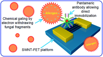
Airborne fungus, including Aspergillus species, is one of the major causes of human asthma. Conventional immunoassay or DNA-sequencing techniques, although widely used, are usually labor-intensive, time-consuming and expensive. In this paper, we demonstrate a sensor for the rapid detection of Aspergillus niger, a well-known allergenic fungal species, using single-walled carbon nanotube (SWNT) field effect transistors (FETs) functionalized with pentameric antibodies that specifically bind to Aspergillus species. This strategy resulted in the real time and highly sensitive and selective detection of Aspergillus due to an electrostatic gating effect from the Aspergillus fungus. This mechanism is in contrast to a previously reported Aspergillus sensor, which was based on mobility modulation from Aspergillus adsorption. Also, our sensor shows a much wider detection range from 0.5 pg mL−1 to 10 μg mL−1 with a lower detection limit of 0.3 pg mL−1. The resulting SWNT-FET was able to selectively detect Aspergillus molds in the presence of more concentrated amounts of other mold species such as Alternaria alternata, Cladosporium cladosporioides, and Penicillium chrysogenum. We expect that our results can be used in real-time monitoring of the indoor air quality of a variety of public facilities for the elderly and children, who are more vulnerable to environmental biohazards.

Introduction

Allergens cause an immunoglobulin E-involved abnormal immune response known as type I hypersensitivity reaction.1 Allergens are widely spread in a variety of different forms such as volatile organic chemicals, fine dust particles, pollens, viruses, fungi, and even foods.2–4 Among fungal allergens, the Aspergillus, Alternaria, Cladosporium, and Penicillium species are the most frequently observed, and most of them produce allergens from their hyphae even before spore germination.5 Although fungi occupy a great portion of officially-recognized allergenic disorders approved by the World Health Organization and International Union of Immunological Societies, and about 19% of patients suffering from respiratory allergic disorders including asthma are allegedly susceptible to the fungal allergens, the working mechanism of allergenic disorder is not fully understood, and no early warning systems for monitoring fungal allergens have been developed.1 In addition, conventional fungi detection methods usually involve sampling of fungal spores and culturing for signal amplification, followed by immunoassays for optical detection, DNA sequencing, and spectroscopic quantification, which are labor-intensive, time-consuming, and costly.6–9 Sensors based on nanostructured materials combined with highly selective biological receptors have been reported, sometimes showing sub-picomolar detection.10 A carbon nanotube-based sensor has actually been introduced for monitoring specific peptide strands and Aspergillus molds.11,12 Also, a cantilever array sensor has been demonstrated to monitor the growth of Aspergillus niger (A. niger).13 However, these previous attempts commonly employed mediator proteins for the correct orientation of the immobilized antibodies. Although nanotube and nanowire-based field-effect transistors have been widely investigated and showed excellent achievements in gas sensing and biosensing applications,11,12,14–16 such an indirect combination of antibodies with the underneath nanotube requires a more complicated fabrication procedure and accordingly causes a bigger sensor-to-sensor variation that may restrict dynamic detection range of the nanotube-based sensors.

Here, we demonstrate a nanoscale sensor for the real-time detection of A. niger with high sensitivity and selectivity by directly combining single-walled carbon nanotube-integrated field effect transistors (SWNT-FETs) with mouse anti-Aspergillus species-specific primary antibodies (WF-AF-1 Abs), which belong to an immunoglobulin M (IgM) class that can be found mostly in pentamer and sometimes in hexamer structures composed of five or six unit antibodies. This IgM antibody selectively binds to galactomannan, a major cell wall component of Aspergillus species.17–20 The activity of the WF-AF-1 Abs was carefully determined using enzyme-linked immunosorbent assay (ELISA) in various temperature conditions. The antibody was then covalently immobilized on the SWNT channel using EDC/NHS chemistry.21–23 Continuous current increase of the SWNT-FET was observed with increasing fungal concentration, which is in contrast to previously reported method using antibodies with mediator proteins.11,12 Furthermore, the detection limit was three orders lower, and the dynamic range was one hundred thousand times wider than previous report.12 We propose that this performance difference is primarily due to the use of structurally more robust IgM antibodies combined with increased Debye length by using low ionic concentration buffer solution.

Experimental section

Reagents

1-Ethyl-3-(3-dimethylaminopropyl)-carbodiimide (EDC) and N-hydroxysuccinimide (NHS) for the activation of carboxylic acid groups of SWNTs were purchased from Aldrich (Milwaukee, USA). WF-AF-1 Abs (MCA 2576, AbD Serotec) for the functionalization of the SWNT channels and fluorescein isothiocyanate (FITC, LNK061F, AbD Serotec) for labelling the WF-AF-1 Abs were purchased from Bio-Rad Laboratories, Inc. A 0.2% (v/v) octadecyltrichlorosilane (OTS) (>90%, Aldrich Chemical Co., Inc.) solution in n-hexane (anhydrous 95%, Aldrich Chemical Co., Inc.) was prepared for aligning and patterning the SWNT channels.

Equipment

Atomic force microscopy (AFM) and scanning electron microscopy (SEM) images were obtained using NTEGRA Prima (TS-150, NT-MDT) and S-4300N (Hitachi High-Technologies Corp.), respectively. The effect of the applied gate voltage, both in air and liquid, was monitored using a probe station equipped with a Keithley 4200 semiconductor parameter analyzer. A wired Pt tip was used as a pseudo-reference electrode. IV characteristics of the SWNT-FETs were recorded using a potentiostat/galvanostat (ref. 600, Gamry Instruments).

SWNT-FET fabrication

A boron-doped p-Si wafer with 300 nm thick thermal oxide (SiO2) was used (resistivity = 1.0–10.0 Ω cm). Electrode pads consisting of 30 nm-thick Au film over 5 nm-thick Cr adhesion layer was formed via photolithography using photoresist (AZ5214) followed by thermal evaporation of metals and lift-off process. Regions of non-polar OTS and SiO2 were patterned between the electrode pads. The OTS patterned substrate was dipped into SWNT (Hanwha Chemical Co., Ltd.) solution to form a SWNT network channel. The SWNTs assembled on the SiO2 regions, avoiding the OTS regions. Additional Cr/Au deposition right on the junction area between the SWNT and the Au electrode allowed better mechanical strength and reduced contact resistance. The final step was to immobilize the Aspergillus species-specific receptors WF-AF-1 Abs on the SWNTs. The entire fabrication processes are shown in Scheme 1. Thirty eight devices were available from a 4′′ p-Si wafer, and each device had two SWNT-FET channels.
image file: c4ra15815f-s1.tif
Scheme 1 Schematic diagram depicting fabrication of the SWNT-FET-based fungi sensors and the antibody-allergen binding. Note that WF-AF-1 Abs were directly immobilized on the carboxylic acid-functionalized SWNT surface via EDC/NHS chemistry.

Direct immobilization of mouse anti Aspergillus species-specific primary antibodies WF-AF-1 Abs

Acid treatment of SWNTs after their growth for catalytic particle removal inevitably over oxidizes backbone carbons of the SWNTs to form many oxygen-containing chemical functional groups such as carboxylic acid groups on the SWNT surface.24 Although the carboxylic acid groups are reactive with amine groups, the amide forming reaction, under normal conditions, could be kinetically unfavourable without a catalyst. As shown in Scheme 1, the EDC/NHS chemistry substitutes the hydroxyl group of the carboxylic acid group with more facile leaving group, i.e., NHS group. Afterwards, the nucleophilic attack of the N-terminal of an antibody on the succinimide-substituted carbonyl carbon was enhanced and formed a strong amide bond. The resulting antibody-immobilized SWNT-FET devices were stored at 4 °C before use.

Strains and growth conditions

The fungal strains used in this study were obtained from the RDA Genebank Information Center of Korea. A. niger KACC 40280, Alternaria alternata (A. alternata) KACC 43544, Cladosporium cladosporioides (C. cladosporioides) KACC 43384, and Penicillium chrysogenum (P. chrysogenum) KACC 44307 were grown at 25 °C for 6 days in malt extract agar. Fungal fragments and spores were harvested with sterile loops and washed twice in phosphate-buffered saline (PBS, pH 7.2). They were resuspended to 1.2 × 10⁷ spores mL−1 (5.7 mg mL−1 in wet weight) in 50 mM carbonate bicarbonate buffer (pH 9.6, Sigma) and ten-fold dilution series of the suspensions were prepared. The fungal concentration was determined by hemocytometer.

Enzyme-linked immunosorbent assay

Indirect ELISA was carried out by coating 200 μL aliquots of different concentrations of A. niger samples in 50 mM carbonate bicarbonate buffer to each well of microplates (NUNC Maxisorp, Nalgene). The plates were incubated overnight at 4 °C, then washed twice with 200 μL PBS buffer with 0.05% Tween-20 (PBST). 200 μL of WF-AF-1 Abs in PBS buffer with 1% BSA (1.0 mg mL−1, 1[thin space (1/6-em)]:[thin space (1/6-em)]1000) was added and the plates were incubated at different temperatures (7, 25, and 37 °C) for two hours. After washing the plates twice with 200 μL PBS, 200 μL HRP conjugated goat-anti-mouse Abs (Bio-Rad) in 1% BSA/PBS (1.0 mg mL−1, 1[thin space (1/6-em)]:[thin space (1/6-em)]1000) was then applied to each well and incubated at various temperatures (7, 25, and 37 °C) for one hour. After washing the plates twice with 200 μL PBST, 100 μL peroxidase substrate (Bio-Rad Laboratories) was added to each well and the plates were incubated for 10 min at room temperature for the blue-green color development. The enzyme reaction was stopped by adding 2% oxalic acid (Sigma-Aldrich). The optical density at 415 nm was read using a microplate reader Victor™ X3 (Perkin Elmer Inc., USA). Fig. 1 shows that more than 70% of the antibodies are still active at 7 °C as compared to that at a higher temperature up to 37 °C, indicating excellent stability of WF-AF-1 Abs at a wide range of temperature.
image file: c4ra15815f-f1.tif
Fig. 1 ELISA optical density measurements for testing antibody activity of WF-AF-1 at 7 °C, 25 °C, and 37 °C.

Fungal cell counting and weighing

To quantify molds, we directly count the fungal spores using hemocytometer and an optical microscope (Eclipse LV100ND, Nikon). Fig. S1 presents the microscope image showing suspended mold spores in buffer solution. We can define the mold spores per unit volume of the mold-suspended solution because grid distance and the well depth of the hemocytometer are given values. However, mass quantity is a more convenient mold quantification method for our experiment since WF-AF-1 Abs binds to both spores and fragments of A. niger. Fungal fragments are prevalently present in biosphere including air and water.25–27 Averaged mass for a spore of A. niger is known as 47 pg.28 To measure such a small mass, a PCR tube container was placed on the weighing plate of an ultra microbalance (XF2U, Mettler Toledo), and as shown in Fig. S2 three more containers which include silica gels were placed in the weighing chamber to protect hygroscopic fungal spores and fragments from moisture. Actual mass of mold could be obtained simply by subtracting the mass of PCR tube container from total mass. Fresh molds grown on agar plates were used to measure the mass. The molds were finally suspended in a PBS solution and diluted to a concentration of 100 μg mL−1.

Simulated clinical test and reversibility experiment

For the mixture experiment in Fig. 5e, we first prepared a primary antibody-immobilized SWNT-FET sensor, and then we first injected 2 μL PBS buffer and waited for current stabilization. Afterwards, we sequentially injected, in 50 s intervals, 2 μL of the four mixtures prepared as follows: 1[thin space (1/6-em)]:[thin space (1/6-em)]1 (v/v) mixture of 10 μg mL−1 C. cladosporioides and 10 μg mL−1 A. alternata; 1[thin space (1/6-em)]:[thin space (1/6-em)]1[thin space (1/6-em)]:[thin space (1/6-em)]1 (v/v) mixture of 10 μg mL−1 C. cladosporioides, 10 μg mL−1 A. alternata, and 10 μg mL−1 P. chrysogenum; 1[thin space (1/6-em)]:[thin space (1/6-em)]1[thin space (1/6-em)]:[thin space (1/6-em)]1[thin space (1/6-em)]:[thin space (1/6-em)]1 (v/v) mixture of 10 μg mL−1 C. cladosporioides, 10 μg mL−1 A. alternata, 10 μg mL−1 P. chrysogenum, and 1 μg mL−1 A. niger; 1[thin space (1/6-em)]:[thin space (1/6-em)]1[thin space (1/6-em)]:[thin space (1/6-em)]1[thin space (1/6-em)]:[thin space (1/6-em)]1 (v/v) mixture of 10 μg mL−1 C. cladosporioides, 10 μg mL−1 A. alternata, 10 μg mL−1 P. chrysogenum, and 10 μg mL−1 A. niger. Note that the first two mixtures contain no A. niger. The last two mixtures contain A. niger of concentrations differing by 10 times. For the reversibility experiment in Fig. 5f, we first prepared a primary antibody-immobilized SWNT-FET sensor, and then we first injected 2 μL PBS buffer and waited for current stabilization. Then, we injected 2 μL of 1 pg mL−1 A. niger and observed the current increase. Afterwards, the current monitoring was stopped momentarily for sample washing. The sample was thoroughly washed with PBS and D.I. water for 5 min. Then, the current monitoring was resumed. This process was repeated for two times.

Results and discussion

Direct immobilization of pentameric WF-AF-1 Abs on the SWNT channel

Scheme 1 describes the preparation procedure of SWNT-FET and the following immobilization of WF-AF-1 Abs on the SWNT channel. Sequential steps finally lead to the production of the Aspergillus sensors that have the SWNT network channel as sensor transducers.29 The SWNTs in the channel selectively assembly on the SiO2 region due to van der Waals interaction, while avoiding the nonpolar OTS regions. Subsequently, WF-AF-1 Abs were immobilized on the SWNT surfaces via EDC/NHS chemistry utilizing carboxyl acid groups on the SWNT surface that are introduced during SWNT growth and cleaning process.24 Note that this immobilization scheme is advantageous compared to the previous Aspergillus sensor using SWNT-FET,12 because by directly attaching the antibody to the SWNT, we can reduce the SWNT-antibody distance, and therefore enhance sensitivity.30

Basic characterization of SWNT-FET

The gap between source and drain electrodes is approximately 10 μm as shown in Fig. 2a. The corresponding SEM image of the SWNT channel having a width of 5 μm proves that a well-oriented SWNT network channel was formed between source and drain. The channel could be formed simply by dipping the OTS-patterned SiO2 substrate into a SWNT dispersed solution, and the SWNTs were selectively assembled on the exposed SiO2 regions while avoiding the nonpolar OTS regions. The IV curve of the SWNT-FET shown in Fig. 2b verifies the Schottky barrier junction featuring rectifying characteristics formed in both ends of the SWNT channel between the semiconducting SWNTs and the deposited gold metal.31,32 To verify the usability of the SWNT-FET as a sensor transducer, we measured the back gate characteristics of the SWNT-FET in air. Fig. 2c shows a typical back gate characteristic of a SWNT-FET. The oxide layer was 100 nm. The SWNT-FET showed a p-type semiconductor behavior, where the conductance decreased with the increase of gate voltage (Vg).33 The typical on–off ratio was ∼9. The apparent low current on/off ratio of our sensor transducers was mainly due to the utilization of as-produced SWNTs, which are composed of mixtures of semiconducting and metallic SWNTs. The carrier mobility μ was calculated to be in the range of 5.6–8.1 cm2 (V s)−1 (details in ESI), which is comparable to previously reported SWNT-FET-based sensors with similar on/off ratio and mobility values.34,35 From the practical point of view, high resistance SWNT-FET devices were carefully screened out on the basis of resistance of the SWNT channel to minimize sensor-to-sensor variation. Fig. 3 summarizes resistance distribution, and devices with resistances levels around several hundred kΩ were used in this experiment.
image file: c4ra15815f-f2.tif
Fig. 2 Electronic properties of SWNT FETs. (a) Detailed images of the 5 μm wide, 10 μm long SWNT channel. (b) An example of the IV characteristic curve of a device with a resistivity of 830 kΩ to confirm the Schottky-barrier junction at both Au-SWNT and SWNT/Au interfaces: scan rate = 50 mV s−1. Note that the resistivity of the devices were measured with assuming linear IV characteristics. (c) Dependence of source current on the applied gate voltage at source-drain voltage Vds = 0.1 V.

image file: c4ra15815f-f3.tif
Fig. 3 Resistance distribution of the SWNT-FET devices.

Binding and functionalization of SWNT-FET

The immobilized WF-AF-1 Abs and their binding to A. niger fragments were confirmed with both AFM and fluorescence microscopy (Fig. 4). We prepared a solution of A. niger fragments by ultra-centrifugation as described in Experimental section. Then, the antibody-immobilized sensor was dipped into solution and left for ∼1 h and then washed extensively with PBS and D.I. water. Fig. 4a and b compare the SWNT channel before and after A. niger fragment binding. The approximate density of the channel determined by counting individual SWNT from the AFM image considering the averaged nanotube length of ∼2 μm is 5 μm−2, which is well above the percolation threshold density.34,35 This density is known to result in similar values of current on/off ratio as our sensor transducers.34 The height profile (Fig. 4c) shows that the fragment binding resulted in an overall increase in the height profile by ∼3 nm due to the fragments. The antibody WF-AF-1 Abs specifically binds to galactomannan, which is present on the surface of the spores and their fragments.25–27 This means that our sensor is sensitive to both spores and fragments of A. niger. We further confirmed the antibody immobilization and fragment binding by introducing FITC-conjugated WF-AF-1 Abs to the sensor (Fig. 4d). The intensive fluorescent radiation along the SWNT channel shows the successful antibody immobilization on the channel and fungal fragment binding. The actual size of spores of Aspergillus species is a few microns,36,37 which is comparable to that of the SWNT channel width (5 μm). Accordingly, it is reasonable to say that most of the radiated emission from the channel area is due to the binding between fungal fragments and the FITC-labelled antibodies.
image file: c4ra15815f-f4.tif
Fig. 4 Verification of antibody immobilization and fungi fragment-antibody binding. AFM images of WF-AF-1 Abs-immobilized SWNT channels (a) before and (b) after the antibody-allergen binding, and (c) corresponding height profiles. (d) Fluorescence microscopy image of FITC-labeled SWNT-FET sensor after fragment binding: excitation wavelength = 465–495 nm (Nikon eclipse LV100ND); exposure time = 60 s.

Sensitivity and selectivity

Current flow through the functionalized SWNT channel was recorded as a function of time with continuous injection of 2 μL of different concentrations of A. niger suspended in 1.5 mM pH 7.2 PBS solution at every 50 seconds (Fig. 5a). A low concentration PBS buffer of 1.5 mM was used to increase the Debye length (∼9 nm around SWNTs).30,38,39 Since the antibody used in this work has a form of IgM, which is physically the largest antibody, Debye length is a crucial factor affecting sensing ability and selectivity. Since IgM has a planar structure with longest diameter of ∼30 nm and thickness of ∼3 nm,40–43 we can expect many IgM antibodies to be immobilized around the SWNTs with random direction while providing multiple binding sites within the Debye length.
image file: c4ra15815f-f5.tif
Fig. 5 Response of A. niger sensor. (a) Real-time response of WF-AF-1 Abs-immobilized SWNT-FET device to consecutively injected A. niger-suspended PBS solution in various concentrations (injection volume = 2 μL). Note that the initial volume of the 1.5 mM PBS solution on the device is 2 μL; (b) corresponding calibration curve; (c) real-time responses of the functionalized SWNT-FET device to sequential injections of different mold-suspended PBS solutions in random order; (d) selective liquid gating effect of our sensor to target and non-target molds. Injection volume and concentration are the same for both A. alternata and A. niger as 5 μL and 1 pg mL−1, respectively. (Vds = 0.1 V; initial volume of 1.5 mM PBS = 10 μL); (e) sequential injection of four different mixtures of fungal solutions. The first two mixtures contain no A. niger, while the last two mixtures contain A. niger of concentrations differing by 10 times; (f) sensor revival test by extensive washing with PBS buffer and water.

The amount of A. niger was carefully weighed using ultra-microbalance before preparing the solutions. Fig. 5b shows that the output current was linearly proportional to the A. niger concentration in the range from 0.5 pg mL−1 to 10 μg mL−1 with an approximate detection limit of 0.3 pg mL−1. This detection limit is three orders lower, and the dynamic range is one hundred thousand times wider than previous report.12 The increase of current with increasing concentration can be explained from electrostatic gating effect of the A. niger spores and fragments. In neutral pH, the surface of A. niger is negatively charged because the isoelectric point of the A. niger is around 2.44 The negatively charged A. niger spores and fragments apply a negative gating effect to the SWNT channel (Fig. 2c, IVg curve), and therefore, the current is increased in the SWNT channel. Note that this current increase behavior is opposite to a previous report using SWNT-FET for the detection of fungi.12 Presumably, their experiment involved mobility decrease of SWNTs by direct binding between fungi and SWNTs.45 We checked the selectivity of our sensor by introducing successively other fungi species to the SWNT sensor. As shown in Fig. 5c, the effect on the current from injections of A. alternata, C. cladosporioides, and P. chrysogenum-suspended solutions are negligible as compared to that of A. niger-suspended solution. Observation of the selective current response of the functionalized SWNT-FET to A. niger can be attributed to specific binding between galactomannan on A. niger surface and the immobilized WF-AF-1 Abs.46 Most cell walls of Aspergillus species including their conidia contain galactomannan.47,48 Table S1 shows normalized current response (IN) of the WF-AF-1 Abs-immobilized SWNT-FET device to various interfering molds. The signal output was enhanced by more than two times with the injection of A. niger in the presence of interfering molds. To verify the sensing mechanism, we used a Pt pseudo-reference electrode for liquid gating.49,50 The IVlg shows hysteresis due to mobile ions in the liquid. Major cause for hysteresis in nanotube and nanowire-based FETs is water molecules or charge trapping at the interface between the solid substrate and nanotube/nanowires.15,51,52 Fig. 5d shows the liquid characteristics before and after the Aspergillus binding. The threshold voltage from the IVlg graph shifted to the right side by about 90 mV without appreciable change in slope when a 5 μL portion of interfering A. niger (1 pg mL−1) was injected. This means the mobility was conserved, and the increased current signal was mainly due to the increased hole carrier concentration from chemical gating by the negatively charged A. niger fragments. Note that our sensor has a wide dynamic range with very low detection limit compared to previous works, and the sensing mechanism is chemical gating which is predictable from the interaction between p-type SWNT-FET and negatively charged A. niger fragments.

The bioaerosol in real world contains many kinds of molds. To show the feasibility of using our sensor in the real-world, we prepared four different simulated clinical samples of molds and performed more detailed specificity experiments (details in Experimental section). Fig. 5e describes response of the SWNT-FET sensor to the simulated clinical samples. The sensor showed no significant response to the first two mixtures containing no A. niger. However, the sensor showed increasing sensor response to the last two mixtures that contain A. niger of concentrations differing by 10 times. To check the feasibility of using our sensors in the real world, we performed reversibility test in low target concentration (Fig. 5f). To show that the reversibility can be performed in principle, we washed the sample with extensive amount of 1 mM pH 7.0 PBS buffer and D.I. water (details in Experimental section). The washed sensor showed a current level close to its initial value, as shown in Fig. 5f. This shows that we can combine our sensor with additional washing system to reverse the sensor response after being exposed to target materials.

Conclusions

Here, we demonstrated a nanoscale sensor for the real-time detection of A. niger with high sensitivity and selectivity by directly combining single-walled carbon nanotube-integrated field effect transistors (SWNT-FETs) with mouse anti Aspergillus species-specific primary antibodies (WF-AF-1 Abs), which belong to an IgM class that has a form of pentamer or hexamer structure composed of five or six unit antibodies. Contrary to previous works, output current in this work increased with increased amount of the target fungal fragments due to the chemical gating effect by the antibody-allergen binding event occurring on the SWNT-FET. The utilization of structurally more robust antibody and increased Debye length due to low buffer ionic concentration appears to allow a wider detection range and lower detection limit for monitoring fungal allergens. Our study can be used to develop a new type of sensor module for an integrated in situ monitoring system of fungal allergens when combined with adequate sampler that collects the airborne fungi into liquid phase.

Acknowledgements

This work was supported by Korea Ministry of Environment as “The Environmental Health Action Program” (grant number: ARQ201303173002), and as “The Conversing Technology Program” (grant numbers: 2013001650001 and ARQ201403075001).

References

  1. R. Crameri, M. Garbani, C. Rhyner and C. Huitema, Allergy, 2014, 69, 176 CrossRef CAS PubMed.
  2. M. Rosmilah, M. Shahnaz, G. Patel, J. Lock, D. Rahman, A. Masita and A. Noormalin, Trop. Biomed., 2008, 25, 243 CAS.
  3. C. Zahradnik, R. Martzy, R. L. Mach, R. Krska, A. H. Farnleitner and K. Brunner, Anal. Bioanal. Chem., 2014, 406, 6827 CrossRef CAS PubMed.
  4. R. J. Bertelsen, C. K. Faeste, B. Granum, E. Egaas, S. J. London, K. H. Carlsen, K. C. Lodrup Carlsen and M. Lovik, Clin. Exp. Allergy, 2014, 44, 142 CrossRef CAS PubMed.
  5. B. J. Green, T. Z. Mitakakis and E. R. Tovey, J. Allergy Clin. Immunol., 2003, 111, 285 CrossRef.
  6. L. Monaci, R. Pilolli, E. De Angelis, M. Godula and A. Visconti, J. Chromatogr. A, 2014, 1358, 136 CrossRef CAS PubMed.
  7. A. Pinto, P. N. Polo, O. Henry, M. C. Redondo, M. Svobodova and C. K. O'Sullivan, Anal. Bioanal. Chem., 2014, 406, 515 CrossRef CAS PubMed.
  8. M. Koeberl, D. Clarke and A. L. Lopata, J. Proteome Res., 2014, 13, 3499 CrossRef CAS PubMed.
  9. M. Svobodova, T. Mairal, P. Nadal, M. C. Bermudo and C. K. O'Sullivan, Food Chem., 2014, 165, 419 CrossRef CAS PubMed.
  10. S. M. Yoo, T. Kang, H. Kang, H. Lee, M. Kang, S. Y. Lee and B. Kim, Small, 2011, 7, 3371 CrossRef CAS PubMed.
  11. J. Oh, G. Yoo, Y. W. Chang, H. J. Kim, J. Jose, E. Kim, J. C. Pyun and K. H. Yoo, Biosens. Bioelectron., 2013, 50, 345 CrossRef CAS PubMed.
  12. R. A. Villamizar, A. Maroto and F. X. Rius, Anal. Bioanal. Chem., 2011, 399, 119 CrossRef CAS PubMed.
  13. N. Nugaeva, K. Y. Gfeller, N. Backmann, H. P. Lang, M. Duggelin and M. Hegner, Biosens. Bioelectron., 2005, 21, 849 CrossRef CAS PubMed.
  14. G. Konvalina and H. Haick, Acc. Chem. Res., 2014, 47, 66 CrossRef CAS PubMed.
  15. Y. Paska and H. Haick, ACS Appl. Mater. Interfaces, 2012, 4, 2604 CAS.
  16. B. Wang, J. C. Cancilla, J. S. Torrecilla and H. Haick, Nano Lett., 2014, 14, 933 CrossRef CAS PubMed.
  17. P. C. Bardalaye and J. H. Nordin, J. Biol. Chem., 1977, 252, 2584 CAS.
  18. T. Nakajima and E. Ichishima, J. Ferment. Bioeng., 1994, 78, 472 CrossRef CAS.
  19. C. M. A. Swanink, J. F. G. M. Meis, A. J. M. M. Rijs, J. P. Donnelly and P. E. Verweij, J. Clin. Microbiol., 1997, 35, 257 CAS.
  20. Y. Tamaki, T. Teruya and M. Tako, Biosci., Biotechnol., Biochem., 2010, 74, 1110 CrossRef CAS PubMed.
  21. G. Krishnamoorthy, R. Selvakumar, T. P. Sastry, S. Sadulla, A. B. Mandal and M. Doble, Mater. Sci. Eng., C, 2014, 43, 164 CrossRef CAS PubMed.
  22. M. D'Este, D. Eglin and M. Alini, Carbohydr. Polym., 2014, 108, 239 CrossRef PubMed.
  23. Y. S. Sohn and Y. K. Lee, J. Biomed. Opt., 2014, 19, 051209 CrossRef PubMed.
  24. A. Suri and K. S. Coleman, Carbon, 2011, 49, 3031 CrossRef CAS PubMed.
  25. R. L. Gorny, T. Reponen, K. Willeke, D. Schmechel, E. Robine, M. Boissier and S. A. Grinshpun, Appl. Environ. Microbiol., 2002, 68, 3522 CrossRef CAS.
  26. A. Pringle, PLoS Pathog., 2013, 9, e1003371 CAS.
  27. T. Reponen, S. C. Seo, F. Grimsley, T. Lee, C. Crawford and S. A. Grinshpun, Atmos. Environ., 2007, 41, 8140 CrossRef CAS PubMed.
  28. N. Nugaeva, K. Y. Gfeller, N. Backmann, M. Duggelin, H. P. Lang, H. J. Guntherodt and M. Hegner, Microsc. Microanal., 2007, 13, 13 CrossRef CAS PubMed.
  29. M. Lee, J. Im, B. Y. Lee, S. Myung, J. Kang, L. Huang, Y. K. Kwon and S. Hong, Nat. Nanotechnol., 2006, 1, 66 CrossRef CAS PubMed.
  30. J. P. Kim, B. Y. Lee, S. Hong and S. J. Sim, Anal. Biochem., 2008, 381, 193 CrossRef CAS PubMed.
  31. Y. He, J. Y. Zhang, S. M. Hou, Y. Wang and Z. P. Yu, Appl. Phys. Lett., 2009, 94, 093107 CrossRef PubMed.
  32. S. Heinze, J. Tersoff, R. Martel, V. Derycke, J. Appenzeller and P. Avouris, Phys. Rev. Lett., 2002, 89, 106801 CrossRef CAS.
  33. M. H. Yang, K. B. K. Teo, W. I. Milne and D. G. Hasko, Appl. Phys. Lett., 2005, 87, 253116 CrossRef PubMed.
  34. V. K. Sangwan, A. Behnam, V. W. Ballarotto, M. S. Fuhrer, A. Ural and E. D. Williams, Appl. Phys. Lett., 2010, 97, 043111 CrossRef PubMed.
  35. E. S. Snow, J. P. Novak, P. M. Campbell and D. Park, Appl. Phys. Lett., 2003, 82, 2145 CrossRef CAS PubMed.
  36. R. Serra, F. J. Cabanes, G. Perrone, G. Castella, A. Venancio, G. Mule and Z. Kozakiewicz, Mycologia, 2006, 98, 295 CrossRef.
  37. T. H. Fang, S. H. Kang, Z. H. Hong and C. D. Wu, Micron, 2012, 43, 407 CrossRef PubMed.
  38. J. P. Kim, B. Y. Lee, J. Lee, S. Hong and S. J. Sim, Biosens. Bioelectron., 2009, 24, 3372 CrossRef CAS PubMed.
  39. J. Wu, K. Gerstandt, H. B. Zhang, J. Liu and B. J. Hinds, Nat. Nanotechnol., 2012, 7, 133 CrossRef CAS PubMed.
  40. E. Stern, R. Wagner, F. J. Sigworth, R. Breaker, T. M. Fahmy and M. A. Reed, Nano Lett., 2007, 7, 3405 CrossRef CAS PubMed.
  41. S. J. Armstrong, M. C. Outlaw and N. J. Dimmock, J. Gen. Virol., 1990, 71, 2313 CrossRef CAS.
  42. B. Chesebro, B. Bloth and S. E. Svehag, J. Exp. Med., 1968, 127, 399 CrossRef CAS.
  43. P. W. Haebel, D. Goldstone, F. Katzen, J. Beckwith and P. Metcalf, EMBO J., 2002, 21, 4774 CrossRef CAS PubMed.
  44. P. Jones, D. Moore and A. P. J. Trinci, J. Gen. Microbiol., 1988, 134, 235 CAS.
  45. I. Heller, A. M. Janssens, J. Mannik, E. D. Minot, S. G. Lemay and C. Dekker, Nano Lett., 2008, 8, 591 CrossRef CAS PubMed.
  46. H. E. Jensen, J. Salonen and T. O. Ekfors, J. Pathol., 1997, 181, 100 CrossRef CAS.
  47. S. Christgau, S. Kauppinen, J. Vind, L. V. Kofod and H. Dalboge, Biochem. Mol. Biol. Int., 1994, 33, 917 CAS.
  48. P. Ademark, R. P. de Vries, P. Hagglund, H. Stalbrand and J. Visser, Eur. J. Biochem., 2001, 268, 2982 CrossRef CAS.
  49. I. Heller, A. M. Janssens, J. Männik, E. D. Minot, S. G. Lemay and C. Dekker, Nano Lett., 2007, 8, 591 CrossRef PubMed.
  50. S. Rosenblatt, Y. Yaish, J. Park, J. Gore, V. Sazonova and P. L. McEuen, Nano Lett., 2002, 2, 869 CrossRef CAS.
  51. W. Kim, A. Javey, O. Vermesh, Q. Wang, Y. Li and H. Dai, Nano Lett., 2003, 3, 193 CrossRef CAS.
  52. K. Bradley, J. Cumings, A. Star, J.-C. P. Gabriel and G. Grüner, Nano Lett., 2003, 3, 639 CrossRef CAS.

Footnote

Electronic supplementary information (ESI) available. See DOI: 10.1039/c4ra15815f

This journal is © The Royal Society of Chemistry 2015
Click here to see how this site uses Cookies. View our privacy policy here.