Molecular insight into the mode-of-action of phosphonate monolayers as active functions of hybrid metal oxide adsorbents. Case study in sequestration of rare earth elements

Gulaim A. Seisenbaeva*a, Inna V. Melnykb, Niklas Hedinc, Yang Chend, Philip Erikssona, Elżbieta Trzopd, Yuriy L. Zub*b and Vadim G. Kesslerb
aDepartment of Chemistry and Biotechnology, Biocenter, Swedish University of Agricultural Sciences, Box 7015, 750 07 Uppsala, Sweden. E-mail: Gulaim.Seisenbaeva@slu.se; Vadim.Kessler@slu.se
bChuiko Institute of Surface Chemistry, National Academy of Sciences of Ukraine, 17, General Naumov Street, Kyiv 03164, Ukraine. E-mail: in.melnyk@gmail.com; yurii.zub@gmail.com
cBerzelii Centre EXSELENT, Department of Materials and Environmental Chemistry, Arrhenius Laboratory, Stockholm University, 106 91 Stockholm, Sweden. E-mail: Niklas.hedin@mmk.su.se
dDepartment of Chemistry, University at Buffalo, SUNY, Buffalo, NY 14260-3000, USA. E-mail: elzbieta@buffalo.edu

Received 30th November 2014 , Accepted 23rd February 2015

First published on 2nd March 2015


Abstract

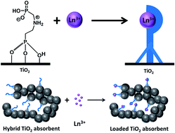
The insight into the molecular aspects of ligand grafting and potential maximal capacity of hybrid organic–inorganic adsorbents bearing phosphonate ligand monolayers as active functions was obtained by single crystal X-ray studies of ligand-functionalized titanium alkoxide complexes. The attachment of molecules occurs generally in the tripodal vertical fashion with the minimal distance between them being about 8.7 Å, resulting in 0.19 nm2 as the minimal surface area per function. In the present experimental work the theoretical loading capacity could almost be achieved for functionalization of mesoporous nanorods of anatase with imino-bis-methylphosphonic acid (IMPA, NH(CH2PO3H2)2) or aminoethylphosphonic acid (AEPA, H2NC2H4PO3H2). The products had the same morphology as the starting material, as was established by SEM and optical microscopy. The size and structure of the individual nanoparticles of the constituting inorganic component of the material were preserved and practically unchanged through the surface modification, as established by powder XRD and EXAFS studies. The surface area of the inorganic–organic hybrids decreased somewhat from the initial ∼250 m2 g−1, on adsorption of AEPA (0.21 mmol g−1) to ∼240 m2 g−1, and on adsorption of IMPA (0.17 mmol g−1) to ∼190 m2 g−1. The ligands were bound effectively to the surface according to TGA, EDS and FTIR analyses and remained in the mono-deprotonated form. The produced hybrid adsorbents had for the selected pH (3.5) high capacities towards adsorption of Rare Earth Element (REE) cations, but with equilibria achieved relatively slowly. The composition of the surface complexes was determined as M[thin space (1/6-em)]:[thin space (1/6-em)]L = 1[thin space (1/6-em)]:[thin space (1/6-em)]1 for IMPA, but varied for the AEPA from 1[thin space (1/6-em)]:[thin space (1/6-em)]3 to 1[thin space (1/6-em)]:[thin space (1/6-em)]1 dependent on the REE, which can be interpreted in terms of charge compensation as the major driving force behind binding. The cation desorption in strongly acidic media for recuperation of the adsorbed REE and the relative capacity of the re-used adsorbent have been quantified.


Introduction

Highly chemically stable and poorly soluble nanostructured transition metal oxides, such as, TiO2 and ZrO2, have already found broad application as adsorbent materials.1,2 A large surface charge3 with a dominating total negative charge in a broad pH interval,4 together with enhanced chemical affinity to highly charged anions makes these materials as efficient adsorbents in immobilization of a broad spectrum of solute species. The adsorbates include heavy metal cations such as uranium and thorium,5 various toxic anions, for example, arsenate [AsO43−]6 and a whole variety of biomolecules carrying carboxylate7 or, especially, phosphate or phosphonate8 anchoring moieties forming generally self-assembled monolayers on the oxide surfaces. These features are attractive for extraction and chromatographic separation of in particular DNA and nucleotides,9 proteins2,10 and potentially also phosphorylated carbohydrates. The adsorption processes rely on the molecular details of the interactions on the surfaces, and nanomaterials with their very large surface-to-volume ratio are especially attractive adsorbents. In the case of TiO2, very large surface-to-volume ratios are available for nanotubes or nanorods produced in high yields by acidification and subsequent exfoliation of layered alkali titanates11 or simply by electrospinning.12 Facile and cost-efficient production of porous titania (anatase) nanorods is possible by rapid hydrothermal synthesis. It employs nanorods of a metal–organic precursor titanium methoxide, Ti(OCH3)4 as starting material.13 The inexpensive and efficient surfactant-free synthesis of nano titania makes related materials attractive alternatives to mesoporous silica adsorbents and magnetic silicas that have otherwise revealed such attractive characteristics as high adsorption capacity and rapid kinetics.14

Titania-based adsorbents can be surface modified for potential uses in various applications using organic monolayers carrying a wide range of organic functionalities. Surface modification allows TiO2 nanoparticles to be stabilized in both aqueous8 and hydrocarbon15 media. They have also been converted into hybrid organic–inorganic materials with principally changed hydrophilicity/hydrophobicity and functionality, applied, for example, in the sorption of CO2 from gas mixtures,16 in separation of hydrophobic dyes from diluted solutions17 and in extraction of heavy metal cations.18 Among those a special attention has recently been focused on Rare Earth Elements (REE). REE, and especially neodymium (Nd) and dysprosium (Dy) are required for production of magnetic materials used both in electronics19 and novel energy production technologies utilizing renewable energy sources,20 and are available as components in ores abundant in the Northern Europe, Iceland, Greenland, USA and Canada. Their production today is, however, almost exclusively concentrated to China, where they are present in easier exploitable forms and where the environmental hazards associated with their extraction and separation (production of huge volumes of acidic wastes and use of huge volumes of organic solvents) are presently not treated with the same care. Also the adsorption of actinide elements accumulating in the nuclear waste and displaying solution behavior analogous to REE became a topic of increased attention in connection with recent disaster at the Fukushima nuclear plant.21 An effort has been reported recently in creation of hybrid mesoporous silica based adsorbents bearing alkyl phosphonate function for selective adsorption of REE.22

Recently it has been proposed to use mesoporous hybrid organic–inorganic materials based on metal oxides modified by a number of commercially available phosphonic acids as adsorbents for selective extraction of REE, and, in particular, of the radioactive elements originating from nuclear fission such as 153Gd.23 These studies were carried out at pH = 2 and the adsorbent showed quite modest capacity (0.005 mmol g−1).

In the present work the structure of the surface layer and the potential maximal adsorption capacity were elucidated with the help of single crystal X-ray studies of relevant molecular models. The results from studies of hybrid adsorbents produced from mesoporous titania nanorods, derived by a precursor-driven approach13 via subsequent straightforward immobilization of aminoethylphosphonic acid AEPA (NH2(CH2)2PO3H2) and imino-bisphosphonic acid IMPA (NH(CH2PO3H2)2) on their surface are discussed.

Experimental

All chemicals and solvents used in this work were obtained from commercial sources and used without further purification. Ti(OCH2CH3)4 (99+%) was purchased from Alfa Aesar. Titanium methoxide Ti(OCH3)4, imino-bis[methylenephosphonic acid], IMPA (CAS number: 17261-34-6), and 2-aminoethyl phosphonic acid, AEPA (CAS number: 2041-14-7), and also, ethanol (200 proof, anhydrous, ≥99.5%) were purchased from Aldrich. The compounds containing titanium were stored and handled in a standard glove-box under a nitrogen atmosphere. The synthesis of the starting mesoporous anatase was carried out by rapid hydrothermal technique as described earlier.13

Synthesis

For surface modification about 1.3 g of the TiO2 nanorods were dispersed in 26 mL of the aqueous solution of the corresponding phosphonic acid (applied in excess, 2 mmol g−1) and kept at room temperature under continuous stirring for 48 h. The product was isolated by sedimentation overnight with subsequent decantation of the solvent and washed by water (3 portions of 26 mL), separated by centrifugation and dried in vacuum during 2 h at room temperature. Yield for TiO2–IMPA was 1.3114 g, TiO2–AEPA – 1.0421 g.

The synthesis of molecular model compounds was carried out either in acetonitrile/acetone mixed solvent to initiate formation of oxo-species via Bradley reaction,24 adding 1 eq. of phosphonic acid per 4 eq. of titanium ethoxide or using solvothermal treatment in ethanol medium as described earlier in ref. 25.

Compound 1, Ti4O(EtO)12(tBuPO3). tert-Butylphosphonic acid (107.8 mg, 0.78 mmol), acetone (265 mg, 4.56 mmol) and acetonitrile (10 mL) were mixed in a round-bottom flask. After stirring the mixture for 1 minute, titanium(IV) ethoxide (1 g, 4.38 mmol) was added to it in a glove box. The mixture was then subjected to reflux for 10 minutes and ethanol (5 mL) was added producing again a clear slightly yellowish solution. The flask was sealed and then cooled down and kept at RT for 72 h. Massive precipitation of colorless crystals of 1 was observed.
Compound 2, Ti10O2(EtO)32(AEP)2, where AEP = 2-aminoethylphoshonate. To a Pyrex glass tube (15 cm in length, 8.14 mm in inner diameter and 11.14 mm in outer diameter) was added titanium(IV) ethoxide (0.10 g, 0.44 mmol), 2-aminoethylphosphonic acid (0.01 g, 0.08 mmol) and ethanol (2.0 mL) in a standard glove-box under a nitrogen atmosphere. The tube was sealed immediately outside the glove-box using a torch, then heated in an oven at 120 °C for 6 days and then slowly cooled to the room temperature in the oven. The mother solution was filtered and left to evaporate in the glove-box. The colorless plate crystals of compound 2 were formed. The crystals of 2 were separated from the solution, washed with ethanol and dried in the glove-box. Yield: 29.5 mg (33.6% based on 2-aminoethylphosphonic acid).

Characterization

The analysis for phosphorus was performed by the certified analytical laboratory of Institute of Organic Chemistry of National Academy of Science of Ukraine at the KFK-2 equipment by burning in Shoniger method (error ± 0.3%). The FT-IR spectra were recorded on a Thermo Nicolet Nexus Fourier-transform infrared spectrometer in the 400–4000 cm−1 range, working in “Nexus Smart Collector” mode with a resolution of 8 cm−1. The samples were ground with solid KBr («Fluka», for IR-spectroscopy). The sample/KBr mass ratio was 1/30. The imaging was made with JSM-6060LA Analytical Scanning Electron Microscope (Jeol, Tokyo, Japan) using secondary electrons at accelerating voltage of 30 kV. To prevent accumulation of positive charges and to receive good contrast, the surfaces of the samples were covered with thin and continuous layers of gold or platinum by cathodic sputtering under dynamic vacuum conditions. The estimation of the amounts of the adsorbed ligand and metals were carried out by a SEM-EDS technique using a Hitachi TM-1000-μ-DeX tabletop scanning electron microscope (with and estimated error ± 0.20 wt%). X-ray powder patterns were obtained using a Bruker SMART APEX-II diffractometer operating with MoKα radiation (λ = 0.71073 Å). The diffraction was registered in rotation mode (1 degree s−1) for powder samples sealed in 0.7 mm Lindeman tubes. Bruker APEX-II and EVA software were used for integration and data treatment. Thermal characteristics of the prepared powders and the adsorption properties of evacuated samples were studied using a Perkin-Elmer Pyris-1 TGA instrument connected for gas analysis with a Perkin-Elmer Spectrum 100 FTIR instrument (error ± 0.02%). The low temperature nitrogen adsorption–desorption isotherms were recorded with a Kelvin 1042 instrument at −196 °C. The samples were pre-degassed at 110 °C for 12 h. The specific surface area was calculated by the BET method,26 and the pore size distribution was determined by the BJH27 using the desorption branch.
EXAFS data collection. Titanium K edge X-ray absorption spectra were recorded at the wiggler beam line I811 at the MAX-Lab, Lund University, Lund, Sweden. The EXAFS station was equipped with a Si {111} double crystal monochromator. The data collection was performed in luminescence mode. Higher order harmonics were reduced by detuning the second monochromator to 30% of maximum intensity at the end of the scans. The energy scale of the X-ray absorption spectra was calibrated by assigning the first inflection point of the K edge of a titanium foil to 4966 eV. The IFEFFIT program package28 was used for the data treatment.
31P nuclear magnetic resonance (NMR) spectroscopy. Direct polarization solid state 31P NMR spectra were recorded under magic angle spinning at 14 kHz at a frequency of 161.95 MHz on a Bruker Avance III spectrometer. A wide-bore superconductive magnet operating at a field of 9.4 T was used. SPINAL (small phase incremental alteration) decoupling of 1H contributions were applied during acquisition. A recycling delay of 100 s was used and 128 scans were acquired for each spectra and an exponential apodization of 50 Hz was applied before Fourier transformation. Liquid phase NMR spectra were recorded with a Bruker Avance 600 MHz SMART Probe spectrometer. The 31P NMR chemical shift was externally calibrated with a solution of concentrated H3PO4.
pH-potentiometric titration of phosphonic acid groups. Measurement of pH was conducted on 420A Scand Inovatra “Orion”. The calibration was performed using standard buffer solutions in accordance with the instructions of the appliance. 0.072 g of sample was poured with 10 cm3 of 0.1 M NaNO3 solution. NaOH 0.024 M solution was added drop wise and under constant stirring to the resulting mixture. After the equilibrium was established, a change in pH was measured (error ± 1%).
REE adsorption studies. Investigation of Y(III), La(III), Nd(III) and Dy(III) sorption from water solutions of their nitrate salts was carried out in static mode (m = 0.05 g (±0.0005), V = 20 cm3, T = 22 °C). The ionic strength was maintained by a 0.1 mol dm−3 NaNO3 solution. Concentrations of metals in the initial solutions and after sorption were determined by direct titration of metal ions by EDTA (method error ± 1%).29 In order to achieve an equilibrium the metal ion sorption time was set to 72 h, as established by the quantitative study of the reaction kinetics. The pH conditions of the suspensions were measured after the sorption. The precipitates were washed one time by 10 cm3 distilled water, and the contents of metals were determined in the filtrate. The difference between the content of metal ions in solution before and after sorption was used to estimate the adsorption (mmol g−1 and mmol L−1). Desorption (recovery) was carried out in a static mode by adding 5 cm3 of a 1 mol dm−3 solution of hydrochloric acid and waiting for 72 h.
Crystallography. The single crystal X-ray diffraction was carried out for compound 1 and 2 respectively at 113 K and 90 K using Bruker SMART APEX-II CCD diffractometers equipped with nitrogen flow apparatus. Data integration was carried out using the program SAINT. Absorption corrections were made with the program SADABS. The structures were solved by direct methods and refined by least-squares methods against F2 using SHELXS-97 and SHELXL-2014.30 Non-hydrogen atoms were refined anisotropically; all hydrogen atoms were placed in geometrically idealized positions and constrained to ride on their parent atoms. Ti4-oxo core is common for two compounds, 1 and 2. In both cases the Ti4-Oxo core shows significant disorder on one of three titanium atoms sites directly connected with RPO3-ligand where R = tetra-butyl and phenyl group respectively. For compound 1 two alternative positions have been identified around Ti(4) atom site and refined with partial occupancies of 52.2% and 47.8%; for compound 2 two alternative positions have been identify in close proximity to the Ti(1) and Ti(3) atom sites. Refinement gave partial occupancies of 65.5% and 34.5% respectively. In addition most of the ethyl groups attached to the surfaces of 1 and 2 have been treated with a disorder model. Regardless of considerable disorder, charge neutrality of 1 and 2, is preserved. Crystal data and details of the data collection and refinement for 1 and 2 are summarized in Table 1.
Table 1 Crystal data and diffraction experiments' details for compounds 1 and 2
Compound 1 2
Chemical composition C28H69O16PTi4 C68H172N2O40P2Ti10
Formula weight 884.40 2199.01
Crystal system Triclinic Triclinic
Space group P[1 with combining macron] P[1 with combining macron]
Z 2 1
a (Å) 10.7705(12) 11.8405(4)
b (Å) 11.6006(12) 13.7396(5)
c (Å) 20.017(2) 17.3724(6)
α (°) 80.035(4) 77.9838(10)
β (°) 89.067(5) 73.2551(10)
γ (°) 63.258(4) 88.8981(11)
V3) 2194.4(4) 2644.56(16)
T (K) 113(2) 90(2)
Θ range (°) 2.42–24.88 2.29–26.71
R1 (I > 4σ(I)) 0.0635 0.0506
wR2 (I > 4σ(I)) 0.1673 0.1151
R1 (all data) 0.1011 0.0761
wR2 (all data) 0.1982 0.1324
Goodness-of-fit 1.012 1.051
Number of parameters 735 821
Unique reflections 9504 [Rint = 0.0347] 10[thin space (1/6-em)]392 [Rint = 0.0414]
Observed reflections (I > 4σ(I)) 6127 7655


Results and discussion

The aim of this study was to provide molecular insights into grafting of the ligands on the surface of hybrid metal oxide adsorbents and develop and evaluate mesoporous adsorbent materials based on nanorods of TiO2 with easily available and quite densely grafted adsorption centers.

The structures of molecular model compounds

In the present study we intended to broaden the family of oxo-alkoxotitanate structures modeling the titania nanoparticles31 and employing both structurally demanding alkyl phosphonate ligands and also the amino alkyl phosphonate ligands to get an insight into the chemical features of grafting these ligands onto metal oxide surface. For realization of this task we chose t-butyl phosphonic acid and aminoethyl phosphonic acid as representatives. Aminoethyl phosphonic acid is poorly soluble in organic solvents and thus an autoclave synthesis was applied. In contrast, t-butyl phosphonic acid has an appreciable solubility even in polar organic solvents and thus we used acetonitrile as medium and acetone as co-solvent to induce formation of oxo-species on short reflux. The latter reaction proceeded with practically quantitative yield. The structure of the obtained derivative 1 (see Fig. 1) turned to be very strongly resembling that of the recently studied phenyl phosphonate analog Ti4O(EtO)12(phenylPO3).25
image file: c4ra15531a-f1.tif
Fig. 1 Molecular structure of Ti4O(EtO)12(tBuPO3), compound 1. H-atoms and atom sites with minor disorder are omitted for clarity.

The RPO3-ligand is attached via three Ti-(μ-O)-P bridges to three titanium atoms Ti(2), Ti(3) and Ti(4); they are asymmetric and unequal with average values of 1.541(5) Å for P–O and 1.987(5) Å for Ti–O contacts in 1 and respectively, 1.543(7) Å for P–O and 2.008(7) Å for Ti–O in 2. All titanium atoms are octahedrally coordinated and connected to the central μ4-O(1) atom. This triangular Ti3O fragment is always present as a building block in the investigated oxo-alkoxo-titanate structures and can be regarded as a model for the Ti3O triangular site exposed on the {100} and {101} faces of the TiO2 nanoparticles with anatase structure resulting from the sol–gel synthesis.

The structure of the derivative of aminoethyl phosphonic acid is closely analogous, involving also a Ti4O(EtO)12(RPO3) unit (see Fig. 2). The presence of an amino group leads to a second coordination of Ti4O(EtO)12(RPO3) units with a Ti2(EtO)8 fragment and allows forming a Ti10-complex. This structural behavior is not unexpected for the titanium alkoxides which readily coordinate primary amines with formation of dimeric cores supported by hydrogen bonding.32 It has to be mentioned that the involvement of the amino group into coordination in the alkoxide model cannot be considered as indication of its possible participation in direct coordination with Ti(IV) centers in the neutral or weakly acidic aqueous medium, in which the adsorbent materials are applied, in the view that it is then coordination-inactive ammonium –NH3+ group that is present there instead of the donor –NH2 one.


image file: c4ra15531a-f2.tif
Fig. 2 Molecular structure of Ti10O2(EtO)32(AEP)2, compound 2. H-atoms and atom sites with minor disorder are omitted for clarity.

An important question dealing with the grafting of ligands onto metal alkoxide surface is related to how densely the groups can be placed on the surface. An answer to this question can be obtained via analysis of the known larger oxo-alkoxo-titanate structures bearing alkyl phosphonate ligands. In fact the size of the earlier investigated larger oligonuclear complexes bearing alkyl phosphonate ligands, for example, Ti25O26(EtO)36(phenylPO3)6 or [Ti26O26(EtO)39(phenylPO3)6]+ ion,25 is of ∼2 nm for the largest dimension (the length of these non-spherical species). The distance between the phosphorus atoms grafted on the surface of these alkyl capped oxide nanoparticles, structurally related to anatase (see ref. 32 for details), is practically constant and constitutes 8.67 Å ≈ 0.87 nm (see Fig. 3). This permits to estimate the maximal theoretically possible coverage of the surface of titania nanoparticles giving minimal surface required per ligand unit to be (0.87/2)2 = 0.19 nm2. This value is slightly lower than the experimental value of 0.21 nm2 per phosphonate ligand for the monolayer coverage of {100} anatase face obtained experimentally (see ref. 8 and refs therein) and permits to calculate maximum loading for the nanoparticle surface to be (1 m2/0.19 10−18 m2)/6.022 1023 mol−1 = 8.77 10−6 mol m−2 = 0.00877 mmol m−2. For the active surface area ca. 100 m2 g−1 for aggregated nanoparticles, the maximal loading would be 0.877 mmol g−1.


image file: c4ra15531a-f3.tif
Fig. 3 Schematic presentation of Ti25O26(EtO)36(phenylPO3)6 according to ref. 29 with indicated independent distances between phosphonate ligands; all other contacts are related by symmetry. H-atoms and ethyl groups on the surface of the Ti25-cluster are omitted for clarity.

The observed loading on the surface of nano adsorbent (please, see below) is in the order of magnitude of one fourth of this value. This can be explained by the fact that the estimated density is related to fully open surface. In a mesoporous matrix with the pores having average size 4 nm and bottleneck connections as is the case with the applied TiO2 material,13 the ligand sizes of AEPA and IMPA ligands, taking into account the van-der-Waals radii, constituting 0.7 and 1.6 nm respectively, would thus be shielding approximately 75% of the available surface, which correlates well with the data obtained in this work.

Synthesis and characterization of hybrid adsorbents

The active functions were introduced into forming hybrid adsorbent by grafting of a set of selected amino phosphonic acids, by direct adsorption of two chosen ligands – AEPA and IMPA. The ceramic yields for the adsorbents were for both ligands ∼70%, and the inorganic–organic hybrid materials were separated by straightforward sedimentation overnight. The deviation from a quantitative yield was ascribed to a partial stabilization in solution of small and non-precipitating nanoparticle aggregates, splitting from the body of nanorods on mechanical treatment (stirring in 72 h), by the adsorbed amino phosphonic ligands. The adsorption of amino phosphonate ligands has already been reported to stabilize dispersions of small non-aggregated metal oxide nanoparticles.8 The effect was more pronounced for AEPA than for IMPA, as it has been observed previously for other oxide systems.8

The powder XRD of the obtained hybrid material distinctly showed that the crystal structure of the nanoparticles (TiO2/anatase) was conserved (see the diffractograms in Fig. 4a). Moreover, even the EXAFS spectra (Fig. 4b), which are highly sensitive to the local ordering in the structure, were practically the same as for bulk anatase TiO2 (ref. 13) and the inorganic-hybrid sorbent TiO2–AEPA. General features of the porous structures were determined by studying the adsorption and desorption of N2 at a temperature of 77 K. The isotherms displayed in Fig. 4c show belongs to the type IV class when using the IUPAC's classification and the mesoporosity is preserved on surface grafting as manifested by similar type IV adsorption isotherms with characteristic hysteresis loop in the region of 0.5–0.8 P/Ps. The hysteresis loop closed at the same pressure for all the sorbents. Pore size distribution is displayed in Fig. 4d.


image file: c4ra15531a-f4.tif
Fig. 4 Texture of initial and modified samples: (a) powder XRD of the samples (anatase – red lines); (b) the Fourier transformed and phase-corrected Ti EXAFS spectra for the starting nanorods (blue line) and the sample obtained by their modification by 2-aminoethyl phosphonic acid, AEPA (red line); (c) N2 adsorption (●) – desorption (○) isotherms of samples TiO2 (1), TiO2–AEPA (2) and TiO2–IMPA (3) and (d) pore size distribution curves of samples TiO2 (1), TiO2–AEPA (2) and TiO2–IMPA (3).

The closure at a relative pressure of 0.45 most probably correspond to the cavitation of N2 (ref. 33) and the pores are probably ink-bottle shaped, which is in agreement with the expected structure built up of partially coalesced uniform-size nanoparticles. The active surface area were somewhat decreased for the modified TiO2, especially for the TiO2–IMPA sample.

The SEM images of the modified TiO2 (see Fig. 5) testify that no significant changes occur in the morphology of the samples as a result of surface grafting of AEPA and IMPA. The size of the primary nanoparticle aggregates were estimated to 60–70 nm for the non-modified TiO2, and for the TiO2–IMPA to 80–90 nm.


image file: c4ra15531a-f5.tif
Fig. 5 SEM images of initial and modified samples: pure TiO2 (a and b), TiO2–AEPA (c and d), and TiO2–IMPA (e and f).

The content of AEPA and IMPA groups grafted on the TiO2 surface is an important characteristic of the obtained materials. These amounts were determined from elementary microanalysis of phosphorus. Consistency checks for these amounts were performed by TGA, EDX analysis, and in case of the TiO2–IMPA sample also from the data on potentiometric titration (the estimated standard deviations for each technique are provided in the Experimental section).

The results of these measurements are summarized in Table 2. It has to be noted that the results of TGA characterized by a small standard deviation have to be evaluated with caution as the thermal transformations at 650 °C and even at 950 °C are not resulting in complete removal of phosphorus. According to EDX, this outcome can be explained by a formation of poorly volatile phosphates (Fig. 6).

Table 2 Content of functional groups and structural-adsorption characteristics of obtained samples
Sample Element analysis data, mass% P Cacid, mmol g−1 from P elem. anal. Δm, % exc. water Cacid, mmol g−1 from TGA EDX data, mass% P Cacid, mmol g−1 from EDX anal. Cacid, mmol g−1 from titration Ssp, m2 g−1 Vs, cm3 g−1 d, nm Cacid, μmol m−2 (unit per nm2)
a Calculated using BJH method.31b Calculated using d = 4Vs/Ssp.
TiO2 247 0.26 4.0a/4.2b
TiO2–AEPA 0.65 0.21 3.96 0.32 0.4 0.13 Not determ. 239 0.24 3.9a/4.0b 0.8 (0.5)
TiO2–IMPA 1.06 0.17 3.83 0.19 0.9 0.15 0.15 188 0.19 3.9a/4.0b 1.8 (1.1)


image file: c4ra15531a-f6.tif
Fig. 6 Thermograms and EDS analysis for the TiO2–phosphonate modified samples.

In total, taking into consideration the results from different techniques and their potential drawbacks and standard deviations, the content of grafted ligands could be estimated for the TiO2–AEPA sample at 0.21 mmol g−1 and for the TiO2–IMPA sample at 0.17 mmol g−1. This correlates reasonably well with the monolayer coverage model earlier evaluated experimentally for dispersions of single nanoparticles subjected to grafting with phosphonic acids8 and derived from the evaluation of the single crystal X-ray studies of the related molecular model compounds (please, see Fig. 1–3 and the discussion above).

An indirect insight into surface coordination of the amino phosphonic acids onto TiO2 is provided by their FTIR spectra (Fig. 8). As reported in literature,34 AEPA can form molecular zwitter-ionic structure in the spectrum of which the vibrational bands relating as to the bending NH3+ modes of the initial acid are located at 1482 cm−1 [δsNH3] and 1643 cm−1 [δasNH3]. During modification of the surface of titanium dioxide by AEPA the phosphonate groups are involved into complex formation and possibly deprotonation, leading for the TiO2–AEPA sample to the shift of the δsNH3 adsorption band to 1510 cm−1 (Fig. 7).


image file: c4ra15531a-f7.tif
Fig. 7 DRIFT spectra for the initial acids and samples bearing grafted ligands.

Intensive absorption band at 1143 cm−1 for AEPA, which can be assigned to νas(PO2) is for the TiO2–AEPA sample shifted to 1122 cm−1. There are similar effects for TiO2–IMPA where the ν(PO) at 1225 cm−1 for pure IMPA is shifted to 1135 cm−1 for TiO2–IMPA (Fig. 8). The spectra of both samples containing grafted ligands (Fig. 5) display a broad line at 500–800 cm−1 characteristic of the initial TiO2 sample, ν(Ti–O), and also weak bands in the 1150–1500 cm−1 region associated with the bending vibrations of the methylene groups in the ligand structures. Medium intensity broad band at ∼1635 cm−1, present apparently due to δ(H2O) vibrations, is masking supposedly the relatively weak δasNH3 bands. The FTIR spectra demonstrate thus that the ligands are definitely located on the surface after grafting and that at least AEPA is present there in zwitter-ionic form, indicating possibility that this acid can be connected to the TiO2 surface with release of only one proton.


image file: c4ra15531a-f8.tif
Fig. 8 Kinetics of Dy3+ ions sorption by samples TiO2–IMPA and TiO2–AEPA.

Additional insight into both grafting of the amino phosphonic acids and their interaction with Ln3+ cations on adsorption from solutions were obtained by solid state 31P NMR spectroscopy. The 31P NMR spectra of the TiO2–IMPA samples all displayed a single peak at a chemical shift of 7.3 ppm (Fig. FS2). Hence, it remained unaffected by adsorption of REE cations, which indicated that the latter are interacting with the ligand predominantly electrostatically. This 31P NMR shift was similar to one of the two narrow lines observed by Bauer et al. for crystalline open-framework materials.35 The 31P NMR lines for the TiO2–IMPA samples were broad as expected from the surface amorphous nature. The full width at half maximum was 9.3, 10, and 10.6 ppm for the TiO2–IMPA, TiO2–IMPA–La3+, and TiO2–IMPA–Y3+. The 31P NMR spectra of TiO2–AEPA contain a dominant peak at ∼20 ppm (18.8–20.8 ppm for the TiO2–AEPA, TiO2–AEPA–La3+, and TiO2–AEPA–Y3+ samples) and also a minor band at a chemical shift of ∼−2 ppm. The chemical shift of pure 2-aminoethanephosphonic acid is 19.1 and 18.9 ppm in solid and liquid state respectively.36 The minor band at a low chemical shift was more prominent for the TiO2–AEPA–Y3+ than for the TiO2–AEPA–La3+ sample, the later had in turn a larger relative intensity of this band than did the TiO2–AEPA.

The reason for appearance of this band might be formation of an AEPA dimer species containing a P–O–P bond. The formation of the latter can potentially be catalyzed by rare earth cations. Building of such dimer species on interaction with sorbents has been observed for AEPA earlier.37,38 The observed 31P NMR peaks are clearly shifted up-field compared to the signal in the fully covalently bound compound 1 (32.0 ppm). This is in excellent correlation with the generally observed trend of strong correlation of the downfield shift with the relative positive charge associated with the magnetized atom: the higher relative positive charge, the stronger positive shift in the NMR signal is observed. In the present case the highest shift 32.0 ppm is observed for compound 1 Ti4O(EtO)12(tBuPO3) (please, see below) with mostly covalent Ti–O–P bonds, followed by the free acids in the solid state (about 20 ppm) and then by more negatively charged ligand adsorbed on the surface, less than 10 ppm.

As indicated by the studies of the structures of molecular model compounds, the grafting occurs in all cases supposedly in a vertical tripod manner, but these tripods can be supposedly of different degrees of protonation. This result correlates reasonably well with the obtained NMR data in this work and those available in literature.39 Reported in literature observation of multiple signals for the phosphonic acids bound to the oxide surfaces under more extreme conditions (higher temperatures and the removal of water under vacuum) indicates most probably simultaneous presence of several coordinated or even condensed forms. The crystallographically studied molecular models reveal often this type of inner-sphere chelated complexes.31 The release of only one proton in the present case on adsorption of an acid might indicate grafting supported by additional hydrogen bonding as hypothesized in ref. 8.

Sorption kinetics of Dy3+ cations from aqueous solutions were investigated for both TiO2–AEPA and TiO2–IMPA (see Fig. 8). The dynamic equilibrium in REE adsorption was achieved for both TiO2–AEPA and TiO2–IMPA within 72 h. Such considerable delay in reaching the equilibrium is usually a feature of mass transport limitations within the materials. These limitations appear logical due to the ink-bottle shaped pores that were indicated in the N2 desorption characteristics in these adsorbents. The Dy3+ adsorption by TiO2–IMPA was faster than on TiO2–AEPA, and was completed to 95% within 24 h.

The sorption isotherms obtained for Y3+, La3+, Nd3+ and Dy3+ cations (see Fig. 9 and Tables TS2 and TS3) belong to the Langmuir type indicating similar sorption mechanisms in all cases. All isotherms had slight kinks in the equilibrium concentration region of 0.05–0.2 mmol L−1. These kinks could speculatively indicate potential conformational changes for the ligands in the surface layer on TiO2 or testify the presence of two different adsorption sites. It may be supposed that for smaller AEPA molecules a more pronounced difference between the adsorption sites on the surface of the material and inside the pores can be observed. As indicated in TS2 and TS3 the sorption of Ln3+ cations caused a pH decrease in solution, which meant that this process was associated with deprotonation of the ligands located on the surface of TiO2. The sorption on TiO2–AEPA (Table TS2) led to a release of one proton per each adsorbed metal ion, while for TiO2–IMPA (Table 2) a release of ∼3 protons was observed (i.e. grafting on the surface of TiO2 was thus in both cases associated with a release of only one proton as was deduced from the data on potentiometric titration). Note that while the Ln3+[thin space (1/6-em)]:[thin space (1/6-em)]L ratio lies for TiO2–AEPA in a relatively broad range 1[thin space (1/6-em)]:[thin space (1/6-em)]2.4 (Dy)–1[thin space (1/6-em)]:[thin space (1/6-em)]4.5 (Y), for the TiO2–IMPA sample it is close to 1[thin space (1/6-em)]:[thin space (1/6-em)]1 [1[thin space (1/6-em)]:[thin space (1/6-em)]0.7(Dy)–1[thin space (1/6-em)]:[thin space (1/6-em)]0.94(Y)] (see Table TS2).


image file: c4ra15531a-f9.tif
Fig. 9 Sorption isotherms of Nd3+, Dy3+, Y3+ and Ln3+ on TiO2, functionalized with aminophosphonic acids.

This implies that in the latter case similar 1[thin space (1/6-em)]:[thin space (1/6-em)]1 complexes were formed, which was in agreement with the released number of protons per each formed complex (on condition that the grafting itself occurs with deprotonation of only one P–OH group). In the view that the sorption occurred at relatively low pH (about 3) the amino groups of the ligands remained protonated, and were not involved in complexation. The composition of the complexes had thus to be driven by the charge compensation principle, leading to L[thin space (1/6-em)]:[thin space (1/6-em)]M3+ ratios 1[thin space (1/6-em)]:[thin space (1/6-em)]1 for IMPA (triply-charged anion (O)2P([double bond, length as m-dash]O)(O–Ti)–CH2NH2+–CH2P([double bond, length as m-dash]O)(O)2) and about 3[thin space (1/6-em)]:[thin space (1/6-em)]1 for AEPA (single-charged anion H3N+–CH2CH2P([double bond, length as m-dash]O)(O–Ti)O). Potential density of OH-groups on the flat anatase surface permits to consider the formation of an LnL3 complex with AEPA.40 This, however, can be questioned for the AEPA in the view that the pH measurements indicate evolution of only one proton on adsorption of Ln3+. An alternative explanation can be that the smaller AEPA ligands are not always available for interactions with the rare earth cations and only formation of complexes with 1[thin space (1/6-em)]:[thin space (1/6-em)]1 composition takes place. Such hinders for complexation may arise because of the surface roughness of applied matrix on the nano level. For the TiO2–IMPA sample the quantity of adsorbed cations was slightly, but noticeably exceeding that to be expected from the exact Ln3+[thin space (1/6-em)]:[thin space (1/6-em)]L = 1[thin space (1/6-em)]:[thin space (1/6-em)]1 ratio (see Table TS2). This might indicate some additional sorption mechanism realized to minor extent, possibly formation of electrostatically bound outer sphere complexes etc.

The results of EDX analysis of the contents of phosphorus and the adsorbed metals for the samples corresponding to final points in the sorption isotherms (Fig. 9) are summarized in Table TS1 together with the L[thin space (1/6-em)]:[thin space (1/6-em)]Ln3+ ratios derived from them. It has to be mentioned that they do not always appear to be in agreement with the data obtained in the sorption experiments. This is especially apparent for the Y3+ cation and the TiO2–AEPA sorbent (see Tables TS2 and TS3). In case of this adsorbent good agreement for the L[thin space (1/6-em)]:[thin space (1/6-em)]Ln3+ ratios is observed only for Nd3+ (3.3, see Table 3), while for La3+ and Dy3+ cations they are relatively close but display an inverted trend (please, compare Table TS3 and TS1). For the TiO2–IMPA samples, the agreement between the L[thin space (1/6-em)]:[thin space (1/6-em)]Ln3+ ratios calculated from EDX analysis for the La3+ and Nd3+ ions is considerably higher (see Table TS1), being close to 2[thin space (1/6-em)]:[thin space (1/6-em)]1, while for the Dy3+ ion it is a bit less, 1.5[thin space (1/6-em)]:[thin space (1/6-em)]1 (see Table TS1). It has to be noted that the EDX data in this case (Table TS1) are apparently less reliable than the data derived from titration experiments (Tables TS2 and TS3): the morphology of the adsorbents is quite complex with pronounced roughness at the nano level, which can lead to scattering of the secondary X-ray radiation used in the analysis and affect the determined relative content of light and heavy elements (P[thin space (1/6-em)]:[thin space (1/6-em)]Ln ratio used for estimation of the complex composition in EDX). The produced adsorbents reveal some visible difference in capacity towards different REE with distinctly better uptake of Dy3+, compared to bigger (La3+ and Nd3+) and much smaller (Y3+) cations. This difference can be caused by matching the size of the cation with the negatively charged coordinated environment provided by the immobilized ligands on the surface. In case of iminodiacetic acid as active adsorption function such effects have recently been revealed and explained with the help of available molecular model structures.14e

Table 3 Sorption–desorption investigation of Dy3+
Sample 1st sorption, mmol g−1 Desorption, mmol g−1 Desorption, % 2nd sorption, mmol g−1 Resorption, %
TiO2–IMPA 0.184 0.175 95 0.119 68
TiO2–AEPA 0.048 0.045 94 0.013 27


The obtained adsorbents show thus quite modest values of static sorption capacity (SSC) and distribution coefficient Q (see Table TS1) compared with the best silica-based mesoporous nanoadsorbents constituting the state-of-the-art in the field.14,41 It is important to note that also the electrostatic/ion exchange mechanism of adsorption revealed in this case is going to provide less selective binding compared to chelating or specifically coordinating ligands.14 Phosphonate adsorption functions are active even in the binding of actinides, potentially limiting application of the developed adsorbents to sources not contaminated heavily with the latter. However, our results are impressively better than those from analogous experiments with hybrid organic–inorganic adsorbents based on ZrTi-0.33 mesoporous adsorbent functionalized with, in particular, amino tris(methylphosphonic) acid: the TiO2–AEPA shows about 10 times and TiO2–IMPA up to 50 times higher adsorption capacity.23 Much lower price of mesoporous titania prepared by the utilized approach makes the produced material potentially interesting for remediation applications. The better efficiency in ligand involvement in adsorption revealed by the TiO2–IMPA material provides it with 3–4 times higher capacity compared with TiO2–AEPA. The kinetics of the adsorption process in the present case was extremely slow due, supposedly to strong aggregation of the applied microparticles. It may, hopefully be improved via sonication of the suspensions, which, however at the same time become more difficult to separate by sedimentation. A possible option for application of the produced materials can be in water remediation, where longer contact with solutions in sedimentation baths is not a hinder.

We have investigated even the perspective of application of the produced adsorbents for recuperation of REE by desorption and also for repeated application. As indicated by the data in the Table 3, desorption of Dy3+-cations by 1 M hydrochloric acid is practically complete (ca. 95%). The uptake of the Dy3+ ions on repeated sorption with the thus cleaned adsorbent turns, however, to be noticeably less efficient – 68% on TiO2–IMPA and only 27% for TiO2–AEPA compared with the original material (see Table 3). It cannot be excluded that especially in case of AEPA even a partial loss of the grafted ligand takes place.

Partial loss of the ligand on desorption can supposedly be avoided using other desorbing reagents than hydrochloric acid. In particular, the application of ammonium sulfate for desorption of REE from solid adsorbents has been proposed.42 In fact, the stability of phosphonate adsorbents in different media has recently attracted much attention. In particular, application of porous and poorly soluble metal phosphonates with enhanced stability in acidic medium has been reported.43

Conclusions

The grafting of phosphonate ligands onto a metal oxide surface occurs apparently in the tripod vertical fashion, the difference between the observed NMR shifts resulting most probably from different protonation. By comparing the data from several experimental methods (FTIR, TGA and EDS analysis) it appeared as the ligands are bound to the TiO2 surface through loss of one proton per ligand.

The investigation of the molecular model compounds permits to set the limit of the area for a phosphonate function for monolayer coverage at 0.19 nm2 for nanoparticle substrate, which is slightly lower than the value observed for monolayer coverage on {100} anatase surface (0.21 nm2).8 The composition of the produced hybrid nano materials was in agreement with this finding, taking into account the shape and size of pores in the applied mesoporous matrix.

A simple and efficient methodology for preparation of hybrid adsorbents based through grafting of amino phosphonate ligands on the surface of mesoporous titanium dioxide was developed. The ligand loading varies for different ligands in the range 0.17–0.21 mmol g−1. The pore volume and active surface area undergo minor decrease as a result of this transformation. The structure and morphology of the initial mesoporous nanomaterial were according to the data of powder XRD and EXAFS studies preserved intact.

The adsorption of REE, Y3+, La3+, Nd3+ and Dy3+, cations from weakly acidic solutions was associated with a considerable decrease in the pH, which indicating an ion exchange mechanism for this process. The complexes contained statistically about 3 phosphonate groups per metal atom corresponding to ML3 composition for TiO2–AEPA and ML composition for TiO2–IMPA adsorbent. This picture is in agreement with the idea about mostly electrostatic nature of interaction in the inner-sphere complexes and that their composition is driven by the charge compensation principle. An alternative explanation for AEPA can be formation of a complex with 1[thin space (1/6-em)]:[thin space (1/6-em)]1 composition with simultaneous shielding of a large part of the smaller ligands because of the surface roughness at the nanometer scale level. The adsorbed metal ions can be released with 95% efficiency by washing the loaded adsorbent with 1 M hydrochloric acid, but the recuperated adsorbent loses at least part of its activity (about 30% for TiO2–IMPA and 70% for TiO2–AEPA). Produced materials may be of interest in extraction of REE and in water remediation applications.

Acknowledgements

The research leading to these results has received funding from the European Community's Seventh Framework Programme ([FP7/2007-2013]) under grant agreement no. 309373. This publication reflects only the author's view, exempting the Community from any liability. Project web site: http://www.eurare.eu. The financial support from Swedish Research Council project Swedish Research Links Program Project “Multifunctional magnetically removable adsorbents” (Grant 2012-9772-98229-17) is gratefully acknowledged. This work was also financially supported by Swedish Research Council Grants “Molecular precursors and molecular models of nanoporous materials” (Grant 2011-3718). Research at the University at Buffalo was supported by the Office of Basic Energy Sciences of the U.S. Department of Energy through grant DE-FG02-02ER15372. We are indebted to Dr Stefan Carlsson at the Max-Lab synchrotron facility for the generous aid with the EXAFS experiments and to Prof. Corine Sandström for the assistance with liquid phase 31P NMR experiments. We express our sincerest gratitude to Prof. Philip Coppens for developing the collaboration in the studies of the molecular models and for fruitful discussions.

Notes and references

  1. X. L. Qu, J. Brame, Q. L. Li and P. J. J. Alvarez, Acc. Chem. Res., 2013, 46, 834 CrossRef CAS PubMed.
  2. Y. Chen, Y. Y. Yi, J. D. Brennan and M. A. Brook, Chem. Mater., 2006, 18, 5326 CrossRef CAS.
  3. B. A. Gregg, Chem. Rev., 2004, 248, 1215 CAS.
  4. T. Preocanin and N. Kallay, Croat. Chem. Acta, 2006, 79, 95 CAS.
  5. M. Bonato, K. V. Ragnarsdottir and G. C. Allen, Water, Air, Soil Pollut., 2012, 223, 3845 CrossRef CAS.
  6. (a) X. H. Guan, J. S. Du, X. G. Meng, Y. K. Sun, B. Sun and Q. H. Hu, J. Hazard. Mater., 2012, 215–216, 1 CrossRef CAS PubMed; (b) T. S. Anirudhan, L. Divya and J. Parvathy, J. Chem. Technol. Biotechnol., 2013, 88, 878 CrossRef CAS; (c) M. I. Danish, I. A. Qazi, A. Zeb, A. Habib, M. A. Awan and Z. Khan, J. Nanomater., 2013, 873694 Search PubMed; (d) Y. C. Tang, C. N. Wu, X. H. Huang, H. P. Zhang, H. Q. Yu, X. Li and Y. Peng, Desalin. Water Treat., 2012, 49, 359 CrossRef CAS.
  7. Q. L. Zhang, L. C. Du, Y. X. Weng, L. Wang, H. Y. Chen and J. Q. Li, J. Phys. Chem. B, 2004, 108, 15077 CrossRef CAS.
  8. R. Pazik, R. Andersson, L. Kepinski, V. G. Kessler, J. M. Nedelec and G. A. Seisenbaeva, J. Phys. Chem. C, 2011, 115, 9850 CAS.
  9. (a) S. Monti and T. R. Walsh, J. Phys. Chem. C, 2011, 115, 24238 CrossRef CAS; (b) H. J. Cleaves, C. M. Jonsson, C. L. Jonsson, D. A. Sverjensky and R. M. Hazen, Astrobiology, 2010, 10, 311 CrossRef CAS PubMed.
  10. E. Topoglidis, A. E. G. Cass, G. Gilardi, S. Sadeghi, N. Beaumont and J. R. Durrant, Anal. Chem., 1998, 70, 5111 CrossRef CAS PubMed.
  11. (a) F. F. Rossetti, M. Textor and I. Reviakine, Langmuir, 2006, 22, 3467 CrossRef CAS PubMed; (b) Y. V. Kolen'ko, K. A. Kovnir, A. I. Gavrilov, A. V. Garshev, J. Frantti, O. I. Lebedev, B. R. Churagulov, G. Van Tendeloo and M. Yoshimura, J. Phys. Chem. B, 2006, 110, 4030 CrossRef PubMed.
  12. J. Choi, A. Ide, Y. B. Truong, I. L. Kyratzis and R. A. Caruso, J. Mater. Chem. A, 2013, 1, 5847 CAS.
  13. G. A. Seisenbaeva, G. Daniel, J. M. Nedelec, Y. K. Gun'ko and V. G. Kessler, J. Mater. Chem., 2012, 22, 20374 RSC.
  14. (a) J. Florek, F. Chalifour, F. Bilodeau, D. Larivière and F. Kleitz, Adv. Funct. Mater., 2014, 24, 2668 CrossRef CAS; (b) S. K. Das, M. K. Bhunia and A. Bhaumik, Microporous Mesoporous Mater., 2010, 128, 34 CrossRef; (c) I. Sierra and D. Pérez-Quintanilla, Chem. Soc. Rev., 2013, 42, 3792 RSC; (d) S. Demirel Topel, E. Polido Legaria, C. Tiseanu, J. Rocha, J. M. Nedelec, V. G. Kessler and G. A. Seisenbaeva, J. Nanopart. Res., 2014, 16, 2783 CrossRef; (e) E. Polido Legaria, S. Demirel Topel, V. G. Kessler and G. A. Seisenbaeva, Dalton Trans., 2015, 44, 1273 RSC.
  15. (a) T. Arita, K. Moriya, T. Yoshimura, K. Minami, T. Naka and T. Adschiri, Ind. Eng. Chem. Res., 2010, 49, 9815 CrossRef CAS; (b) B. Faure, G. Salazar-Alvarez, A. Ahniyaz, I. Villaluenga, G. Berriozabal, Y. R. De Miguel and L. Bergström, Sci. Technol. Adv. Mater., 2013, 14, 023001 CrossRef.
  16. (a) F. J. Song, Y. X. Zhao, H. L. Ding, Y. Cao, J. Ding, Y. F. Bu and Q. Zhong, Environ. Technol., 2013, 34, 1405 CrossRef CAS PubMed; (b) F. J. Song, Y. X. Zhao, H. L. Ding, Y. Cao, J. Ding, Y. F. Bu and Q. Zhong, Appl. Surf. Sci., 2013, 268, 124 CrossRef CAS.
  17. (a) N. Sridewi, Y. F. Lee and K. Sudesh, Int. J. Photoenergy, 2011, 2011, 597854 CrossRef; (b) J. J. Li, J. T. Feng and W. Yan, J. Appl. Polym. Sci., 2013, 3231 CrossRef CAS.
  18. S. Abbasizadeh, A. R. Keshtkar and M. A. Mousavian, Chem. Eng. J., 2013, 220, 161 CrossRef CAS.
  19. X. Q. Cao, J. Mater. Sci. Technol., 2007, 23, 15 CAS.
  20. A. J. Hurd, R. L. Kelley, R. G. Eggert and M. H. Lee, MRS Bull., 2012, 37, 405 CrossRef.
  21. S. Schneider, C. Walther, S. Bister, V. Schauer, M. Christl, H. A. Synal, K. Shozugawa and G. Steinhauser, Sci. Rep., 2013, 3, 2988 Search PubMed.
  22. I. V. Melnyk, V. P. Goncharyk, N. V. Stolyarchuk, L. I. Kozhara, A. S. Lunochkina, B. Alonso and Y. L. Zub, J. Porous Mater., 2012, 19, 579 CrossRef CAS.
  23. C. S. Griffith, M. De Los Reyes, N. Scales, J. V. Hanna and V. Luca, ACS Appl. Mater. Interfaces, 2010, 2, 3436 CAS.
  24. G. A. Seisenbaeva and V. G. Kessler, Nanoscale, 2014, 6, 6229 RSC.
  25. Y. Chen, E. Trzop, J. D. Sokolow and P. Coppens, Chem.–Eur. J., 2013, 19, 16651 CrossRef CAS PubMed.
  26. S. Brunauer, P. H. Emmett and E. Teller, J. Am. Chem. Soc., 1938, 60, 309 CrossRef CAS.
  27. E. P. Barrett, L. G. Joyner and P. P. Halenda, J. Am. Chem. Soc., 1951, 73, 373 CrossRef CAS.
  28. M. Newville, J. Synchrotron Radiat., 2001, 8, 322 CrossRef CAS PubMed.
  29. G. Schwarzenbach and H. A. Flaschka, Complexometric Titrations, Methuen, London, 1967, p. 360 Search PubMed.
  30. G. M. Sheldrick, Acta Crystallogr., Sect. A: Found. Crystallogr., 2008, 64, 112 CrossRef CAS PubMed.
  31. P. Coppens, Y. Chen and E. Trzop, Chem. Rev., 2014, 114, 9645 CrossRef CAS PubMed.
  32. U. Schubert, Acc. Chem. Res., 2007, 40, 730 CrossRef CAS PubMed.
  33. P. I. Ravikovitch and A. V. Neimark, Langmuir, 2002, 18, 9830 CrossRef CAS.
  34. M. L. Roldán, A. E. Ledesma, A. B. Raschi, M. V. Castillo, E. Romano and S. A. Brandán, J. Mol. Struct., 2013, 1041, 73 CrossRef.
  35. S. Bauer, H. Müller, T. Bein and N. Stock, Inorg. Chem., 2005, 44, 9464 CrossRef CAS PubMed.
  36. R. K. Harris, L. H. Merwin and G. Hägele, Magn. Reson. Chem., 1989, 27, 470 CrossRef CAS.
  37. J. E. Cockburn and A. P. Williams, J. Chromatogr. A, 1982, 249, 103 CrossRef CAS.
  38. N. Bibent, T. Charpentier, S. Devautour-Vinot, A. Mehdi, P. Gaveau, F. Henn and G. Silly, Eur. J. Inorg. Chem., 2013, 2350 CrossRef CAS.
  39. G. Guerrero, P. H. Mutin and A. Vioux, Chem. Mater., 2001, 13, 4367 CrossRef CAS.
  40. C. B. Mendive, T. Bredow, A. Felthof, M. A. Blesa and D. Bahnemann, Phys. Chem. Chem. Phys., 2009, 11, 1794 RSC.
  41. (a) M. R. Awual, T. Kobayashi, Y. Miyazaki, R. Motokawa, H. Shiwaku, S. Suzuki, Y. Okamoto and T. Yaita, J. Hazard. Mater., 2013, 252–253, 313 CrossRef CAS PubMed; (b) M. R. Awual, T. Kobayashi, H. Shiwaku, Y. Miyazaki, R. Motokawa, S. Suzuki, Y. Okamoto and T. Yaita, Chem. Eng. J., 2013, 225, 558 CrossRef CAS.
  42. G. A. Moldoveanu and V. G. Papangelakis, Hydrometallurgy, 2013, 131–132, 158 CrossRef CAS.
  43. A. Dutta, A. K. Patra and A. Bhaumik, Microporous Mesoporous Mater., 2012, 155, 208 CrossRef CAS.

Footnote

Electronic supplementary information (ESI) available: Tables with the summary of the data of TGA, EDS, potentiometric and elementary microanalysis and the 31P NMR spectra. CCDC 1027996 and 1027997. For ESI and crystallographic data in CIF or other electronic format see DOI: 10.1039/c4ra15531a

This journal is © The Royal Society of Chemistry 2015