Bo
Kou
ab,
Jiaxiao
Zhang
b,
Xu
Huai
b,
Xingguo
Liang
c and
Shou-Jun
Xiao
*a
aState Key Laboratory of Coordination Chemistry, School of Chemistry and Chemical Engineering, Nanjing National Laboratory of Microstructures, Nanjing University, Nanjing 210093, Jiangsu, China. E-mail: sjxiao@nju.edu.cn
bSchool of Materials Science and Engineering, Nanjing Institute of Technology, Nanjing 211167, China
cSchool of Food Science and Engineering, Ocean University of China, Qingdao 266003, China
First published on 9th December 2014
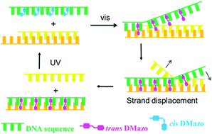
Tethered with bistable 2′,6′-dimethylazobenzene (DMazo) via a glycerol linker, an artificial 35 nt-long DNA has performed photoresponsive hybridization and reversible light-driven strand displacement.
Light as a clean, safe, renewable and nonpolluting energy, is an ideal external trigger to actuate DNA nanodevices. Asanuma et al. introduced azobenzene to DNA via a threoninol backbone (briefly as azo-DNA), and reported the photo-reversible duplex association and dissociation simply by switching the cis–trans configurations of azobenzene.13–15 They also applied this azo-DNA to photoresponsive DNA-tweezer and DNA-capsule successfully.16,17 However most works on light-driven association and dissociation of the threoninol-inserted azo-DNA duplex with its native complement were set on strands less than 20 bases in length, which might be the strand length limit for efficient association and dissociation. The threoninol-inserted azo-DNA longer than 20 nts would attenuate the hybridization match of the trans-azo-DNA to its native complement for duplex formation and decrease the trans-to-cis photoisomerization efficiency under UV light and thus lead to less dissociation of the artificial DNA duplex. We have recently reported a glycerol-inserted azo-DNA, which remarkably elevated the trans-to-cis photoisomerization efficiency in the hybrid duplex at room temperature.18 But the glycerol-inserted azo-DNA is less thermo-stable at excited cis-form, which limits the application where bistable states are needed in a dynamic system.
Since the half-life time of cis-2′,6′-dimethylazobenzene (DMazo) inserted in DNA via a threoninol backbone is about 8 times long as non-substituted azobenzene,19 we introduced DMazo into DNA via a glycerol linker (Fig. 1, labelled as DMazo–gDNA) to improve its cis-form thermo-stability. Measured with UV absorption changes at 340 nm, the half-life time of cis-form DMazo–gDNA lasted for 24 h at 60 °C (Fig. S2 in ESI†), 24 times long as our previously reported glycerol-inserted azo-DNA.18 The photoresponsive hybridization and light-driven DSD of DMazo–gDNA were further investigated by using a 35 nt-long DNA as shown in Scheme 1.
![]() | ||
Fig. 1 Structural comparison of azobenzene to 2′,6′-dimethylazobenzene-modified DNA tethered via glycerol linker (abbreviated as DMazo–gDNA). |
![]() | ||
Scheme 1 Schematic illustration of (a) photoresponsive hybridization and (b) light-driven DNA strand displacement of DMazo–gDNA. |
To quantify the degree of hybridization, the photoresponsive DNA and the complementary strand were tagged with DABCYL (4-(4-dimethylaminophenylazo)benzoic acid) (D16D in Scheme 1, representing the 35 nt DMazo–gDNA) and FAM ((6-fluorescein-6-carboxamido)-hexanoate) (Fbc in Scheme 1) respectively. The association and dissociation of the duplex were indicated with quenching and emission of the fluorescence of FAM as demonstrated in Scheme 1a, and the results were shown in Fig. 2 (more details see ESI†). The circled curve represents the association percentages under visible light irradiation, while the squared curve the association percentages under UV light irradiation, corresponding to different temperatures. The melting curves (Fig. S3 in ESI†) indicated that the association and dissociation of a duplex of D16D and its complement bc (D16D/bc) could be switched by light between 79.5 °C and 39 °C, the melting temperatures (Tm) of the same D16D/bc duplex at trans- and cis-forms respectively. Thus at 37 °C (below Tm of cis-form), UV light irradiation could only dissociate 11% of the total duplex. Obviously, below 50 °C, the dissociation percentage is less than 50%, which is lower than required for photo-switching (generally, a dissociation percentage more than 70% is required). At 60 °C, the visible light rapidly induced the duplex's formation and a Forster energy transfer occurred between FAM and DABCYL, which quenched 90% fluorescence; while the UV light dissociated approximately 87% of the total duplex quickly. At 70 °C, closing to the Tm of D16D/bc duplex at trans-form, both association and dissociation can be switched perfectly and quickly. The photoresponsive hybridization efficiencies (the difference of association percentages between visible and UV light irradiation) can be simply obtained as 7%, 47%, 77%, and 76% at 37 °C, 50 °C, 60 °C, and 70 °C separately by correspondent subtraction of squared point from circled point. To sum, the optimal bistable hybridization/dissociation states for Vis/UV switching can be executed efficiently in a temperature window of 50 to 70 °C.
D16D: GADAGDTGDACDATDGGDAGDACDGTDAGDGGDT ADTTDGADATDGADGGG-DABCYL-3′ (D represents DMazo in the sequence codes); Fbc: 5′-6-FAM-CCCTCATTCAATACCCTAC GTCTCCATGTCACTTC; cb29: ACATGGAGACGTAGGGTATT GAATGAGGG; cb26: TGGAGACGTAGGGTATTGAATGAGGG; cb23: AGACGTAGGGTATTGAATGAGGG; cb20: CGTAGGGTA TTGAATGAGGG (sequence bc is adopted from ref. 11).
Since the cis-form DMazo–gDNA duplex demonstrates much lower Tm than its correspondent native DNA duplex, it is thermodynamically feasible that under UV light native DNA is preferred to hybridize with the complementary strand rather than DMazo–gDNA. While under visible light DMazo–gDNA will displace the native strand, because the trans-form DMazo–gDNA duplex demonstrates higher Tm and thus stronger binding than its correspondent native DNA duplex (Fig. S3 in ESI†). Thus a photoresponsive DSD system including DMazo–gDNA and native strands with different lengths is designed as in Scheme 1b. Taking a native 20 nt-long DNA (cb20) as an example to illustrate the DSD: when D16D was added to the pre-hybridized Fbc/cb20 duplex, the fluorescence of the solution decreased gradually over tens of minutes to hours, indicating D16D replaced cb20 to hybridize with Fbc gradually. The slow fluorescence quenching in the DSD process was different from the rapid quenching in the case only two strands of Fbc and D16D were present. The DSD experiments of the Fbc/cb20/D16D system were executed at 4 different temperatures: 37, 50, 60, and 70 °C, and the results were shown in Fig. 3. The displacement degree can be assayed by the difference of Fbc/D16D association percentages at a certain temperature in the presence of cb20. From Fig. 3, we can simply calculate the displacement degrees at 37 °C, 50 °C, 60 °C, and 70 °C as 57%, 77%, 81%, and 86% respectively. From Fig. 2, we know that at lower temperatures UV irradiation is not efficient to switch azobenzene from trans- to cis-form and thus to dissociate D16D from the duplex. However, in the DSD system, with the help of its competitor strand of cb20, the dissociation becomes easier. It should be pointed out that, when photo-switched at a higher temperature than the Tm of Fbc/cb20 (65 °C in Fig. S3†), 70 °C as an example, the reversible fluorescence emission and quenching occurred immediately (Fig. S6 in ESI†), because of the complete dissociation of Fbc/cb20 and the complete hybridization of Fbc/D16D. Recycling experiments with UV/visible light switching were implemented without distinct efficiency decay which ensured the reversible light irradiation (Fig. S6 in ESI†).
A toehold exchange is necessary for any traditional DSD to initiate the strand displacement followed by a branch migration process subsequently. It was reported that different toehold length led to different displacement rate ranging over several magnitudes.11 We selected 4 native DNA strands with 20, 23, 26 and 29 bases (cb20–cb29 in Scheme 1) to expose toeholds of different lengths to accomplish the DSD process at 50 °C. As shown in Fig. 4, D16D displaced about 79% and 72% of the native strand of cb20 (squared curve) and cb23 (circled curve) in 10 min respectively. While increasing the competing native strand length to 26 nts (cb26), thus shortening the toehold, the DSD speed declined and the final displacement percentage decreased to 67% (triangled curve), 12% lower than in the cb20 displacement system. When the competing strand length increased to 29 nts (cb29), it had not reached an equilibrium even after 256 min, and D16D displaced only 37% of cb29 (the upset-triangled curve). The non-equilibrium phenomena for Fbc/cb29/D16D might be explained as that a short toehold less than 6-mer cannot stabilize the hybrid protruding duplex, and the inserted glycerol DMazo interrupts the branch migration process dynamically. Compared to the same native sequence DSD, DMazo–gDNA do need a longer toehold to initiate the light-driven DSD.
We further confirmed the light-driven DSD by non-denaturing polyacrylamide gel electrophoresis, with Fbc/cb26/D16D as an example in Fig. 5 (another example of Fbc/cb23/D16D is in Fig. S7†). Due to the inserted DMazo, D16D shows slower mobility, Lane 3 under visible light and Lane 4 under UV light, than the native Fbc at Lane 2. The cis-form D16D at Lane 4 shows a little bit slower mobility than the trans-form D16D at Lane 3. All of cb26, D16D and Fbc are present in Lane 5 under UV irradiation and Lane 6 under visible light irradiation respectively. In Lane 6, the strong band of Fbc/trans-D16D duplex demonstrates the minimum mobility, the displaced cb26 shows the maximum mobility (corresponding to Lane 1), while two residual bands in the middle are certified as Fbc/cb26 duplex and trans-D16D respectively (corresponding to Lane 7 and Lane 3), due to partial DSD. Under UV irradiation, two strong bands of cis-D16D (corresponding to Lane 4) and Fbc/cb26 (corresponding to Lane 7) are observed in Lane 5, while two weak bands are correspondent to the residues of Fbc/trans-D16D and cb26 respectively. According to the above results, light-driven DSD has been proven to be realized in our working system.
In conclusion, DMazo was introduced into the photoresponsive DNA by glycerol linker. The cis-form of DMazo has 20 times longer half life time than that of non-substituted azobenzene. The duplex formed by trans-DMazo–gDNA (trans-D16D) and the 35 nt-long native complementary strand (Fbc) demonstrates a higher melting temperature than that of the 35 nt-long native duplex. The optimal bistable hybridization/dissociation states of the duplex of D16D/Fbc can be simply switched efficiently by Vis/UV light irradiation in a temperature window from 60 to 70 °C. An application of the light-driven strand displacement was executed with D16D to replace its competitor short strands of cb20, cb23, cb26, for binding to Fbc, under visible light; and the reversible replacement can be done under UV light irradiation. During the displacement process, an intermediate, the metastable triple strand described in Scheme 1, will determine the branch migration rate, and thus the DSD dynamics. The light-driven reversible DNA strand displacement does not need a “fuel strand” further in the backward direction and thus no waste duplex strands are produced, which is a “reversible approach”. In contrast, the strand displacement can only step forward by addition of a longer “fuel strand” for the traditional native system, which is a “one-way forward approach”. Such light-driven reversible DNA strand displacement can be recycled many times with little efficiency-decay (see Fig. S6 in ESI†). However there are also drawbacks for the photoresponsive DSD system, where the displacement process is slower than with the same native strands, and a longer toehold is needed for initiation of DSD than with the same native strands. Nevertheless, this “green” light-driven DNA strand displacement system promises a bright prospect in DNA actuation and nanodevices.
Footnote |
† Electronic supplementary information (ESI) available: Experimental details, DNA melting curves, fluorescence spectra and additional PAGE gel electrophoresis. See DOI: 10.1039/c4ra15449e |
This journal is © The Royal Society of Chemistry 2015 |