Reece D. Gatelya,
Holly Warrena,
Mattia Scardamagliab,
Tony Romeoc,
Carla Bittencourtb and
Marc in het Panhuis*ad
aSoft Materials Group, School of Chemistry, University of Wollongong, Wollongong, NSW 2522, Australia. E-mail: panhuis@uow.edu.au
bChimie des Interactions Plasma Surface, CIRMAP, University of Mons, Place du Parc 20, 7000 Mons, Belgium
cElectron Microscope Centre, AIIM Facility, University of Wollongong, Wollongong, NSW 2522, Australia
dIntelligent Polymer Research Institute, ARC Centre of Excellence for Electromaterials Science, AIIM Facility, University of Wollongong, Wollongong, NSW 2522, Australia
First published on 10th February 2015
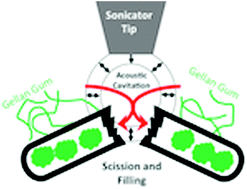
The preparation and characterization of carbon nanofibre–gellan gum composite materials is presented. Electron microscopy analysis reveals that nanofibres are affected by sonolysis, i.e. fibre length reduces, while filling occurs. Spectroscopic analysis suggests that the nanofibres are modified during the preparation of the dispersions. It is shown that despite these effects, composite materials prepared using a short period of sonolysis (4 min) exhibit robust conductivity, strain at failure and Young's modulus values of 35 ± 2 S cm−1, 20 ± 1% and 1.3 ± 0.3 MPa, respectively.
The improvement of the mechanical21,22 and electrical23,24 characteristics of materials by incorporation of conducting carbon fillers is an active area of research. However, it is well-known that carbon fillers can be difficult to (homogeneously) disperse in aqueous solutions due their hydrophobicity and van der Waals interactions.25 This disperse-ability issue has been successfully addressed by using dispersants (e.g. surfactants, polymers) in combination with sonolysis methods.26–28 However, one of the drawbacks of sonolysis is that it can lead to damage to the carbon filler29,30 and/or the dispersant.31 In general, this results in a detrimental effect on the properties of the composite material.29,32–35 For example, it has been shown that extensive sonolysis (21 hours) reduced the average CNT length from 3.5 μm to less than 0.5 μm.35 This reduction in length was coupled with a significant decrease in the conductivity of the resulting CNT network. Furthermore, the detrimental effect of sonolysis on the molecular mass of polymers is well-known.36–41
Here we investigate the effect of sonolysis on the properties of composite materials prepared by dispersing vapour grown carbon nanofibres (VGCNFs) with the biopolymer gellan gum. Gellan gum (GG, Scheme 1) is a linear, anionic, water soluble biopolymer which is derived from the bacteria Sphingomonas elodea (formerly Pseudomonas elodea or Auromonas elodea42). The repeating unit of the polymer is a tetrasaccharide, which consists of two residues of D-glucose and one of each residues of L-rhamnose and D-glucuronic acid. It is well-known for its applications in food technology ever since it was approved by the European Union and the United States Food and Drug Administration nearly two decades ago.43 More recently, it has been demonstrated that gellan gum is an efficient dispersant for conducting carbon fillers such as carbon nanotubes, graphene and VGCNFs.44–47
![]() | ||
Scheme 1 Schematic representation of the molecular repeat structure of low-acyl gellan gum. M+ indicates cationic counterions (e.g. Na+). |
VGCNFs are a conducting carbon filler material which were first manufactured in 1889 as a potential replacement for filaments in glow lamps.48 Their structure was first elucidated in 1952 using electron microscopy, which showed stacks of highly graphitised carbon forming a tubular shape.49 VGCNFs are produced by a catalytic thermal chemical vapour deposition technique with a floating catalyst.50 This method produces two characteristic structures, (i) ‘stacked cup’ (or ‘herringbone’) structure, which looks similar to a series of graphite cups without bases stacked on top of each other, and (ii) a ‘stacked deck’ (or ‘parallel’ structure) which is a series of multiple concentric tubes of graphitised carbon (similar to those observed for multi-walled CNTs), but at a slight (approx. 4°) angle.51 These structures are subsequently heat treated to remove (most of) the amorphous carbon outer layer and further improve their physical, mechanical, and electrical properties.52 It has been shown that conductivity and mechanical strength of the nanofibres is enhanced through heat treatment at 1500 °C.53
VGCNF composite materials have been produced using poly(caprolactone), poly(urethane), poly(ethylene) and epoxy resins.54–61 For example, recently it was demonstrated that shape memory properties of VGCNF–epoxy composite materials were enhanced by chemical functionalisation of VGCNFs.54 Other potential applications include the use of VGCNFs as constituents in electromagnetic interference shielding materials as discussed in a recent review article.62
In this paper, VGCNF–GG dispersions and composite (free-standing thin films) materials are prepared using sonolysis, vacuum filtration and evaporative casting. XPS/Raman spectroscopy, scanning electron microscopy, electrical and mechanical analysis was used to determine the effect of sonolysis on VGCNF graphitisation, average length, extent of opening, degree of filling, electrical conductivity, Young's modulus and ductility.
Homogeneous dispersions were prepared by adding 100 mg of VGCNFs (Pyrograf Products, PR24-LHT, Batch info: PS1345 Box 8, HT 170, diameter up to 200 nm) to 10 mL GG solution (3 mg mL−1) and applying horn sonolysis using a digital sonicator (Branson Digital Sonifier, power output 6 W, 0.5 s pulse, 0.5 s break between pulses). The microtip horn (Consonic, diameter 3.175 mm) was held 1 cm off the base of a 20 mL glass sample vial (diameter 25 mm).
Additional free-standing films were prepared by evaporative casting. Briefly, as-prepared dispersions were poured into a plastic petri-dish (diameter 55 mm) and allowed to dry under controlled conditions (21 °C, 50% RH) in a temperature–humidity chamber for up to 24 hours. The resulting films were then carefully removed from the substrate to produce free-standing films.
X-ray photoelectron spectroscopy (XPS) was carried out in UHV conditions (base pressure in the 10−9 mbar range) using a spectrometer (VERSAPROBE PHI 5000, Physical Electronics), equipped with a hemispherical electron analyzer and monochromatic Al Kα X-ray excitation source. The energy resolution was 0.7 eV. All binding energies were calibrated to the C1s peak at 284.6 eV. The XPS spectra were deconvoluted into different chemical surroundings using commercially available software (CASA-XPS).
Apart from removing the other types of carbon, sonolysis also resulted in opening the VGCNFs. For example, four VGCNFs can be identified in Fig. 1d, of which the ‘herringbone’ structure is not damaged, but at least one of the three ‘parallel’ structures is open ended. Furthermore, we made the interesting observation that two of these VGCNFs appear to be filled. The enlarged view in Fig. 1e clearly shows evidence of a filled VGCNF. Quantitative image analysis of TEM micrographs revealed that approximately 1/3 of the imaged VGCNFs appeared to be either completely or partially filled. However, analysis of dispersions prepared using longer sonication times (e.g. 30 min) revealed that most of the imaged fibres were either filled and/or opened (sheared). For example, the VGCNFs in Fig. 1g have been opened and are filled, whereas one of the fibres shown in Fig. 1h has not been opened (and is therefore not filled). In addition, other fibres (such as the one shown in Fig. 1i) revealed a fibre with the ‘parallel’ section filled and an undamaged (non-filled) ‘herringbone’ section. Examination of the TEM images shows that fibre damage and degree of filling increased with increasing sonication time.
Quantitative TEM analysis showed that the fraction of filled VGCNFs increased from 33% (after 2 min of sonolysis) to 85% after 30 min of sonolysis. This is further evidence that the filling effect is most likely due to the opening of the VGCNFs, i.e. whenever a fibre is damaged as a result of sonolysis, it is filled with the surrounding dispersant (gellan gum). We suggest that this is a result of capillary forces, as has been previously observed for carbon nanotubes.3,63
The effect of the wall thickness was also examined quantitatively. It was found that after 2 minutes of sonolysis the VGCNFs with thinner walls were more likely to be sheared and filled, whereas the VGCNFs with the thicker walls were less likely to have been sheared. Fig. 1e shows two fibres with different wall thicknesses; the top one is thicker and undamaged, whereas the bottom one is thinner and has been sheared and filled. This effect was personified after 30 min sonication; as previously mentioned, all VGCNFs that had been sheared were completely filled. It was found that all fibres with thin walls had been sheared, whereas fibres with thinner walls were less likely to be damaged. Fig. 1h shows a filled fibre next to an unfilled fibre, and it can be seen that the right hand fibre was unfilled and not sheared, but has a significantly thicker wall compared to the fibre on the left, which has been sheared and filled.
SEM analysis of free-standing films (Fig. 2) was used to assess the effect of sonication on the average length of the VGCNFs. After 2 min of sonication, it was found that the ends of the VGCNFs were reasonably circular in shape (Fig. 2c), whereas after 30 min sonication the ends appeared to be more deformed and more ellipsoidal in shape (Fig. 2d). At present it is not clear if the deformation is a direct result of sonolysis or an in-direct effect due to fracturing. Further research is necessary to confirm this.
The length of the fibres decreased with increasing sonication times. For example, for free-standing films prepared by evaporative casting, the average length decreased from 3.2 μm (2 min sonication) to 1.5 μm (30 min sonication), see Fig. 2e. This data exhibited a power-law (y = mx−b) dependence with b = 0.29 ± 0.01. Similar results were obtained for films prepared by the vacuum filtration process (data not shown, power law exponent b = 0.22 ± 0.01). This shortening through sonolysis is attributed to acoustic shearing as a result of inertial cavitation. The effect of this length reduction on the electrical characteristics is discussed in the next section.
Films prepared by evaporative casting were used since they retain all of the VGCNF and the gellan gum materials present in the dispersion. In contrast, it is well-known that during the vacuum filtration process (to produce Buckypapers) some proportion of both the dispersant and carbon fillers are removed.
The current–voltage (I–V) characteristics of all free-standing films (tested under controlled ambient conditions) exhibited ohmic behaviour, i.e. linear I–V characteristics. The total resistance (RT) of the films was calculated from the I–V characteristics and plotted against film length (L) (Fig. 3a). The conductivity was then evaluated by fitting the RT versus L data to:41,45
RT = L/(Acσ) + RC, | (1) |
![]() | ||
Fig. 3 Electrical characterisation of free-standing composite films prepared by evaporative casting. (a) Total resistance (21 °C, 50% RH) as a function of film length for films prepared by evaporative casting of dispersions sonicated for 4 (diamonds) and 30 (circles) minutes. (b) Electrical conductivity as a function of sonication time. (c) Conductivity as a function of average VGCNF length as determined by SEM analysis. Straight lines in (a), (b) and (c) are linear fits to eqn (1) and power law fit with exponent 0.8. |
This behaviour is linked to the sonication-induced reduction in VGCNF length and can be explained as follows (similar to arguments used for carbon nanotubes as detailed in ref. 35); the VGCNFs form a percolative network in which the resistance is determined by a combination of the resistances along each of the VGCNFs and the junctions between the VGCNFs. It is relatively straightforward to determine which of these the dominant effect is. A reduction in the length of the VGCNFs is coupled with an increase in the number of junctions. Therefore, if the junction resistance is the determining factor in the conductivity of the film then it has been shown that the conductivity should follow a power law dependence on the length of the fibres.35 In other words, conductivity decreases with increasing length of the fibres if junction resistance is dominant, while conductivity is independent of fibre length if junction resistance is negligible. Fig. 3c shows that the conductivity as a function of fibre length follows a power law with exponent 0.8. Hence, the junctions between the VGCNFs dominate the electrical behaviour of the films.
Tensile testing was performed on free-standing films prepared by evaporative casting to assess the effect of sonication on the mechanical characteristics (Fig. 4a). The Young's modulus decreased linearly with increasing sonication time from 1.3 ± 0.3 MPa (4 min sonication) to 0.21 ± 0.07 MPa (20 min sonication), see Fig. 4b. This indicated that the films became more ductile with increasing sonication time, i.e. the films failed at a higher strain but lower stress. Previous research32,41 has attributed this to a combined effect of damage to the polymer (shortening of the polymer chain length with increasing sonolysis) and the carbon filler (reduction in length as discussed above). The combined effect of this is a reduction in the Young's modulus and the tensile strength of the composite.
It is clear that the reduction in the mechanical and electrical properties must be taken into account when using horn sonolysis. Our results indicate that 4 min of horn sonolysis results in composite materials which exhibit robust conductivity (35 ± 2 S cm−1) and are mechanical sound. These values are comparable to conductivity values achieved for gellan gum composite materials with multi-walled carbon nanotubes (50 ± 5 S cm−1), but (as expected) lower then with single-walled carbon nanotubes (110 ± 15 S cm−1).46 Due to the smaller amount of sonication time composite films with VGCNF have better mechanical properties compared to films prepared with carbon nanotubes.
As Raman has a depth of analysis large enough to probe the entire VGCNF and gellan gum does not exhibit suitable characteristic bands it is unlikely that this spectroscopic technique provides information about the effect of filling. To better understand the effects of sonication on the interactions between VGCNF and gellan gum in the dispersions, samples were characterised using XPS analysis. Fig. 6 shows the typical XPS survey spectra recorded from unsonicated and sonication-treated VGCNFs (2 and 30 minutes, shown in figure). From the three spectra we can identify 2 main peaks which change their ratio: one at 284.4 eV, which is associated with photoelectrons emitted from carbon 1s core level and a second peak at about 533 eV generated by photoelectron emitted by O1s core level.
More information on the changes due to the sonication treatment can be understood from closer analysis of the C1s and O1s XPS spectra recorded on the different samples (Fig. 7a and b). The pristine C1s spectrum (grey dotted line at the bottom of Fig. 7a) centred at 284.4 eV has the typical asymmetric line shape of photoelectrons emitted from carbon atoms participating in sp2 bonds. This asymmetry is associated with the many-electron response to the sudden creation of a photohole.65
This spectrum also shows a second component at 290.6 eV associated with the electron energy loss due to π-plasmon excitations. The dispersion of the VGCNFs in gellan gum clearly results in a growing shoulder at the high binding energy side of the sp2 peak. Fig. 7c shows an example of the results of the curve fittings performed to explain the spectra of the sonicated samples. The modifications produced by the sonication treatment can be identified by a broad structure that peaks at 288 eV. This structure was previously attributed to photoelectrons emitted from carbon atoms belonging to carbon functional groups singly and/or doubly bound to one or two oxygen atoms.66
To reproduce the C1s peak recorded after 8 min of sonication (Fig. 7c), 6 components were used. Only three components (1, 2 and 6, Fig. 7c) are required for fitting the spectrum of pristine (unsonicated) VGCNF: in addition to the asymmetric sp2 peak at 284.4 eV, two Gaussian functions were used to reproduce the other features observed in the pristine spectrum. One Gaussian at 285.0 eV is associated with photoelectrons emitted from carbon atoms at sp3 bonds in amorphous carbon. During VGCNF synthesis, competing pathways can lead to amorphous carbon formation rather than to crystalline graphitic nanofibres (as discussed in electron microscopy section, see also Fig. 1b and c). The other Gaussian, centred at 290.6 eV, corresponds to the π-plasmon excitations. The three other Gaussian components (3 to 5, Fig. 7c) are related to oxygen containing functional groups present in gellan gum, and centered at 286.1 eV (hydroxilic), 287.2 eV (carbonylic) and 288.6 eV (carboxylic). It is clear from Fig. 7a that the broad structure at 288 eV decreases with increasing sonication. In addition, the O1s peak (previously observed for modified gellan gum67) is also decreasing with increasing sonication time (Fig. 7b). In other words, the amount of oxygen containing groups is decreasing near the surface of the VGCNFs as shown in Fig. 7d. The relative amount of oxygen in the unsonicated sample was evaluated to be 2%.
XPS is predominantly a surface technique (about 8 nm depth of analysis). Combined with the diameter of the VGCNF this would suggest that XPS can only provide data about the surface of the VGCNF and not the interior. Hence, it is suggested that the observed decrease in the O1s spectra (Fig. 7b) and the oxygen containing groups in the C1s spectra (Fig. 7d) can be explained by the reduction of gellan gum on, or near the surface of VGCNF due to filling of the VGCNF with gellan gum. Electron microscopy results (Fig. 1) appear to be in support of this suggestion, with the fibres becoming filled to a larger extent with increasing sonication time.
The effect of sonolysis on vapour grown carbon nanofibers in gellan gum composite materials has been investigated. Finding the minimum time for sonication is a typical first step when producing dispersions containing carbon nanostructures, however rarely is the effect of sonolysis on the properties of the constituents and the resulting materials studied in detail.
It was found that the average length of the VGCNFs was halved with just 30 minutes of low energy sonication. Electron microscopy analysis revealed that the VGCNFs were opened, shortened and filled. Spectroscopy analysis revealed that sonication treatment resulted in modification of the VGCNFs.
Our investigations revealed that the electrical characteristics reduced (from 35 ± 2 S cm−1 to 25 ± 2 S cm−1) due to sonication-induced reduction in length of the carbon nanofibers. In contrast, it is likely that the reduction in mechanical characteristics is mostly due to the effect of sonolysis on polymer chain length.
This paper demonstrates that despite the sonication-induced opening, filling and modification of carbon nanofibers composite materials that are mechanical robust and electrically conducting can be easily prepared by limiting the amount of horn sonolysis (to just 4 min).
Methods for the filling of carbon nanotubes are well established, there has been limited research regarding the filling of carbon nanofibers.62,68 One of the remaining challenges is to prepare filled tubular carbon nanostructures with properties suitable for selective drug delivery (e.g. nanosized needles) and for autonomic healing of polymeric materials (e.g. load-bearing biomedical materials). It is suggested that filled VGCNFs offer great opportunities for addressing these challenges.
This journal is © The Royal Society of Chemistry 2015 |