DOI:
10.1039/C4RA14984J
(Paper)
RSC Adv., 2015,
5, 30303-30309
In situ crosslinkable hydrogels formed from modified starch and O-carboxymethyl chitosan
Received
21st November 2014
, Accepted 6th March 2015
First published on 9th March 2015
Abstract
An in situ hydrogel based on oxidation cholesterol starch (OCS) and O-carboxymethyl chitosan (CMCT) that is completely devoid of potentially cytotoxic small molecule cross-linkers and does not require complex manoeuvres or catalysis has been formulated and characterized. The network structure was created by Schiff base formation. The mechanical properties, internal morphology and swelling ability of the injectable hydrogel were examined. Rheological measurements demonstrated that increasing the concentration of the monomer improved the storage modulus. SEM showed that the hydrogel possessed a well-defined porous structure. In addition, the Schiff base reaction was acid sensitive. Under acid conditions, the hydrogel could hydrolyse quickly compared with high pH conditions. Doxorubicin (DOX) was used as a model drug to investigate the control and release properties of the hydrogel. The cytotoxic potential of the hydrogel was determined using an in vitro viability assay with L929 cells as a model and the results revealed that the hydrogel was non-cytotoxic.
1. Introduction
As a group, cancers account for approximately 13% of all deaths each year and have attracted more and more attention.1 The death rates have been increasing primarily due to an aging population and lifestyle changes in the developing world.2 The common pharmaceuticals for curing tumors are doxorubicin,3–5 dactinomycin6–8 and daunorubicin.9–11 However, due to the toxicity and side effects of these drugs for normal tissues and cells,12–15 development of a suitable transport method to transmit them to the right place is necessary. Many kinds of drug-carriers have arisen to fit this requirement.16–18 The typical carriers for anti-tumours drug are nanoparticles,19,20 nanogels,21,22 micelles23,24 and hydrogels.25,26 Although these transporters display good performance, the low loading capacity of the nano-based particles and their loss during biological cycle processes limits their application. Meanwhile, the three-dimensional structure of the hydrogels means that surgery is necessary to use them in people with solid tumours. The appearance of in situ hydrogels opens a new way to solve these problems.27
Injectable hydrogels are based on the idea that a certain biomaterial can be injected as a liquid, and then form a solid gel in situ.28 Injectable hydrogels are of particular interest because drugs, proteins, and cells can be easily incorporated into polymer solutions prior to administration and the hydrogels form in situ for wounded tissues.29 The drugs, proteins or cells can easily diffuse around the tumor. Therefore, injectable hydrogels have vast application prospects in drug/cell delivery and tissue engineering.30 In consideration of the biomedical application of injectable hydrogels, natural polymers are the favoured material to construct such a hydrogel compared with synthetic polymers due to their biocompatibility and biodegradability. Those polymers include starch, chitosan, hyaluronic acid, cellulose, xanthan gum, gelatin, and so on. Among them, starch, which is an entirely biodegradable biopolymer and is the cheapest, has been used to fabricate hydrogel materials for biomedical applications.31 Chitosan is another biopolymer, which is a linear polysaccharide composed of randomly distributed β-(1–4)-linked D-glucosamine (deacetylated unit) and N-acetyl-D-glucosamine (acetylated unit). The amino group in chitosan has a pKa value of ∼6.5, which leads to protonation in acidic to neutral solution with a charge density dependent on pH and the degree of deacetylation. It has been widely utilized in gene therapy,32 cell regulation,33 and tissue regeneration.34
Generally, in situ hydrogels can be synthesized via physical or chemical approaches. Compared to physical methods, the chemical approach may produce hydrogels with stable covalent bonds. The typical in situ chemical approach includes Schiff base reactions,35 Michael addition reactions,36 ionic interactions37 or photo-crosslinking reactions.38 Among those approaches, Schiff bases are imines formed by the condensation of aldehydes or ketones with primary amines. They are degradable via hydrolysis, and the stability of these bonds decreases as the pH decreases.39 The reversible, dynamic nature of the Schiff base means that it is utilized in preparing a wide range of in situ hydrogels with integrated properties, including pH-sensitivity,40 self-healing,41 biocompatibility and degradability.42
Recently, we have reported a novel kind of injectable hydrogel which was synthesized using core–shell starch nanoparticles with aldehyde groups and poly(vinyl amine) (PVAM) with primary amines via a Schiff base reaction. The starch nanoparticles were prepared with the addition of cholesterol groups and aldehyde groups, which assemble to form core–shell structures.43 Compared to linear starch with aldehyde groups, the introduction of the hydrophobic cholesterol group offered the hydrogel enhanced mechanical properties. However, the high-cost and synthetic nature of PVAM may be a drawback of such hydrogels for application in biomedical fields. Herein, a new injectable hydrogel composed of O-carboxymethyl chitosan and core–shell starch nanoparticles was synthesized via the Schiff base reaction. The mechanical and swelling properties of the hydrogel were investigated in detail. Moreover, doxorubicin hydrochloride, as a model drug, was used to evaluate the controlled-release properties of the hydrogel. In addition, the 3-(4,5)-dimethylthiahiazo(-z-y1)-3,5-di-phenytetrazoliumromide (MTT) assay was also conducted to investigate the cytotoxic activity against the L929 cell line. The combination of in situ gelation, feasible mechanical properties, good water uptake ability, well-controlled release behavior and excellent biocompatibility make the polysaccharide-based hydrogel applicable in various biomedical fields.
2. Experimental
2.1. Materials
Cholesterol-modified oxidation starch (OCS) was obtained according to a previously published method43 (the degree of cholesterol substitution was 0.01; the degree of oxidation was 46%). Chitosan (with a degree of deacetylation of 93%) and chloroacetic acid (98%) were purchased from Aladdin Chemistry Co, Ltd. Doxorubicin hydrochloride (DOX) was purchased from Beijing Zhongshuo Pharmaceutical Technology Development Co, Ltd. All other chemicals were obtained from Beijing Chemical Factory and used as received.
2.2. Synthesis of O-carboxymethyl chitosan (CMCT)
CMCT was synthesized as per a procedure described in the literature.44 Chitosan powder (10 g) was suspended in 100 mL of isopropyl alcohol and the resulting slurry was stirred in a 500 mL flask at room temperature. 25 mL of 10 N aqueous NaOH solution, divided into five equal portions, was then added to the stirred slurry over a period of 25 min. The alkaline slurry was stirred for an additional 30 min. Subsequently, chloroacetic acid (60 g) was added in five equal portions, at 1 min intervals. Heat was then applied to bring the reaction mixture to a temperature of 60 °C and stirring at this temperature was continued for 3 h. Afterward, the reaction mixture was filtered and the filtered solid product (CMCT) was thoroughly rinsed with methanol. The resultant CMCT was dried in an oven at 45 °C.
The degree of substitution of carboxymethyl groups on 6-O- and 3-O- could be determined from the ratio of peak areas of the carboxymethyl group (O–CH2COOD, 4.40–4.05 ppm) and the –CH3 group (CH3CO–, 2.0 ppm) in the 1H-NMR spectrum in Fig. 1, and it was found to be 0.60.
 |
| Fig. 1 The 1H-NMR spectrum of CMCT in D2O. | |
2.3. Preparation of the hydrogel
CMCT (50 mg) and OCS (50 mg) were separately dissolved in water (1 mL) to form respective 5% (w/w) solutions (for the CMCT solution, it was necessary to limit the concentration to 5%; if >5%, the viscosity of the solution was high and it was not easy to inject by syringe) which were stored at 5 °C. The hydrogel was prepared by mixing the CMCT and OCS solutions in an equal volume ratio using a syringe with a “T” structure. Finally, the mixture was kept at 37 °C for complete gelation.
2.4. Determination of gelation time
The gelation time of the hydrogel when varying the concentration of OCS were estimated using a vial tilting method. The concentration of CMCT was constant at 5% (w/w), and the concentrations of OCS were 2.5% (w/w), 5.0% (w/w), 7.5% (w/w) and 10% (w/w). All the solutions were sealed with parafilm and kept at 37 °C for 1 h before testing. The testing was started immediately after mixing the two components, and continued until no flow was observed for at least 30 s when a vial containing the hydrogel was inverted at 37 °C.45
2.5. Characterization
1H-NMR spectra were recorded using a Bruker AV400 spectrometer (Germany) operating at room temperature, with the sample dissolved in D2O. FTIR characterization was performed on a Perkin-Elmer Spectrum 100 spectrometer (USA), and the data were collected with 32 scans at a resolution of 2 cm−1 at room temperature. The lyophilized hydrogel samples were characterized by scanning electron microscopy (SEM) with an accelerating voltage of 5.0 kv using a model XL 30 ESEM (Philips).
2.6. Rheological measurements
To investigate the mechanical properties of the resultant hydrogels, dynamic frequency sweep tests were conducted on a US 302 rheometer (Anton Paar) in the linear viscoelastic region. The frequency applied to the hydrogel sample was increased from 0.1 to 100 rad s−1. The data were collected under a controlled strain γ of 1%. G′ is an elastic component of the complex modulus for measurement of the gel-like behavior of a system, whereas G′′ is a viscous component of the complex modulus and is a measure of the sol-like behavior of the system.
2.7. Swelling experiments
The hydrogel samples were immersed into various PBS solutions and distilled water at 37 °C. The degree of swelling (SW) was determined using the following equation:
where M0 represents the weight of the dry hydrogel and Mt represents the weight of the hydrogel at equilibration time. All the experiments were carried out with three samples and the reported data are average values.
2.8. Release of DOX from the hydrogel
For the DOX release experiments, DOX (2.0, 1.0 or 0.5 mg mL−1) was dissolved in an OCS solution in phosphate-buffered saline (PBS, pH = 7.4, 5.0 or 3.0) and then reacted with CMCT to form DOX-loaded hydrogel. Freeze-dried hydrogel samples were immersed into PBS solution, sealed in a dialysis bag (Mw cutoff: 3.5 kDa) and incubated in the release medium (25 mL) at 37 °C under oscillation at 45 rpm. At selected time intervals, the buffer solution outside the dialysis bag was removed for UV-Vis analysis and then replaced with fresh buffer solution. The released amount of DOX was determined from the absorbance at 480 nm with the help of a calibration curve of DOX in the same buffer. Then, the accumulative weight and relative percentage of the released DOX were calculated as a function of incubation time.
2.9. Cell line
L929 (mouse fibroblast cells) was chosen for cell tests. L929 cells were supplied by the Medical Department of Jilin University, China. L929 cells were cultured in Dulbecco’s modified Eagle’s medium (DMEM, GIBCO) supplemented with 10% heat-inactivated fetal bovine serum (FBS, GIBCO), 2 mM L-glutamine, 100 U mL−1 penicillin and 100 μg mL−1 streptomycin (Sigma), and the culture medium was replaced once very day.
2.10. In vitro cytotoxicity of the supramolecular hydrogel
L929 cells were used to evaluate the biocompatibility of the hydrogel by MTT assay. The cells were seeded in a 96-well plate at a density of 10
000 cells per well in 100 μL of DMEM. Then, CMCT, OCS and CMCT/OCS solutions were added to the wells with mixing. Three parallel wells for each sample were used at a specific concentration. After co-incubation with L929 cells for 24 h, 48 h and 72 h, 20 μL of MTT solution in PBS (5 mg mL−1) was added to each well and the plate was incubated for another 4 h at 37 °C. After that, the medium containing MTT was removed and 150 μL of DMSO was added to each well to dissolve the MTT-derived formazan crystals. Finally, the plates were shaken for 10 min, and the absorbance of the formazan product was measured at 492 nm using a microplate reader.
3. Results
3.1. Synthesis of CMCT/OCS hydrogel
In our process, carboxymethyl groups were introduced into the backbone of the chitosan to improve the hydrophilicity under physiological conditions. The O-carboxymethyl chitosan (CMCT) was synthesized successfully through the etherification reaction between chitosan and chloroacetic acid. The degree of substitution of carboxymethyl groups was 0.60, as determined from the 1H-NMR spectrum.
Meanwhile, starch was successively decorated with cholesterol groups and aldehyde groups, as described previously. This modification sustains a certain hydrophilic–hydrophobic balance for the starch macromolecules so that nanoparticles with a well-defined core–shell structure were obtained, as shown in Fig. 2. The core–shell structure offered the starch hydrogel enhanced mechanical properties and water absorbability. The degree of substitution of cholesterol groups (DS, i.e. the number of cholesterol groups per glucopyranose unit) used in this study was 0.01. The degree of oxidation of the starch was 46%.
 |
| Fig. 2 The schematic representation of the hydrogel formation process. | |
After injecting the CMCT and OCS solutions out of the syringe, the hydrogel could be formed via in situ cross-linking by Schiff base reactions within minutes (Table 1). The ability of the Schiff base reaction to respond to pH gave the hydrogel reversibility and degradability, which make it a suitable material for utilization in the field of control and release.46
Table 1 The characteristics of hydrogels formed with different weight ratios between CMCT solutions and OCS solutions at 37 °C. The gel represents hydrogel
Samples |
CMCT/OCS (w%/w%) |
tgels (s) |
Products |
1 |
5.0/2.5 |
500 |
Gel |
2 |
5.0/5.0 |
210 |
Gel |
3 |
5.0/7.5 |
90 |
Gel |
4 |
5.0/10 |
30 |
Gel |
The structure of the hydrogel was confirmed by FTIR characterization. As shown in Fig. 3, the peak of the aldehyde groups at 1730 cm−1 disappeared after the reaction. At the same time, the intensity of the hemiacetal peak at 1100 cm−1 decreased and became a shoulder after the reaction. This demonstrated that the consumption of the aldehydes was most likely by a reaction with the amines. However, the imine peak (1665 cm−1) wasn’t detectable and might have been masked by the multiple absorbance peaks at this wave-number.47 Indeed, the –NH2 groups of CMCT along with water adsorbed on the polysaccharide in this area show broad and strong peaks at this wave-number.
 |
| Fig. 3 The FTIR of the samples of OCS, CMCT and hydrogel. | |
3.2. Rheological analysis
For the in situ formed hydrogel, rheological analysis was a powerful and accurate method to investigate viscoelastic properties.48 Frequency sweep experiments were performed 24 h after the gel was formed with the protection of parafilm at 37 °C. As shown in Fig. 4A, with the concentration of the OCS solution increasing, the storage modulus (G′) of the hydrogel increased. When the concentration of OCS reached 7.5% (w/w), the modulus achieved its maximum. After that, there was no significant change in the storage modulus if the content of OCS increased from 7.5% (w/w) to 10% (w/w). It is worthy of note that, as calculated in the experimental part, the degree of oxidation of the OCS was about 46% and the degree of substitution of carboxymethyl groups in the CMCT was 0.6. Therefore, the aldehyde groups and amine groups at this mass ratio didn’t show an equal molar ratio. However, the data demonstrated that the hydrogel at this mass ratio exhibited the biggest storage modulus. This phenomenon could be explained as follows: there exists a balance between the aldehyde groups and the hemiacetal groups, and when the Schiff base reaction was occurring, not all of the hemiacetal groups were reacted with amine groups, while the aldehyde groups were all consumed, as has been confirmed from the FTIR spectra in Fig. 3.
 |
| Fig. 4 (A) The modulus of the CMCT/OCS based hydrogel. The concentration of the CMCT solution before mixing was 5% (w/w) in all the samples. The concentration of the OCS solution was varied. (B) The morphology of the hydrogel at 5/5% (w/w) before swelling and (C) after swelling. | |
Next, we take samples number 2 to analyse the relation between rheology performance and its network structure with consideration of the anti-infection property of the chitosan. For sample 2 in Fig. 4A, a typical hydrogel character was exhibited. Both the storage modulus and the loss modulus increased slightly with increasing frequency. This means that the hydrogel forms a well-defined pore structure.49 In order to confirm this hypothesis, SEM was adopted to observe the morphology of the dried hydrogel. As shown in Fig. 4B, the dried hydrogel shows a regular pore dispersion with a 20 μm pore diameter. In order to understand well the inter-structure of the hydrogel and eliminate the interference from residual monomers, the morphology of the hydrogel after swelling in distilled water was also observed. As shown in Fig. 4C, the pore diameter became large and the pore dispersion was still well-defined. This demonstrated that a well-defined cross-linking network was formed in the hydrogel.50
3.3. Swelling analysis
The swelling behavior of a hydrogel plays an important role in its practical applications. Fig. 5 presents the swelling properties of the CMCT/OCS hydrogel in distilled water and various PBS solutions. It was 78 times in the distill water and with increasing pH from 3.0 to 7.4, the degree of swelling increased from 8.5 to 23 accordingly. In this process, the osmotic pressure played an important role in affecting the degree of swelling of the hydrogel. The volume of the hydrogel would be changed if a concentration gradient exists, and the bigger the concentration gradient, the more obvious the volume change. Therefore, the hydrogel in water exhibited the highest degree of swelling compared with those in PBS solutions. Moreover, due to the electrostatic interactions among the chains, the pH value exhibited a significant effect in regulating the change of the volume of the hydrogel.43 At a low pH value, the gel, which contains a number of unreacted amino groups, shrank in the presence of a large number of cations along with displaying declining water capacity. When the pH value increased, the gel swelled due to the reduction of the cations in the solution. It is also worthy of note that the hydrolysis of the imine groups in the hydrogel might affect the degree of swelling in an acid environment, leading to a decline in the degree of swelling.
 |
| Fig. 5 Water absorption capacity of the hydrogel (sample 2) in various PBS solutions and distilled water. | |
3.4. In vitro release of DOX
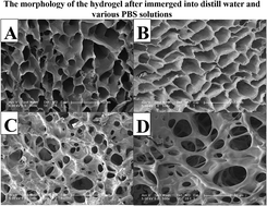
Hydrogels, which are very similar to biological tissue, are a useful tool for transmitting a target drug to the right place.51 Here, DOX was used as a model drug to investigate the control and release properties of the hydrogel. As shown in Fig. 6 and its inset, there was no dramatic initial burst release for the drug-loaded hydrogel under different pH conditions, and the hydrogel presented pH-sensitive release profiles. The lower the pH value, the faster the drug was released. The hydrogel showed sustained release of DOX at pH = 7.4, and no more than 30% of the DOX was released in 15 days. In the same time period, about 70% of the DOX was released at pH = 5.0 and pH = 3.0. We attributed this to the hydrolysis of the Schiff bases.52 The imine group is fragile under acid conditions and the stability of these bonds decreases as the pH decreases. In order to confirm this hypothesis, SEM was used as a powerful tool to investigate the morphology of the hydrogel during the hydrolysis process. As seen in Fig. 7, the hydrogel had a regular multi-pore structure before exposure to various PBS solutions. When the hydrogel was immersed into the PBS solution with pH = 7.4 for 24 h, the pore diameter and dispersion did not exhibit any obvious change and the pores were separated from each other by walls. However, when the hydrogel was immersed into PBS with pH = 5.0, there appeared vast differences in the morphology compared with the morphology of the hydrogel immersed into PBS at pH 7.4 and distilled water. The pores became irregular and the separating walls were eroded. Moreover, the erosion was more obvious for the hydrogel immersed into pH = 3.0. This further demonstrated that the imine groups were more easily hydrolysed under acid conditions. Therefore, the control and release behaviours could be explained as follows: DOX was restricted in the passageway of the gel and diffused to the outside in a one-dimensional direction in PBS at pH 7.4. With decreasing pH, the imine groups were hydrolysed and the networks were eroded, so the barriers of the pores were disappearing, resulting in the fast release of DOX in the acid environment.
 |
| Fig. 6 The release behavior of the hydrogel (sample 2) with different buffers. Inset: DOX release during the first 24 h. | |
 |
| Fig. 7 The morphology of the hydrogel after immersion into various solutions for 24 h. (A) Distilled water; (B) PBS with pH = 7.4; (C) PBS with pH = 5.0 and (D) PBS with pH = 3.0. | |
Moreover, various doses of DOX were also studied for the control and release. Although the doses of DOX were different, the behaviour of release for the DOX was similar in various PBS solutions. This demonstrates that the control and release behaviors of DOX in the hydrogel were independent of the concentration of DOX. The uniform dispersion of the pores and the hydrolysis of the imine groups in solution might play a key role during this process. Therefore, the drug-loaded hydrogel has potential for biomedical applications, since tumor tissues are known to be acidic.
3.5. Cell cytotoxicity
The cytotoxicity potential, as reflected by the viability of cells encapsulated in the hydrogel, was evaluated by MTT assay. Cells were grown in the presence of 200 μL cylindrical CMCT/OCS gels for up to 3 days, with medium changes every 24 hours. Cell viability was examined at 1, 2 and 3 days and some of the results are depicted in Fig. 8A. No significant difference was found between the cells incubated with the hydrogel and the control, suggesting that the hydrogel did not have any adverse effect on cell growth; this could be inferred as material non-cytotoxicity.
 |
| Fig. 8 (A) The cell viability of the L929 cells inside of the hydrogel after 3 days, where the concentrations of CMC and OCS were 0.5 mg mL−1. (B) The cell viability of L929 cells after 3 days with varying concentrations of the polymer. (C) The cell morphologies in saline after 72 h for the control. (D) The cell morphologies after 72 h in the hydrogel, where the concentrations of CMC and OCS were 1 mg mL−1. | |
In order to further confirm the non-cytotoxicity of the hydrogels, we take the concentration as a factor to evaluate the cell viability. Fig. 8B shows that all the CMCT and OCS solutions and hydrogels induced only minimum decreases in relative cell viability over the concentration range from 0.0313 to 1 mg mL−1 after 3 days of cell exposure. The relative viability of the L929 cells was 0.85 ± 0.05 for 1 mg mL−1 CMCT solution, 0.86 ± 0.04 for 1 mg mL−1 OCS solution and 0.92 ± 0.05 for the representative hydrogel, indicating that none of the hydrogels exhibited significant cytotoxicity. Moreover, we also observed the cell morphologies with an optical microscope when the concentrations of OCS and CMCT were each 1 mg mL−1, as shown in Fig. 8C and D. If we increased the OCS concentration from 1 mg mL−1 to 2 mg mL−1, the relative viability of the L929 cells decreased due to the toxicity of the aldehyde groups.
All in all, the hydrogel may have potential for biomedical applications, although more thorough testing with human cell lines, as well as in vivo testing, would be required to confirm this potential.
4. Conclusions
We demonstrated a method to synthesize an in situ hydrogel based on natural polysaccharide derivatives. The starch-based nanoparticles and modified chitosan cross-linked in situ to form a hydrogel via Schiff base reactions. The dried hydrogel has a well dispersed pore structure and the swelling ratio in PBS solutions increased with increasing pH. Moreover, we used DOX as a model drug to simulate the controlled-release properties of the hydrogel. The results showed that the DOX-loaded hydrogel prefers to release DOX in an acid environment, and the curve of the release wasn’t dependent on the loading amount of the drug. Further, the cytotoxic potential of the hydrogel was determined by an in vitro viability assay using L929 cells as a model, and the results reveal that the hydrogel is non-cytotoxic. The hydrogel could be a promising drug carrier for biomedical applications.
Acknowledgements
Financial support from the National Natural Science Foundation of China (grant nos 51321062 and 51103150) is gratefully acknowledged.
Notes and references
- G. L. Davis, J. Dempster, J. D. Meler, D. W. Orr, M. W. Walberg, B. Brown, B. D. Berger, J. K. O’Connor and R. M. Goldstein, Proceedings-Baylor University Medical Center, 2008, 21, 266 Search PubMed.
- A. Jemal, F. Bray, M. M. Center, J. Ferlay, E. Ward and D. Forman, Ca-Cancer J. Clin., 2011, 61, 69–90 CrossRef PubMed.
- W. Arap, R. Pasqualini and E. Ruoslahti, Science, 1998, 279, 377–380 CrossRef CAS.
- F. Greco, M. J. Vicent, S. Gee, A. T. Jones, J. Gee, R. I. Nicholson and R. Duncan, J. Controlled Release, 2007, 117, 28–39 CrossRef CAS PubMed.
- X. Yang, J. J. Grailer, I. J. Rowland, A. Javadi, S. A. Hurley, D. A. Steeber and S. Gong, Biomaterials, 2010, 31, 9065–9073 CrossRef CAS PubMed.
- R. E. Slayton, R. C. Park, S. G. Silverberg, H. Shingleton, W. T. Creasman and J. A. Blessing, Cancer, 1985, 56, 243–248 CrossRef CAS.
- B. Langholz, J. M. Skolnik, J. S. Barrett, J. Renbarger, N. L. Seibel, A. Zajicek and C. A. Arndt, Pediatr. Blood Cancer, 2011, 57, 252–257 Search PubMed.
- C. R. Hill, M. Cole, J. Errington, G. Malik, A. V. Boddy and G. J. Veal, Clin. Pharmacokinet., 2014, 53, 741–751 CrossRef CAS PubMed.
- C.-Y. Wang, M. W. Mayo and A. S. Baldwin, Science, 1996, 274, 784–787 CrossRef CAS.
- S. M. Taghdisi, P. Lavaee, M. Ramezani and K. Abnous, Eur. J. Pharm. Biopharm., 2011, 77, 200–206 CrossRef CAS PubMed.
- L. Zhang, H.-J. Yao, Y. Yu, Y. Zhang, R.-J. Li, R.-J. Ju, X.-X. Wang, M.-G. Sun, J.-F. Shi and W.-L. Lu, Biomaterials, 2012, 33, 565–582 CrossRef CAS PubMed.
- D. E. Brenner, P. H. Wiernik, M. Wesley and N. R. Bachur, Cancer, 1984, 53, 1042–1048 CrossRef CAS.
- D. Galaris, A. Georgellis and J. Rydström, Biochem. Pharmacol., 1985, 34, 989–995 CrossRef CAS.
- D. M. Green, J. Z. Finklestein, P. Norkool and G. J. D’Angio, Cancer, 1988, 62, 270–273 CrossRef CAS.
- B. N. Zordoky, A. Anwar-Mohamed, M. E. Aboutabl and A. O. El-Kadi, Drug Metab. Dispos., 2011, 39, 1440–1450 CrossRef CAS PubMed.
- F. Levi-Schaffer, A. Bernstein, A. Meshorer and R. Arnon, Cancer Treat. Rep., 1982, 66, 107–114 CAS.
- T. Kimura, I. Fujita, N. Itoh, N. Muto, T. Nakanishi, K. Takahashi, J. Azuma and K. Tanaka, J. Pharmacol. Exp. Ther., 2000, 292, 299–302 CAS.
- L. Eiriksson, T. Wells, H. Steed, A. Schepansky, V. Capstick, P. Hoskins, J. Pike and K. Swenerton, Gynecol. Oncol., 2012, 124, 553–557 CrossRef CAS PubMed.
- I. Brigger, C. Dubernet and P. Couvreur, Adv. Drug Delivery Rev., 2002, 54, 631–651 CrossRef CAS.
- L. Brannon-Peppas and J. O. Blanchette, Adv. Drug Delivery Rev., 2012, 64, 206–212 CrossRef PubMed.
- M. Oishi and Y. Nagasaki, Nanomedicine, 2010, 5, 451–468 CrossRef CAS PubMed.
- N. V. Nukolova, H. S. Oberoi, S. M. Cohen, A. V. Kabanov and T. K. Bronich, Biomaterials, 2011, 32, 5417–5426 CrossRef CAS PubMed.
- N. Nasongkla, E. Bey, J. Ren, H. Ai, C. Khemtong, J. S. Guthi, S.-F. Chin, A. D. Sherry, D. A. Boothman and J. Gao, Nano Lett., 2006, 6, 2427–2430 CrossRef CAS PubMed.
- N. Rapoport, Prog. Polym. Sci., 2007, 32, 962–990 CrossRef CAS PubMed.
- H. D. Han, C. K. Song, Y. S. Park, K. H. Noh, J. H. Kim, T. Hwang, T. W. Kim and B. C. Shin, Int. J. Pharm., 2008, 350, 27–34 CrossRef CAS PubMed.
- C. Chun, S. M. Lee, C. W. Kim, K.-Y. Hong, S. Y. Kim, H. K. Yang and S.-C. Song, Biomaterials, 2009, 30, 4752–4762 CrossRef CAS PubMed.
- H. T. Ta, C. R. Dass and D. E. Dunstan, J. Controlled Release, 2008, 126, 205–216 CrossRef CAS PubMed.
- M. Kurisawa, J. E. Chung, Y. Y. Yang, S. J. Gao and H. Uyama, Chem. Commun., 2005, 4312–4314 RSC.
- J. Li, X. Ni and K. W. Leong, J. Biomed. Mater. Res., Part A, 2003, 65, 196–202 CrossRef PubMed.
- B. Jeong, Y. H. Bae and S. W. Kim, J. Controlled Release, 2000, 63, 155–163 CrossRef CAS.
- C. Elvira, J. F. Mano, J. San Roman and R. L. Reis, Biomaterials, 2002, 23, 1955–1966 CrossRef CAS.
- H. T. Ta, C. R. Dass, I. Larson, P. F. Choong and D. E. Dunstan, Biomaterials, 2009, 30, 4815–4823 CrossRef CAS PubMed.
- S. M. Richardson, N. Hughes, J. A. Hunt, A. J. Freemont and J. A. Hoyland, Biomaterials, 2008, 29, 85–93 CrossRef CAS PubMed.
- N. Boucard, C. Viton, D. Agay, E. Mari, T. Roger, Y. Chancerelle and A. Domard, Biomaterials, 2007, 28, 3478–3488 CrossRef CAS PubMed.
- H. Tan, C. R. Chu, K. A. Payne and K. G. Marra, Biomaterials, 2009, 30, 2499–2506 CrossRef CAS PubMed.
- C. Hiemstra, L. J. van der Aa, Z. Zhong, P. J. Dijkstra and J. Feijen, Macromolecules, 2007, 40, 1165–1173 CrossRef CAS.
- E. Ruel-Gariépy and J.-C. Leroux, Eur. J. Pharm. Biopharm., 2004, 58, 409–426 CrossRef PubMed.
- K. A. Smeds and M. W. Grinstaff, J. Biomed. Mater. Res., 2001, 54, 115–121 CrossRef CAS.
- E. Cordes and W. Jencks, J. Am. Chem. Soc., 1962, 84, 832–837 CrossRef CAS.
- T. Wang, M. Turhan and S. Gunasekaran, Polym. Int., 2004, 53, 911–918 CrossRef CAS.
- B. Yang, Y. Zhang, X. Zhang, L. Tao, S. Li and Y. Wei, Polym. Chem., 2012, 3, 3235–3238 RSC.
- N. Xiao, H. Liang and J. Lu, Soft Matter, 2011, 7, 10834–10840 RSC.
- Y. Li, C. Liu, Y. Tan, K. Xu, C. Lu and P. Wang, Carbohydr. Polym., 2014, 110, 87–94 CrossRef CAS PubMed.
- X.-G. Chen and H.-J. Park, Carbohydr. Polym., 2003, 53, 355–359 CrossRef CAS.
- H. Kuang, H. He, Z. Zhang, Y. Qi, Z. Xie, X. Jing and Y. Huang, J. Mater. Chem. B, 2014, 2, 659–667 RSC.
- Y. Zhang, L. Tao, S. Li and Y. Wei, Biomacromolecules, 2011, 12, 2894–2901 CrossRef CAS PubMed.
- A. Serrero, S. Trombotto, P. Cassagnau, Y. Bayon, P. Gravagna, S. Montanari and L. David, Biomacromolecules, 2010, 11, 1534–1543 CrossRef CAS PubMed.
- Y.-F. Tang, Y.-M. Du, X.-W. Hu, X.-W. Shi and J. F. Kennedy, Carbohydr. Polym., 2007, 67, 491–499 CrossRef CAS PubMed.
- M. Malkoch, R. Vestberg, N. Gupta, L. Mespouille, P. Dubois, A. F. Mason, J. L. Hedrick, Q. Liao, C. W. Frank and K. Kingsbury, Chem. Commun., 2006, 2774–2776 RSC.
- P. X. Ma and J.-W. Choi, Tissue Eng., 2001, 7, 23–33 CrossRef CAS PubMed.
- T. Billiet, M. Vandenhaute, J. Schelfhout, S. Van Vlierberghe and P. Dubruel, Biomaterials, 2012, 33, 6020–6041 CrossRef CAS PubMed.
- K. D. Yao, T. Peng, M. X. Xu, C. Yuan, M. F. Goosen, Q. Q. Zhang and L. Ren, Polymer. Int., 1994, 34, 213–219 CrossRef CAS.
|
This journal is © The Royal Society of Chemistry 2015 |
Click here to see how this site uses Cookies. View our privacy policy here.