Fabrication and electromagnetic wave absorption of novel Ni0.4Zn0.2Mn0.4Ce0.06Fe1.94O4–carbonyl iron composites

Liu Yuan*, Liu Xiang Xuan, Li Rong and Li Ying
Xi'an Research Institute of High Technology, No. 603 Faculty, Xi'an 710025, China. E-mail: liuyuanbixue@163.com

Received 19th November 2014 , Accepted 27th January 2015

First published on 27th January 2015


Abstract

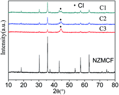
Ni0.4Zn0.2Mn0.4Ce0.06Fe1.94O4 (NZMCF)–carbonyl iron (CI) composites were fabricated using metal organic chemical vapor deposition (MOCVD) and were used as electromagnetic wave (EMW) absorbing materials. X-ray diffraction, scanning electron microscopy and energy dispersive spectrometry show that the NZMCF–CI composites were successfully prepared. Optimizing the weight ratio of CI would likely cause the composites to attain the EM parameters necessary in EMW absorbing materials. When the weight content of CI in the composites was approximately 27% (labeled as C2), the minimum reflection loss (RL) was −32.4 dB at 15.6 GHz and the corresponding thickness was 2.0 mm. The RL of C2 exceeds −10 dB from 6.6 to 18 GHz for an absorber thickness of 2.8 mm, which covers part of the C-band (3.8–8.2 GHz) and the entire X-band (8.2–12.4 GHz) and Ku-band (12.4–18 GHz).


1. Introduction

Electromagnetic wave (EMW) radiation has become a new type of environmental pollution due to the rapid development of the electronic industries.1 Its impact on all electronic equipment via interference and harm to human health have gradually attracted a lot more research attention.2 Currently, the means of protection against jamming and EM shielding mainly used anti-electromagnetic interference, making research into absorbing materials a hot issue.3–7 A high-performance absorbent is the core in basic research of absorbing materials.

NiFe2O4 has been of considerable interest for many years due to its appropriate magnetic properties, chemical stability and low cost.8–10 However, its magnetic and electronic properties must be adjusted to satisfy the requirements of different applications. In an attempt to improve the EMW absorption properties of NiFe2O4, Mn–Zn and Ce substituted NiFe2O4 were prepared in our previous study,11–13 which exhibited better EMW absorption compared with NiFe2O4. However, a large thickness (d > 4 mm) and narrow absorption bandwidth were also found to be an intrinsic disadvantage for EMW absorption. Carbonyl iron (CI) has been extensively applied in absorbing materials because of its simple preparation process, low cost, large magnetic loss, and strong absorbance ability.14–16 Moreover, CI shows good dielectric properties at high frequencies, and adjusting the content in the composites can effectively regulate the complex permittivity.17

In this article, Ni0.4Zn0.2Mn0.4Ce0.06Fe1.94O4 (NZMCF) was initially synthesized using a sol–gel method. There are no reported experimental results on the EM properties of the composites, which have CI deposited on the surface of NZMCF. This special structure and interface interaction between the core and shell favor EMW absorption.18 In addition, a novel fabrication process using metal organic chemical vapor deposition (MOCVD) to obtain a novel composite materials was developed, which used high purity Fe(CO)5 and NZMCF as raw materials and high purity N2 as the carrier gas. The influence of the CI shell on the crystal structure, morphology, complex permittivity and complex permeability, and EMW absorption properties of the particles were investigated.

2. Experimental

2.1 Synthesis of samples

2.1.1 Synthesis of NZMCF. According to NZMCF, stoichiometric amounts of Ni(NO3)2, Zn(NO3)2, Mn(NO3)2, Ce(NO3)3 and Fe(NO3)3 were dissolved in distilled water and an appropriate amount (1[thin space (1/6-em)]:[thin space (1/6-em)]1 molar ratio of citric acid to NO3−1) of citric acid was added to complex the metal ions completely. The pH of the solution was maintained at 7.0 by the slow addition of ammonia solution (25 wt%). The solution was evaporated at 80 °C until a viscous gel was formed. The gel was air dried at 120 °C, ignited in air, and burned into dendritic powders. Finally, the precursor powder was sintered at 1200 °C for 3 h (heating and cooling rate of 5 °C min−1, natural cooling after the temperature had dropped to 300 °C).
2.1.2 MOCVD process step. Experimental procedure is as follows: 50 g of NZMCF powder and 15 ml of iron pentacarbonyl [Fe(CO)]5 were added to a reactor and the evaporator, respectively. N2 was passed into the system making sure that all the air in the tube was purged out of the system and then the N2 supply was turned off while closing the valve between reactor and evaporator. When the NZMCF and Fe(CO)5 were heated to a temperature 180 °C and 80 °C, the value between reactor and evaporator was opened, while a flow of N2 30 ml min−1 to Fe(CO)5 steam was blown into the reactor. To obtain a uniform coating, the reactor was vigorously and continuously stirred mechanically. The flow of N2 was controlled by using a gas flow meter. Using a HH-SA digital thermostat oil bath pot to heat the Fe(CO)5. The reactor piping was coated with a layer of insulation sleeve to prevent gaseous Fe(CO)5 condensation. The Fe(CO)5 steam blowing time was 10 min, 30 min, and 50 min. The final sample was cooled to room temperature under N2 protection. The weight content of CI in the composites was approximately 16%, 27%, and 33%, which was determined by comparing the weights of specimens before and after the experiment. The composites were labeled as C1, C2, and C3.

2.2 Measurement of properties

The phase structure of the powder was identified using X-ray diffraction (XRD; D-max-IIB, Japan). Scanning electron microscopy (SEM) was recorded on a VEGA II XMU INCA scanning electron microscope and was employed for morphological analysis. INCA 7718 spectroscopy (EDS) was used to analyze the distribution of elements in the sample. The EM parameters were measured and determined using a HP-8720ES vector network analyzer (2–18 GHz) and Agilent coaxial transmission airline. The frequency dependence of complex permeability and permittivity for each composite specimen was then obtained using the Agilent 85071E material measurement software, which is based on Weir's reflection–transmission formulation. Samples used for EM parameter measurements were prepared by dispersing powders into paraffin wax at a mass fraction of 60% and then pressing the mixtures into a compact toroidal shape with outer and inner diameters of 7.0 and 3.0 mm, respectively.5 The RL of the prepared absorbers versus frequency was studied using an HP 8510B vector network analyzer and standard horn antennas in an anechoic chamber. The absorbing agent powders (C2, 60 wt%) were dispersed into epoxide resin via adding solvent and using high energy ultrasonic treatment for 30 min. Furthermore, the hardener was added into the mixtures followed by stirring at 1000 rpm for 10 min. Finally, the composite materials were fabricated on an aluminum substrate with a standard size.5

3. Results and discussion

3.1 XRD analysis

Fig. 1 shows the XRD patterns of NZMCF and NZMCF–CI composites. The characteristic XRD peaks in the NZMCF spectrum were compared with the characteristic XRD peaks of the sample spectrum using the powder diffraction card database (JCPDS 71-1269). NZMCF anastomosis was very good, no peaks corresponding to impurities were found. Sharp XRD peaks were found for the crystal plane. The relative intensity and the crystalline form tend to be complete, indicating that pure NZMCF powder was generated. After the CI was coated on the surface of NZMCF, all the samples exhibited diffraction peaks belonging to NZMCF and the α-Fe phase (JCPDS 06-0696), which indicates that in addition to NZMCF and the α-Fe phase no other impurity phase was generated. As the CI amount increases, the characteristic diffraction peaks of NZMCF were gradually weakened and α-Fe at 2θ = 44.5°, (110) crystal plane orientation of growing. This indicated that the CI content in the NZMCF–CI composites gradually increases with time.
image file: c4ra14846k-f1.tif
Fig. 1 XRD pattern of NZMCF and NZMCF–CI composites.

3.2 SEM analysis

The SEM images of NZMCF and the NZMCF–CI composites are shown in Fig. 2. Fig. 2a shows that the SEM images of NZMCF powder have sharp edges and corners, are polyhedral in shape, and gather in small groups. The CI particles (Fig. 2b) are spherical in shape with diameters ranging from 1 μm to 4 μm. When the reaction time was short, a small amount of Fe(CO)5 vapor was added in the reaction, such that the NZMCF surface cannot be completely covered by CI (see Fig. 2c). The particles that were prepared at 30 min and 50 min stack on top of each other due to magnetic attraction between the particles and are uniformly coated with CI, as illustrated in Fig. 2d and e. In order to more directly observe the CI shell, epoxy resin (EP) was used for cold mounting followed by sanding and polishing. SEM was used to observe the cross section of C2 and C3, as illustrated in Fig. 2f and g. It can be seen in Fig. 2f and g that the NZMCF particles are coated with a layer of continuous white shell and their average thickness were about 0.36 μm and 0.78 μm, which were measured using an electronic ruler. The EDS line scan pattern through the C2 section is marked A in Fig. 2h. The Fe, Ni, Mn, Zn, Ce, and O distribution in the cladding layer indeed confirmed that the white shell was CI.
image file: c4ra14846k-f2.tif
Fig. 2 SEM images of NZMCF and NZMCF–CI composites (a) NZMCF, (b) CI, (c) C1, (d) C2, (e) C3, (f) cross section of C2, (g) cross section of C3, (h) EDS line scan of C2.

Fig. 3a and b are the surface and section SEM observations, which show the microstructure of the coating filled with 60% C2 composites. It was observed that the fillers were well dispersed in the EP matrix and no significant porosity was noticed.


image file: c4ra14846k-f3.tif
Fig. 3 SEM image of the microstructure of the coating surface (a) and section (b).

3.3 Complex permittivity and permeability

Fig. 4 shows the complex permittivity and permeability of pure NZMCF, pure CI and the NZMCF–CI composites. It can be clearly seen that the complex permittivity and permeability of the NZMCF–CI composites was larger than those found for pure CI, indicating that the absorbing properties of the composite have been significantly improved. Fig. 4a and b show the real (ε′) and imaginary (ε′′) parts of the permittivity of the specimens plotted as a function of frequency, respectively. The ε′ values of the NZMCF–CI composites were larger than that found for NZMCF and increased with an increasing weight ratio of CI in the composites. Moreover, an inverse change was found in the ε′′ values of the NZMCF–CI composites. When the mass fraction of CI was increased by 27%, the ε′ average values increased from 7.01 to 7.93, while the ε′′ average values increased from 2.30 to 2.88, indicating that the dielectric loss of the NZMCF–CI composites have been improved. The experiments showed that as the reaction time rises, the composite with CI content increases, which form a surface layer, and greatly increases the conductivity of the composites and contributes to the enhancement of permittivity.19 The resonance behavior of the complex permittivity is typically sourced from the space charge, dipole, ionic, and electron polarizations.20 However, dipole polarization has a dominating function in the metal matrix composite and could operate at higher frequencies compared with that of the space charge polarization. Ionic and electronic polarizations generally operate at THz and PHz.20 Therefore, the complex permittivity should be derived from the dipole polarization in the NZMCF–CI composites. Electrons transfer freely in the surface layer and accumulate on the interface layer between CI and NZMCF due to the CI shell, which forms a border capacitor and generates electric dipole polarization, and this structure will further increase its interfacial polarization, which makes the complex permittivity of NZMCF–CI composite increase.
image file: c4ra14846k-f4.tif
Fig. 4 Complex permittivity (a and b) and permeability (c and d) of NZMCF, CI and NZMCF–CI composites.

The real (μ′) and imaginary (μ′′) parts of the permeability are shown in Fig. 4c and d, respectively. It can be seen that the permeability of the coated samples was improved to some extent when compared with the original NZMCF. The μ′ of the coated samples slowly decreased as frequency increased and higher μ′ values were found in the composites with higher CI contents. In contrast, the μ′′ of NZMCF–CI was almost uniformly larger than that of pure NZMCF over the range of 2–18 GHz, and first increased then decreased with an increase in CI content. In general, for ferrite magnetic materials, the microwave magnetic loss originates mainly from the domain wall resonance and natural ferromagnetic resonance.21 Domain wall resonance is usually exhibited in the low-frequency region (<2 GHz). However, resonance due to the spin rotational component occurs at high-frequency regions. Therefore, the resonance peak observed may be attributed to natural resonance.

3.4 EMW absorption properties

The reflection loss (RL) was calculated using transmission line theory,22,23 and can be expressed by the following equations:
 
image file: c4ra14846k-t1.tif(1)
 
image file: c4ra14846k-t2.tif(2)
where Zin is the normalized input impedance of a metal-backed microwave absorption layer, Z0 = 377 Ω is the intrinsic impedance of free space, εr and μr are the relative complex permittivity and permeability, respectively, f is the frequency of the EMW, d is the thickness of an absorber, and c is the velocity of light in vacuo.

Fig. 5 shows the RL of pure NZMCF powder, CI powder and the NZMCF–CI composites at 2 mm thickness in the frequency range of 2–18 GHz. The EMW absorption properties of the NZMCF–CI composites were much better than pure NZMCF powder and CI power. The RL is sensitive to the CI content in the NZMCF–CI composites. From the reflectivity curves obtained, the position of the reflection peak loss moved to lower frequency regions with increasing CI content. The accessibility of CI can significantly improve the absorption properties of the NZMCF powders.


image file: c4ra14846k-f5.tif
Fig. 5 EMW absorbing properties of NZMCF, CI and NZMCF–CI at a sample thickness of 2 mm.

Absorbing materials with a good effect must meet two conditions: first EMW must penetrate into the interior of the material as much as possible, which is a good matching characteristic, the other is the material must be able to consume the incoming EMW, which have a large attenuation constant α.24 Fig. 6 shows both the magnetic loss and dielectric loss, which contribute to microwave absorption (tan[thin space (1/6-em)]δε = ε′′/ε′, tan[thin space (1/6-em)]δμ = μ′′/μ′). As shown in Fig. 6, the magnetic loss first increases then decreases with increasing CI content in the composites. The improvement in magnetic loss of the NZMCF–CI composites was suspected to originate from the efficient combination of NZMCF and CI. In general, the EMW absorption of a material is considered to be related to the balance between their magnetic loss and dielectric loss. C2 showed better interaction than others, indicating a better matching characteristic.


image file: c4ra14846k-f6.tif
Fig. 6 Dielectric loss and magnetic loss factor of (a) NZMCF particles and NZMCF–CI composites (b) C1, (c) C2, and (d) C3.

The attenuation constant can be expressed as: image file: c4ra14846k-t3.tif.24 The attenuation constant of the NZMCF, CI and NZMCF–Fe particles are shown in Fig. 7. After CI film was grown on the surface of NZMCF, its attenuation constant increased significantly. This means the absorbing performance of NZMCF–CI is better than pure NZMCF and CI. It can be clearly seen that C2 and C3 have the largest attenuation constants when compared with the other materials.


image file: c4ra14846k-f7.tif
Fig. 7 Attenuation constant of the NZMCF particles, CI particles and NZMCF–CI composites.

In order to more directly study the EMW absorption performance of C2, the relationship between RL and frequency at a thickness of 1.0 mm to 3.0 mm was calculated and shown in Fig. 8a. As the thickness increases, the reflectance peak gradually moves to a lower frequency. It can be seen from Fig. 8a that the minimum RL was −32.4 dB at 15.6 GHz with a corresponding thickness of 2.0 mm and for a coating thickness of 2.8 mm, an RL exceeding −10 dB was obtained in the frequency range of 6.6–18 GHz, which covers part of the C-band (3.8–8.2 GHz) and the entire X-band (8.2–12.4 GHz) and Ku-band (12.4–18 GHz).


image file: c4ra14846k-f8.tif
Fig. 8 EMW absorbance properties of NZMCF–CI with different thicknesses. (a) C2, (b) C3.

The EMW absorbance properties of C3 with different thickness are illustrated in Fig. 8b. It can be clearly concluded that C2 has better EMW absorbing properties than C3. Although the RL of C3 at 2 mm thickness shows a wide absorption bandwidth, the entire minimum RL of C3 is larger than C2. Overall, C2 has the best EMW absorbing properties when compared to C3.

In order to verify the accuracy of the results, a comparison of the measured RL of the NZMCF–CI coating at thickness d = 2.8 mm and the calculated results are illustrated in Fig. 9. Although the measured results and calculated values slightly differ, both measured and theoretically calculated results have similar curve patterns and absolute values. The minimum reflection loss at 10.2 GHz was nearly −22.8 dB for the measured results, while the minimum reflection loss of −26.3 dB can be obtained at 10 GHz for the calculated results. The difference between the calculated and measured values can be attributed to the surface irregularity of the absorber samples and the differences in the actual thickness of the layers.


image file: c4ra14846k-f9.tif
Fig. 9 Comparison of measured and calculated reflection loss values for NZMCF–CI (d = 2.8 mm).

4. Conclusions

NZMCF–CI composites were successfully prepared using MOCVD. After the CI film was deposited on the NZMCF surface, its EM parameters undergo significant changes. NZMCF–CI composites exhibited excellent EMW absorption properties in the frequency range of 2–18 GHz. When the weight content of CI in the composites was approximately 27% (labeled as C2), a minimum reflection loss (RL) was −32.4 dB at 15.6 GHz with a corresponding thickness of 2.0 mm. The RL of C2 exceeds −10 dB from 6.6 to 18 GHz for an absorber thickness of 2.8 mm.

References

  1. Y. Liu, X. X. Liu, R. Li, W. Wen and X. J. Wang, RSC Adv., 2015, 5, 8713 Search PubMed.
  2. T. Giaimakopoulou, A. Oikonomou and G. Kordas, J. Magn. Magn. Mater., 2004, 271, 224–229 CrossRef PubMed.
  3. H. L. Lv, G. B. Ji, M. Wang, C. M. Shang, H. Q. Zhang and Y. W. Du, RSC Adv., 2014, 4, 57529–57533 RSC.
  4. M. Zong, Y. Huang, Y. Zhao, X. Sun, C. H. Qu, D. Luo and J. B. Zheng, RSC Adv., 2013, 3, 23638–23648 RSC.
  5. Y. Liu, X. X. Liu and X. J. Wang, J. Alloys Compd., 2014, 584, 249 CrossRef CAS PubMed.
  6. P. Bhattacharya, S. Dhibar, G. Hatui, A. Mandal, T. Das and C. K. Das, RSC Adv., 2014, 4, 17039–17053 RSC.
  7. S. Hazra, B. K. Ghosh, H. R. Joshi, M. K. Patra, R. K. Jani, S. R. Vadera and N. N. Ghosh, RSC Adv., 2014, 4, 45715–45725 RSC.
  8. T. Giannakopoulou, L. Kompotiatis, A. Kontogeorgakos and G. Kordas, J. Magn. Magn. Mater., 2002, 246, 360–365 CrossRef CAS.
  9. J. Jacob, M. A. Khadar, A. Lonappan and K. T. MathewA, Bull. Mater. Sci., 2008, 36, 847–851 CrossRef PubMed.
  10. J. G. S. Duque, E. A. Souza, C. T. Meneses and L. Kubota, Phys. B, 2007, 398, 287–290 CrossRef CAS PubMed.
  11. Y. Liu, X. X. Liu, X. J. Wang and X. Chen, J. Chin. Ceram. Soc., 2013, 41, 755 Search PubMed.
  12. Y. Liu, X. X. Liu, X. J. Wang, Z. Y. Zhang, R. Li and L. Guo, Chin. J. Nonferrous Met., 2013, 23, 168 CAS.
  13. Y. Liu, X. X. Liu and X. J. Wang, Adv. Appl. Ceram., 2015, 114, 82 CrossRef CAS PubMed.
  14. Z. T. Zhu, X. Sun, H. R. Xue, H. Guo, X. L. Fan, X. C. Pan and J. P. He, J. Mater. Chem. C, 2014, 2, 6582–6591 RSC.
  15. M. S. Kim, Y. D. Liu, B. J. Park, C. Y. You and H. J. Choi, J. Ind. Eng. Chem., 2012, 18(2), 664–667 CrossRef CAS PubMed.
  16. X. M. Ni, Z. Zheng, K. Xiao, L. Huang and L. He, Mater. Chem. Phys., 2010, 120(1), 206–212 CrossRef CAS PubMed.
  17. Y. L. Cheng, J. M. Dai, D. J. Wu and Y. P. Sun, J. Magn. Magn. Mater., 2010, 322(1), 97–101 CrossRef CAS PubMed.
  18. H. Wang, Y. Y. Dai, W. J. Gong, D. Y. Geng, S. Ma, D. Li, W. Liu and Z. D. Zhang, Appl. Phys. Lett., 2013, 102, 223113 CrossRef PubMed.
  19. S. Ramo, J. R. Whinnery and T. V. Duzer, Fields and Waves in Communication Electronics, Wiley, New York, 1984 Search PubMed.
  20. T. Lacrevaz, B. Fléchet, A. Farcy, J. Torres, M. Gros-Jean, C. Bermond, T. T. Vo, O. Cueto, B. Blampey, G. Angénieux, J. Piquet and F. de Crécy, Microelectron. Eng., 2006, 83(11–12), 2184–2188 CrossRef CAS PubMed.
  21. M. Z. Wu, Y. D. Zhang, S. Hui, T. D. Xiao, S. H. Ge, W. A. Hines, J. I. Budnick and G. W. Taylor, Appl. Phys. Lett., 2002, 23, 4404 CrossRef PubMed.
  22. R. C. Che, L. M. Peng, X. F. Duan, Q. Chen and X. L. Liang, Adv. Mater., 2004, 16, 401–405 CrossRef CAS.
  23. M. R. Meshram, N. K. Agrawal, B. Sinha and P. S. Misra, J. Magn. Magn. Mater., 2004, 271, 207–214 CrossRef CAS PubMed.
  24. B. Lu, H. Huang, X. L. Dong, X. F. Zhang, J. P. Lei, J. P. Sun and C. Dong, J. Appl. Phys., 2008, 104(11), 114313 CrossRef PubMed.

This journal is © The Royal Society of Chemistry 2015