
Hierarchical flower-like Bi2WO6 hollow microspheres: facile synthesis and excellent catalytic performance†
Peigen Zhangab,
Jian Zhanga,
Anjian Xieab,
Shikuo Liab,
Jiming Songa and
Yuhua Shen*ab
aSchool of Chemistry and Chemical Engineering, Anhui University, Hefei 230601, P. R. China. E-mail: yhshen@ahu.edu.cn; Tel: +860551 63861475
bLab for Clean Energy & Green Catalysis, Anhui University, Hefei 230601, P. R. China
First published on 20th February 2015
Abstract
A one step method has been developed for the fabrication of hierarchical flower-like bismuth tungstate (Bi2WO6) hollow spheres via a solvothermal process. The size of these microspheres is about 1.5 μm, and the shells are composed of nanosheets with a thickness of about 15 nm. The product has a specific surface area of 95 m2 g−1. The formation mechanism of flower-like Bi2WO6 is proposed, which involves the nucleation and formation of nanoparticles followed by their self-assembly to microspheres, oriented growth, Ostwald ripening and transformation into flower-like hollow microspheres. The Bi2WO6 hollow spheres exhibit excellent visible light catalytic efficiency for the degradation of rhodamine B (RhB), up to 98% within 50 min. The efficiency remains at 92% after five photodegradation cycles due to the hierarchical hollow structure and large surface area.
Introduction
Recently, photocatalysts have attracted a considerable amount of interest because they play a very important role in solving environmental and pollution problems, employing solar energy for the catalytic degradation of organic pollutants. Therefore, the development of environmentally friendly and highly efficient photocatalysts has aroused great interest. These photocatalysts should have the following three characteristics: they should absorb sunlight with an appropriate energy band, they should promote a longer lifetime of electron holes and they should possess a conduction (valence) band with a more negative (positive) electrode potential. Researchers have developed a series of narrow band semiconductor photocatalytic materials, such as WO3, Fe2O3 and CdS etc.,1 to make more effective use of solar energy. For the purpose of treating environmental organic pollutants, the synthesis of visible light catalysts has become a hot topic and is becoming more and more relevant via the development of more effective and convenient approaches.Bi2WO6 is a layered Aurivillius oxide2 with a narrow width of band gap. It can be excited by visible light, improving the utilization of solar energy. Research shows that Bi2WO6, owing to its appropriate valence band and thermodynamic properties, can decompose water and can be employed in the degradation of chloroform, acetaldehyde and other harmful substances under visible light,3 indicating the high photocatalytic efficiency of Bi2WO6. As we know, the morphology and structure of photocatalysts have a direct effect on their photocatalytic activity. In the past years, there have been some reports of Bi2WO6 photocatalysts with different morphologies synthesized by different methods. For instance, Li et al.4 obtained layered Bi2WO6 microspheres by utilizing PVP as the template; Amano et al.5 synthesized spark Bi2WO6 microspheres in aqueous solutions with low pH values; Chen et al.6 prepared Bi2WO6 microspheres by an inorganic salts-assisted hydrothermal method; and Shang et al.7 fabricated Bi2WO6 nanocages with unique morphologies by using carbon spheres as a template. Compared with the hard template method, the soft template method has some advantages, such as the inexpensive and simple processing and ease of recycling, which decrease costs in industrial applications. So it is important to synthesize Bi2WO6 photocatalysts by choosing the proper soft template.
As we all know, ethylene glycol, a non-aqueous solvent with an –OH group, has a good water or alcohol solubility, which makes it easy to handle. It is widely used in the synthesis of monodisperse metal and metal oxide nanoparticles, and also as a soft template for highly ordered structural materials.8 Ethanol forms a hydrogen bonding network easily due to the presence of the hydroxyl group. Therefore, we use a mixture of ethylene glycol and ethanol as both solvent and template to synthesize highly ordered flower-like porous and hollow Bi2WO6 microspheres by a solvothermal method. The as-prepared product possesses high stability and excellent photocatalytic activity for the photodegradation of RhB. The formation and growth mechanism of hierarchical flower-like Bi2WO6 is worth exploring, which is done through the observation of intermediate products obtained at different reaction stages. The hollow Bi2WO6 catalyst will have potential applications in water treatment.
Experimental
Materials
Bismuth nitrate pentahydrate (Bi(NO3)3·5H2O), sodium tungstate dihydrate (Na2WO4·2H2O), rhodamine B and ethanol were supplied by Shanghai Chemical Reagent Co. Ltd (P. R. China). Ethylene glycol (EG) was obtained from Sinopharm Chemical Reagent Co. Ltd (P. R. China).All reagents were of analytical grade and were used without further purification. Milli-Q water (Millipore Corp., with resistivity of 18.2 MΩ cm) was employed for all experiments.
Synthesis of flower-like hollow Bi2WO6 microspheres
Flower-like Bi2WO6 microspheres were synthesized via a solvothermal method. Bi(NO3)3·5H2O (2.5 mmol) and Na2WO4·2H2O (1.25 mmol) were each separately dissolved in 12.5 mL of ethylene glycol. Then, the Na2WO4·2H2O solution was added dropwise into the Bi(NO3)3·5H2O solution with magnetic stirring, which was followed by the addition of absolute ethanol (25 mL). After stirring for 30 minutes, the mixed solution was transferred into a 100 mL Teflon lined autoclave and kept at 160 °C for 12 h. The Teflon lined autoclave was then naturally cooled down to room temperature. The products were isolated by centrifugation and washed several times with doubly distilled water and absolute ethanol. Finally, the products were dried at 60 °C for 24 h in a vacuum oven and stored. Some experiments were carried out to control the morphologies of the samples, in which several reaction parameters such as the reaction time or the volume ratio of EG to ethanol (VEG![[thin space (1/6-em)]](https://www.rsc.org/images/entities/char_2009.gif) :VE) were altered while other reaction conditions were kept constant.
:VE) were altered while other reaction conditions were kept constant.
Characterization
Transmission electron microscopy (TEM) measurements were carried out on a JEM model 100SX electron microscope (Japan Electron Co.) operated at an accelerating voltage of 80 kV. Field emission scanning electron microscopy (FESEM) measurements were taken with a Hitachi S4800 scanning electron microscope. The phase structure and phase purity of the as synthesized products were examined by X-ray diffraction (XRD), using a MAP18XAHF instrument, with the X-ray diffractometer using Cu-Kα radiation (λ = 1.5406 Å) at a scan rate of 6° min−1. The accelerating voltage and applied current were 30 kV and 25 mA, respectively (MAC Science, Japan). Energy-dispersive spectra (EDS) were examined on a JEM-2010F high-resolution transmission electron microscope equipped with an EDS detector working at an accelerating voltage of 200 kV. UV-vis diffuse reflectance spectra of the samples were obtained using a UV-vis solid spectrometer (Shimadzu SolidSpec-4100) by using BaSO4 as a reference. Room temperature PL spectra were recorded on an F-4500 at room temperature (Japan Electron Co.). The specific surface area was estimated using the Brunauer–Emmett–Teller (BET) equation based on the nitrogen adsorption isotherm obtained with a Micromeritics ASAP 2020 Analyzer (Surface Area and Porosity System).Photocatalytic activity of the products
The decolorization of a model pollutant, RhB dye, was chosen to evaluate the photocatalytic activity of the flower-like hollow Bi2WO6 microspheres under visible light. 50 mg of Bi2WO6 was dispersed in 50 mL of RhB solution (1.0 × 10⁻⁵ mol L−1). The solution was stirred for 30 min to ensure the catalyst was completely dispersed and a balance of adsorption and desorption of the catalyst in dark conditions was reached. The photocatalytic experiment was carried out in a self-designed reactor, using a 500 W xenon lamp equipped with a 420 nm cut-off filter to provide visible light irradiation, which was placed 6 cm above the reaction slurry. The concentration changes of the RhB in the photodegradation process were measured by a UV-vis spectrophotometer. In each recycling experiment, the flower-like hollow Bi2WO6 microspheres were separated from solution after the photocatalytic degradation of RhB, then washed with ethanol and deionized water three times respectively before being redispersed in the same concentration and volume of RhB solution for another cycle. Some control experiments were carried out to study the degradation of RhB. Several reaction parameters, pH values or concentration of the RhB solution for instance, were varied while other reaction conditions remained constant.Results and discussion
Fig. 1a shows the EDS spectrum of the product obtained from the solvothermal reaction time of 12 h when the volume ratio of EG to ethanol is 1:1. The peaks of Bi, O and W elements are found. The atomic ratio of the three elements is 1.96:1:5.91, which is close to the composition of Bi2WO6. Fig. 1b shows the XRD pattern of the as-prepared product. The eight diffraction peaks at 2θ of 28.3°, 36.0°, 47.2°, 55.9°, 58.6°, 68.6°, 75.9° and 78.3° can be indexed as the monoclinic phase of Bi2WO6 with the crystal planes of (103), (202), (220), (303), (107), (400), (109) and (307), respectively, which is completely consistent with the standard card (JCPDS no. 26-1044, a = 5.480 Å, b = 5.480 Å, c = 11.500 Å). The sharp diffraction peaks indicate the high crystallization of the as-prepared Bi2WO6. Since there are no other diffraction peaks or impurity phases, the product of the solvothermal method is confirmed to be high purity.
 | ||
| Fig. 1 (a) EDS spectrum and (b) XRD pattern of the product obtained at 12 h (VEG:VE = 1:1). | ||
Fig. 2a and b show the SEM images of the product at the reaction time of 12 h. We can see that Bi2WO6 nanoparticles are spherical with a flower-like morphology, and the size of microspheres is relatively uniform with a diameter of about 1.5 μm. The broken microsphere indicates that the microspheres are hollow. The higher magnification image (Fig. 2b) shows that the petal structure of the Bi2WO6 microspheres is assembled by many nanosheets with an average thickness of about 15 nm, and some pores are present in the microsphere shell because of the cross-linking of nanosheets. The SEM images demonstrate that the structure of the Bi2WO6 microspheres is porous as well as hollow. The hollow structure can also be verified by the TEM images in Fig. 2c and d. The contrast of the darker parts and lighter parts indicates the hollow structure. From Fig. 2d, we know that the darker parts composed of nanosheets are the shell with an average thickness of 250 nm, and the lighter parts in the center of the microsphere show the hollow part with a diameter of about 800 nm. The observed results from TEM and SEM both prove that the as-synthesized products are porous flower-like hollow microspheres.
 | ||
| Fig. 2 (a) FESEM image of Bi2WO6 prepared at 12 h (VEG:VE = 1:1), (b) the higher magnification image of (a), (c and d) TEM images of the as-prepared Bi2WO6. | ||
To further investigate the formation mechanism of the highly ordered self-assembled hollow microspheres, we analyzed the morphology and phase of the products at different reaction stages.
Fig. 3 shows SEM images of the samples with the reaction times of 1 h, 4 h, and 8 h, respectively. At 1 h, the sample exhibits a sphere shape with a diameter of about 150 nm (Fig. 3a). When we extend the reaction time to 4 h, the nanoparticles assembled into uniform microspheres with a rough surface (Fig. 3b and c) and diameter of about 1.2 μm. When the reaction time stretches to 8 h, the outer layer nanoparticles of the microspheres gradually grow into nanosheets, whose thickness is about 10 nm (Fig. 3d and e). The broken microspheres in Fig. 3d indicate the formation of the hollow structure. At the reaction time of 12 h, a highly ordered porous and hollow microspherical structure with a larger diameter (Fig. 2) is obtained by the Ostwald ripening process. The XRD patterns shown in Fig. 3f demonstrate the crystallinity of the samples improves with the increase of the reaction time.
 | ||
| Fig. 3 SEM images of the samples at the reaction time of (a) 1 h, (b and c) 4 h, and (d and e) 8 h, (f) XRD patterns of the samples obtained at different reaction stages. | ||
Fig. 4 shows the possible formation and growth mechanism of the flower-like hollow Bi2WO6 microspheres. First, Bi(NO3)3·5H2O and Na2WO4·2H2O are dissolved in ethylene glycol and ethanol. When the two solutions are mixed, no precipitation is found (Fig. S1†) due to the coordination or hydrogen bonds between Bi3+ or WO42− and the –OH of ethylene glycol and ethanol.2,9 At the initial stage of the solvothermal reaction, Bi3+ and WO42− are released slowly under the high temperature and high pressure conditions, leading to the nucleation and growth of the Bi2WO6 nanoparticles via the ionic reaction10 (Fig. 3a). With the increase of reaction time, these nanoparticles gradually self-assemble into large spheres with rough surfaces (Fig. 3b and c) to minimize the interfacial energy.11 Then an Ostwald ripening process takes place, the smaller nanoparticles in the center of the large spheres dissolve into the solution. Meanwhile, an oriented growth process also occurs with the formation of nanosheets on the external surfaces of the large spheres (Fig. 3d and e).12,13 Finally, the hierarchical flower-like Bi2WO6 microspheres with uniform nanosheets are observed in Fig. 2.
 | ||
| Fig. 4 Schematic illustration of the probable formation mechanism of hierarchical flower-like Bi2WO6 hollow spheres. | ||
Fig. 5a shows the UV-visible diffuse reflectance spectrum of the product obtained at 12 h (VEG:VE = 1:1). The absorption edge of the flower-like hollow microspheres is estimated to be about 480 nm, indicating that a red shift of the peak occurred and the absorption range of the flower-like hollow microspheres expanded to the visible region, compared to the commercial Bi2WO6 (ref. 14).
 | ||
| Fig. 5 (a) UV-vis diffuse reflectance spectrum and (b) (αhν)2 vs. hν plot of the hollow Bi2WO6 microspheres prepared at 12 h (VEG:VE = 1:1). | ||
The basic equation for the optical absorption band edge of the crystal semiconductor is in accordance with eqn (1):15
| (αhv)2 = A(hv − Eg)n | (1) |
N2 adsorption–desorption isotherms and pore size distribution (inset) are employed to evaluate the surface area of the flower-like hollow Bi2WO6 microspheres (Fig. 6). Hysteresis is clearly seen in the N2 adsorption–desorption curve, indicating that the products are porous, which is consistent with the observed morphology of the flower-like hollow Bi2WO6 microspheres. The specific surface area of the hollow microspheres is 95 m2 g−1 and the pore volume is 0.97 cm3 g−1, both of which are larger than those reported previously.17,18 The pore sizes of 36 nm and 92 nm may be caused by the space between the nanosheets. The flower-like hollow microspheres with large specific surface area are beneficial to increase the contact efficiency with organic contaminants to improve the photocatalytic activity.
 | ||
| Fig. 6 N2 adsorption–desorption isotherm curves and pore size distribution (inset) of the hierarchical flower-like Bi2WO6 product. | ||
The photocatalytic activity of the synthesized hierarchical flower-like hollow Bi2WO6 microspheres in the degradation of RhB is evaluated under visible light at room temperature. The characteristic absorption peak of RhB at 554 nm is used to monitor the catalytic process. Fig. 7a shows the temporal evolution of the photocatalytic degradation of RhB solution. With the increase of visible light irradiation time, the intensity of the absorption peak of RhB decreases gradually along with the blue shift of the peak. The inset in Fig. 7a shows the dynamic variety of RhB solution color with the degradation time. It is clearly seen that the RhB solution changes gradually from pink to colorless due to the step by step demethylation, gradually generating N,N,N-three ethyl rhodamine, N,N-three ethyl rhodamine, N-ethyl rhodamine and rhodamine. Finally, the conjugate structure is converted into CO2 and H2O.19,20 Fig. 7b presents the photocatalytic efficiency of the as-synthesized product for the degradation of RhB. It is worth mentioning that the catalytic efficiency of the flower-like hollow Bi2WO6 microspheres is 98% for the degradation of RhB under visible-light irradiation within 50 min. This results from the large specific surface area of the flower-like porous shell and the hollow structure of the interior, which could provide many binding sites for effective catalysis. Fig. 7c reveals that the catalytic efficiency of the as-synthesized hollow Bi2WO6 microspheres is still more than 92% after five photocatalytic cycles, indicating the stability of the flower-like hollow catalyst.
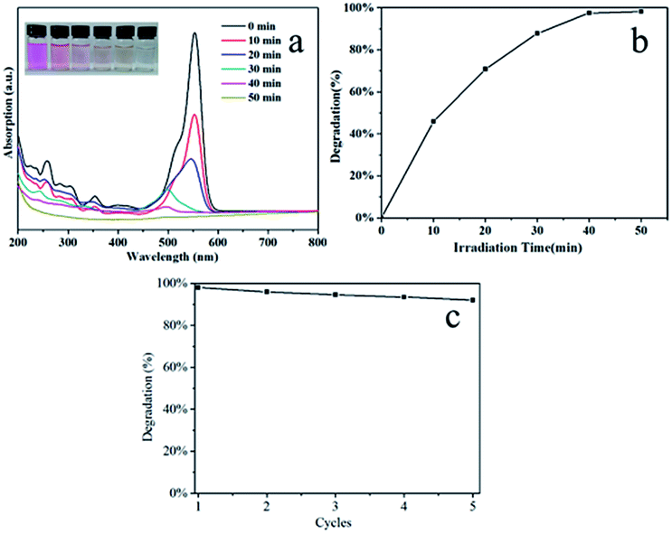 | ||
| Fig. 7 (a) The temporal evolution of the absorption spectra of the RhB solution (inset shows the corresponding time-dependent color change of RhB solution), (b) degradation rate of RhB in the presence of hierarchical flower-like hollow Bi2WO6 under visible-light irradiation, (c) reusability of the hollow Bi2WO6. | ||
Fig. 8a–c show the SEM images of the samples prepared at the reaction time of 12 h with different volume ratios of EG to ethanol. It is seen that the morphologies of the samples are sensitive to content of both solvents. When the volume ratio of EG to ethanol is 1:0, irregular morphologies of Bi2WO6 can be observed (Fig. 8a). Hollow spheres with a rough surface are detected in Fig. 8b (VEG:VE = 2:1). The hierarchical flower-like Bi2WO6 hollow microspheres formed at VEG:VE = 1:1 are shown in Fig. 2. When the volume ratio of EG to ethanol is 0:1, blocks are observed (Fig. 8c). From the above discussion, we conclude that EG and ethanol both play important roles in the formation of products with different morphologies. These samples are further used for the photocatalytic degradation of RhB. It is found that the hierarchical flower-like hollow Bi2WO6 prepared at a VEG/VE of 1:1 possesses the best catalytic activity (Fig. 8d).
 | ||
| Fig. 8 SEM images of the Bi2WO6 prepared at the reaction time of 12 h with different volume ratios of EG to ethanol: (a) 1:0, (b) 2:1, (c) 0:1, (d) comparison of the degradation rates of RhB (1.0 × 10⁻⁵ mol L−1) in the presence of the prepared samples shown in Fig. 8a–c under visible light for 50 min. | ||
Fig. 9a shows the influence of pH value on the degradation of RhB in the presence of the hierarchical flower-like hollow Bi2WO6 (VEG:VE = 1:1). As the pH value changes from 4 to 6.5, the photocatalytic degradation rate of RhB increases. When the pH value is more than 8, the degradation rate of RhB decreases. The possible reason for this is that the Bi2WO6 can be easily hydrolysed into H2WO4 and Bi2O3 in the acidic solution.21 In the alkaline solution, the amount of OH− anions lead to the catalyst surface being negatively charged and the RhB is turned into anions due to the dissociation of –COOH groups. The repulsion of Bi2WO6 and RhB makes the adsorption between them reduced and impedes the degradation reaction. Another possible reason may be the formation of Bi3.84W0.16O6.24 in the alkaline solution, which possesses little photocatalytic activity for the degradation of RhB.22 From Fig. 9b, it is clearly observed that the highest degradation rate of RhB is 98% under visible-light irradiation at the pH value of 6.5. We conclude that the photocatalytic degradation rate of RhB could be controlled by the pH value of the solution.
 | ||
| Fig. 9 Degradation rate of RhB (1.0 × 10⁻⁵ mol L−1) in the presence of hierarchical flower-like hollow Bi2WO6 in solutions with different pH values: (a) time-dependent, (b) the maximum value under visible-light irradiation for 50 min. | ||
The influence of the initial concentration (C0) on the degradation rate of RhB in the presence of hierarchical flower-like hollow Bi2WO6 is observed in Fig. 10. The experimental data are fitted by applying a pseudo-first-order model (lnC/C0 = −kt) to verify the reaction rate constant (k) of the degradation of RhB. The k values are 0.0841 min−1, 0.014 min−1 and 0.00255 min−1 for the initial RhB concentrations of 1.0 × 10⁻⁵ mol L−1, 5.0 × 10⁻⁵ mol L−1 and 1.0 × 10⁻⁴ mol L−1, which proves that the initial concentration of RhB has a significant effect on the degradation rate of RhB. The degradation rate of RhB is higher when the initial concentration is lower. The above first-order linear relationship can be explained by the Langmuir–Hinshelwood model:21
 | (2) |
 | ||
| Fig. 10 First-order plots for the degradation of RhB at various initial concentrations in the presence of hierarchical flower-like hollow Bi2WO6. | ||
K refers to the adsorption equilibrium constant; C is the concentration of RhB solution at the reaction time of t. For dilute solutions of KC ≤ 1, because of the weak adsorption of RhB on the surface of the catalyst, eqn (2) can be expressed as follows:
| r = kKC | (3) |
Therefore, the degradation reaction of RhB by Bi2WO6 apparently follows first-order kinetics in the Langmuir–Hinshelwood model due to the choice of low concentrations of RhB solution in our experiment.
Fig. S2† shows the influence of the catalyst dose on the degradation of RhB (1.0 × 10⁻⁵ mol L−1, 50 mL). From Fig. S2b,† we can know that k values increase as the catalyst content is varied from 10 mg to 70 mg. According to eqn (3), the degradation time of the same amount of RhB will be reduced, which can be observed in Fig. S2a.† The possible reason may be that, at lower initial concentrations, more photocatalytic active sites can be supplied to absorb more photons, which promote the formation of more electron and hole pairs,23 leading to the increase of degradation rate.
Conclusions
Hierarchical flower-like Bi2WO6 hollow microspheres are synthesized. The hollow Bi2WO6 microspheres are assembled by many nanosheets with a thickness of about 15 nm. The larger specific surface area of the hollow microspheres is favorable for the adsorption of RhB. The hollow Bi2WO6 microspheres possess excellent photocatalytic activity as well as reusability for the degradation of RhB under visible light. We also find the morphologies of Bi2WO6 are affected by the volume ratio of EG to ethanol. The photocatalytic degradation rate of RhB is related to the pH value of the solution and the morphology of Bi2WO6. This study puts forward a simple and convenient method for fabricating a porous and hollow Bi2WO6 photocatalyst, providing the possibility for Bi2WO6 photocatalysts to be used for treating polluted water.Acknowledgements
This work is supported by the National Nature Science Foundation of China (no. 91022032, 21173001, and 21371003).Notes and references
- H. J. Zhang, G. H. Chen and D. W. Bahnemann, J. Mater. Chem., 2009, 19, 5089–5121 RSC.
- Z. Zhang, W. Wang, D. Jiang and J. Xu, Appl. Surf. Sci., 2014, 292, 948–953 CrossRef CAS PubMed.
- Y. Zhang and Y. J. Xu, RSC Adv., 2014, 4, 2904–2910 RSC.
- Y. Y. Li, J. P. Liu, X. T. Huang and G. Y. Li, Cryst. Growth Des., 2007, 7, 1350–1355 CAS.
- F. Amano, K. Nogami, R. Abe and B. Ohtani, J. Phys. Chem. C, 2008, 112, 9320–9326 CAS.
- Z. Chen, L. W. Qian, J. Zhu, Y. P. Yuan and X. F. Qian, CrystEngComm, 2010, 12, 2100–2106 RSC.
- M. Shang, W. Z. Wang and H. L. Xu, Cryst. Growth Des., 2009, 9, 991–996 CAS.
- C. L. Yan and D. F. Xue, J. Phys. Chem. B, 2006, 110, 11076–11080 CrossRef CAS PubMed.
- S. P. Hu, C. Y. Xu and L. Zhen, Mater. Lett., 2013, 95, 117–120 CrossRef CAS PubMed.
- G. Tian, Y. Chen, W. Zhou, K. Pan, Y. Dong, C. Tian and H. Fu, J. Mater. Chem., 2011, 21, 887–892 RSC.
- C. C. Yec and H. C. Zeng, J. Mater. Chem. A, 2014, 2, 4843–4851 CAS.
- X. Wang, L. Chang, J. Wang, N. Song, H. Liu and X. Wan, Appl. Surf. Sci., 2013, 270, 685–689 CrossRef CAS PubMed.
- C. Xu, X. Wei, Y. Guo, H. Wu, Z. Ren, G. Xu, G. Shen and G. Han, Mater. Res. Bull., 2009, 44, 1635–1641 CrossRef CAS PubMed.
- Y. Yan, Y. Wu, Y. Yan, W. Guan and W. Shi, J. Phys. Chem. C, 2013, 117, 20017–20028 CAS.
- J. Zhang, Y. Wang, S. Li, X. Wang, F. Huang, A. Xie and Y. Shen, CrystEngComm, 2011, 13, 5744–5750 RSC.
- B. Zhao, M. Wang, L. Lin, Q. Zeng and D. He, Ceram. Int., 2014, 40, 5831–5835 CrossRef CAS PubMed.
- Y. Liu, H. Tang, H. Lv, Z. Li, Z. Ding and S. Li, Ceram. Int., 2014, 40, 6203–6209 CrossRef CAS PubMed.
- G. Zhu, J. Liang, M. Hojamberdiev, S. Aldabe Bilmes, X. Wei, P. Liu and J. Zhou, Mater. Lett., 2014, 122, 216–219 CrossRef CAS PubMed.
- Y. Li, J. Liu and X. Huang, Nanoscale Res. Lett., 2008, 3, 365–371 CrossRef CAS.
- W. Xiong, Q. Zhao, X. Li and D. Zhang, Catal. Commun., 2011, 16, 229–233 CrossRef CAS PubMed.
- H. b. Fu, C. S. Pan, W. Q. Yao and Y. F. Zhu, J. Phys. Chem. B, 2005, 109, 22432–22439 CrossRef CAS PubMed.
- C. Xu, X. Wei, Z. Ren, Y. Wang, G. Xu, G. Shen and G. Han, Mater. Lett., 2009, 63, 2194–2197 CrossRef CAS PubMed.
- H. b. Fu, W. Q. Yao, L. W. Zhang and Y. F. Zhu, Mater. Res. Bull., 2008, 43, 2617–2625 CrossRef CAS PubMed.
Footnote |
| † Electronic supplementary information (ESI) available. See DOI: 10.1039/c4ra14782k |
| This journal is © The Royal Society of Chemistry 2015 |