Use of multifunctional phosphorylated PAMAM dendrimers for dentin biomimetic remineralization and dentinal tubule occlusion

Tianda Wanga, Sheng Yangab, Lei Wanga and Hailan Feng*a
aDepartment of Prosthodontics, Peking University School and Hospital of Stomatology, Beijing, China. E-mail: kqfenghl@bjmu.edu.cn; Tel: +86-10-82195232
bChongqing Key Laboratory of Oral Diseases and Biomedical Sciences, College of Stomatology, Chongqing Medical University, Chongqing, China

Received 17th November 2014 , Accepted 7th January 2015

First published on 8th January 2015


Abstract

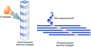
The disequilibrium between demineralization and remineralization of teeth, especially of dentin, may lead to serious consequences like dental caries that are considered to affect people's quality of life. The employment of biomimetic analogs of proteins to duplicate biomineralization, which is a well-regulated process mediated by extracellular matrix proteins, may provide new insights to solve these problems. Here we report the use of a modified multifunctional dendrimer, synthesized by the introduction of phosphate groups via a Mannich-type reaction onto poly(amidoamine) (PAMAM) dendrimers, to biomimetically remineralize dentin. The phosphorylated PAMAM dendrimers were demonstrated to act, along with an amorphous calcium phosphate stabilizing agent, polyacrylic acid (PAA), as biomimetic analogs of noncollagenous proteins to induce the remineralization of demineralized dentin. Phosphorylated PAMAM dendrimers treated demineralized dentin discs were immersed in a remineralizing solution containing PAA for up to 7 days. The success of this remineralization was examined using attenuated total reflection Fourier-transform infrared spectroscopy (ATR-FTIR), X-ray diffraction (XRD), energy-dispersive X-ray spectroscopy (EDS) and electron microscopy. These results showed that demineralized dentinal collagen fibrils were successfully phosphorylated by the treatment of phosphorylated PAMAM dendrimers and embedded with calcium-deficient hydroxyapatite after remineralization. The surfaces of demineralized dentin discs were covered with newly induced crystals and the patent dentinal tubules were occluded. A good biocompatibility was also determined. Thus, phosphorylated PAMAM dendrimers could be applied as a minimally invasive method of management of dentin caries, employed to improve the resin–dentin bonding stability and also be used in the treatment of dentin hypersensitivity.


1. Introduction

Teeth are always mentioned as the most heavily mineralized tissues in human body. Composed of both organic phase and inorganic phase, dentin, as one of the main components of teeth and a hard tissue with excellent rigidity and strength, is of great importance in oral functions. During the entire life of an individual, demineralization and remineralization of teeth, especially of dentin, coexist. The disequilibrium between these two aspects may lead to serious consequences like dental caries, which are considered to affect people's quality of life.1 Dental caries is caused by bacterial metabolism. Although fluoride-releasing restorative materials are used to deal with this condition by remineralizing affected enamel, remineralization of dentin is more difficult to be achieved and still needs the development of innovative method.2 Resin–dentin bonding is another major reason for dentin demineralization. Short bonding durability may arise from the degradation of resin infiltrated collagen matrix caused by enzymes like matrix metalloproteinases (MMP) at the bonding interfaces.3 As the collagen fibrils are exposed by etching with acids or acidic resin monomers derived from self-etching primers/adhesives, remineralization of demineralized collagen fibrils is thus worth of application in improving dentin bonding stability.

To date, two main strategies have been carried out to remineralize demineralized dentin. One depends on the heterogeneous nucleation which was induced by seed crystallites arising from partially demineralization. Although employed in many aspects including the remineralization of carious dentin, such a traditional ion-based strategy cannot be used in locations where seed crystallites are absent.4–6 The second remineralizing strategy based on the bottom-up mechanism imitating the biomineralization of human bone and dentin.7 Dentin is a highly complex composite consisting of 50 weight percent (wt%) inorganic mineral, 40 wt% extracellular matrix (ECM) and 10 wt% aqueous fluids.8 Besides type I collagen (90 wt%) which provides the three-dimensional structural framework for dentin biomineralization, ECM is composed of acidic noncollagenous proteins (NCPs), which are rich in acidic amino acids such as glutamic acid and aspartic acid.9–11 By existing in ECM and attaching to collagen fibrils, NCPs can regulate the biomineralization of dentin by inducing the nucleation and growth of hydroxyapatite (HAP), the main component of dentin. The employment of bottom-up strategy is thus aimed at mimicking the functions of NCPs and achieving the biomimetic remineralization of dentin, using NCPs analogs like polyaspartic acid and polyacrylic acid. Dendrimers are also expected to have the ability to duplicate the functions of NCPs and induce the remineralization of dentin through a bottom-up process.

Dendrimers are a class of monodisperse, highly ordered polymeric macromolecules that are composed of multiple branches radiating from one core molecule to terminal groups.12,13 The special structural constituents make it possible to control the molecular weight and size of dendrimers during the synthetic process. Due to their easily functionalized terminal groups and biomimetic properties, these molecules, including poly(amidoamine) (PAMAM) dendrimers, are now widely investigated as artificial proteins in many fields, especially in biomimetic mineralization.14 In previous studies of our group, an amphiphilic PAMAM dendron with aspartic acids on the periphery and an aliphatic chain at the focal point was successfully synthesized and employed to regulate the crystallization process of HAP.15 Also, PAMAM dendrimers with their terminal groups modified with glutamic acid were demonstrated to have the potential to regulate the growth of calcium phosphate in a double diffusion system.16 Here, we describe the creation of functionalized, phosphorylated PAMAM dendrimers that act as biomimetic analogs of the NCPs to induce the remineralization of demineralized dentin. The terminal groups of the third generation (G3.0) PAMAM dendrimers were functionalized by converting the amine groups to phosphate groups, which were essential to the biomineralization process, via a Mannich-type reaction.17 The ability of these phosphorylated PAMAM dendrimers in remineralizing demineralized dentin was thus tested.

2. Materials and methods

2.1 Synthesis of phosphorylated PAMAM dendrimers

The phosphorylated PAMAM (P-PAMAM) dendrimers were synthesized following the method developed by Punyacharoennon et al.17 Briefly, 1 mL of a 500 mg mL−1 G3.0 PAMAM dendrimer aqueous solution (2.316 mmol amine groups) and 500 mg of phosphorous acid (6.097 mmol) were dissolved in 5.8 mL of distilled water in a 100 mL round-bottom flask. After 2.5 mL of concentrated hydrochloric acid was added, the mixture was heated to 40 °C and kept at this temperature for 30 min. Then, 710 μL of a 40% (w/v) formaldehyde solution was added dropwise with slow stirring and the reaction was kept at 40 °C for an additional 2 h. After neutralized with 25% (w/v) ammonium hydroxide, the reaction mixture was dialyzed using dialysis tubing (molecular weight cut-off: 1000 Da) for 48 h and lyophilized using a freeze dryer (FreeZone 4.5, Labconco Corporation, MO, USA) for 24 h. The mechanism of this reaction is shown in Fig. (1A). The amino groups on the dendrimer are converted into phosphate groups via a Mannich-type reaction. The yield of P-PAMAM dendrimers was 225.5 mg. Attenuated total reflection Fourier-transform infrared spectroscopy (ATR-FTIR) was used to determine whether the terminal groups of the G3.0 PAMAM dendrimers had been successfully functionalized. Spectra were obtained using a 6700 FT-IR spectrophotometer with a diamond attenuated total reflection set-up (Thermo Fisher Scientific Inc., Waltham, MA, USA). The product was also analysed by nuclear magnetic resonance spectroscopy (1H NMR) using a Brucker 300 MHz spectrometer. The percentage of grafting was obtained through the NMR result.
image file: c4ra14744h-f1.tif
Fig. 1 (A) Mechanism of G3.0 PAMAM dendrimer phosphorylation via Mannich-type reaction, (B) the ATR-FTIR spectra of G3.0 PAMAM dendrimers and phosphorylated G3.0 PAMAM dendrimers and (C) the 1H-NMR spectrum of phosphorylated G3.0 PAMAM dendrimers.

2.2 Phosphorylation and remineralization of demineralized dentin discs

2.2.1 Ethics statement. The protocol was approved by the Ethics Committee of Peking University School and Hospital of Stomatology focusing on the use of human body material in medical research (approval number: PKUSSIRB-2012019).
2.2.2 Preparation of dentin discs. Extracted caries-free human third molars were collected, after written informed consent was obtained. The teeth were cleaned thoroughly and stored in 0.5% thymol at 4 °C for no longer than one month prior to their use.18

Dentin discs, each with a thickness of approximately 1.0 mm, were prepared by making two parallel cuts perpendicular to the long axis of each tooth, above the cement-enamel junction (CEJ), using a low-speed water cooled diamond saw (Isomet, Buehler Ltd., Lake Bluff, IL, USA). Each disc was carefully prepared and inspected to ensure that they were free of coronal enamel or pulpal exposures. Then the coronal surfaces of the dentin discs were polished with 400, 800 and 1200 grit carbide polishing papers under running water. The smear layer was removed by means of ultrasonication using an ultrasonic cleaner (FS20, Fisher Scientific Co., Pittsburgh, PA, USA) in distilled water for 30 s. After the backs of the dentin discs were coated with an acid-resistant varnish, these discs were demineralized with 0.5 M neutral ethylene diamine tetraacetic acid (EDTA) solution at room temperature for 72 h under shaking and rinsed with distilled water. ATR-FTIR was carried out to qualitatively characterize the demineralization degree of these dentin discs, which were dehydrated in an ascending ethanol series (70–100%).

2.2.3 Phosphorylation of demineralized dentin discs. Phosphorylation treatment was carried out by immersing the demineralized dentin discs into 5 mg mL−1 P-PAMAM dendrimer solution at 37 °C for 24 h under shaking. ATR-FTIR was used after these dentin discs were to confirm whether the demineralized dentin discs were successfully phosphorylated using P-PAMAM dendrimers.
2.2.4 Biomimetic remineralization. A remineralizing solution composed of 1.50 mM calcium, as CaCl2, and 0.90 mM phosphate, as KH2PO4 with the pH adjusted to 9.5, was used in the subsequent experiments.19 0.28 mM low molecular weight polyacrylic acid (PAA; Sigma-Aldrich; MW = 1800) was added as an amorphous calcium phosphate (ACP) stabilizing agent.20,21 Sixteen phosphorylated demineralized dentin discs were used in the remineralization experiments, which were performed by immersing each disc into 50 mL of remineralizing solution at 37 °C for 7 days. The remineralizing solution was changed every two days. After remineralization, the dentin discs were washed three times with deionized water, dehydrated using an ascending ethanol series (70–100%), and then, as the final chemical dehydration step, immersed in hexamethyldisilazane, which was allowed to evaporate slowly.20 To characterize the remineralization effects, these dentin discs were firstly checked by ATR-FTIR and X-ray diffraction (XRD) and were then characterized by field emission scanning electron microscopy (FE-SEM) and energy-dispersive X-ray spectroscopy (EDS). XRD measurements were carried out with an X-ray diffractometer (D/MAX 2400, Rigaku, Japan). The data were collected in the 2θ range from 15–50 degrees at a scan rate of 2 degrees per min. After the dentin discs were dehydrated, the surface topography of the dentin discs was observed by SEM (JSM-6301F, JEOL, Japan) with a beam voltage of 15 kV. SEM images obtained were compared to those of positive control samples consisting of unphosphorylated discs that were immersed in the same remineralizing solution and negative control samples consisting of unphosphorylated discs that were immersed in remineralizing solution without PAA, both for 7 days. The pH of the solutions used was adjusted to 9.5 to favor the formation of ACP and prevent the formation of octacalcium phosphate (OCP).22

2.3 The stabilizing abilities of P-PAMAM on ACP

The effects of different concentrations (10, 20, 50, 100 and 500 mg mL−1) of P-PAMAM in the stability of ACP were examined. Solutions of 4.5 mM CaCl2·2H2O, 2.1 mM K2HPO4, 0.02% (w/v) sodium azide and P-PAMAM dendrimers in Tris buffer saline (TBS) were prepared in 20 mL amounts.23 The solutions were placed in an incubator at 37 °C for a seven-day period, and optical density measurements at a fixed wavelength of 650 nm were taken at 24, 48, 72, 96, 120, 144 and 168 hours using a UV-1800, UV-vis spectrophotometer (Shimadzu, Columbia, MD, USA). Calcium phosphate solution without P-PAMAM dendrimers served as the negative control and deionized water was used as the baseline.

Transmission electron microscopy (TEM) was used to examine the characteristics of the calcium phosphate precipitates formed in solutions mentioned above with different concentrations of P-MAMAM. According to our preliminary experiments, twenty millilitre aliquots of solutions with P-PAMAM at concentrations of 10 and 500 mg mL−1 were centrifuged and each lot of precipitate was re-suspended in 50 mL of deionized water. Two millilitre of each suspension were transferred to carbon-formavar-coated copper grids, air-dried, and examined using a Hitachi H9000 (Hitachi, Japan) at 80 keV.

2.4 Cytotoxicity assay

The quantitative cytotoxic assay with the tetrazolium salt MTT (3-(4,5-dimethylthiazol-2-yl)-2,5-diphenyl tetrazolium bromide) was carried out to determine the cytotoxicity of P-PAMAM dendrimers. Human dental pulp stem cells (HDPSC) were seeded (5000 cells per well) in 96-well cell culture plates (Corning, Inc., Corning, NY, USA) and were grown in Dulbecco's Modified Eagle's Medium (DMEM) supplemented with 20% fetal bovine serum (FBS), 2% penicillin/streptomycin and 1% glutamine in a 37 °C humidified incubator and 5% CO2 atmosphere. After the cells were attached, the culture medium was replaced with fresh culture medium containing different concentrations of P-PAMAM dendrimers or unmodified dendrimers. The culture medium was replaced with MTT solution after the cells were cultured with dendrimers for 24 hours. Formazan formed in live cells cultured with MTT for an hour was dissolved with the addition of dimethyl sulfoxide (DMSO). Cytotoxicity was determined by measuring the absorbance of the solutions at 562 nm using a Synergy H1 microplate reader (BioTek Instruments Inc., Burlington, VT, USA) and the half maximal inhibitory concentration (IC50) values of phosphorylated and unmodified PAMAM dendrimers on HDPSC were obtained.

3. Results

3.1 Synthesis of phosphorylated PAMAM dendrimers

A Mannich-type reaction consists of an amino alkylation of an acidic proton placed next to a carbonyl functional group, using formaldehyde and ammonia or any primary or secondary amine. This reaction was carried out at low pH to introduce phosphate groups onto the terminal amine groups of the PAMAM dendrimers, as shown in Fig. (1A).17

Fig. (1B) shows the FTIR spectra of unmodified and phosphorylated PAMAM dendrimers. The spectrum of the phosphorylated PAMAM dendrimers exhibited new peaks assigned as phosphate groups around 1000 cm−1 (1087, 1068 and 980 cm−1), revealing the successful phosphorylation.

Fig. (1C) show the 1H NMR spectra of phosphorylated PAMAM dendrimers. 1H NMR (300 MHz, RT, D2O): δ = 3.429 (2H, –NHCH2CH2NH2); δ = 3.239–3.249 (4H, –NHCH2CH2CO–); δ = 3.063 (7H, –NHCH2CH2N–); δ = 2.780–2.810 (1H, –NHCH2CH2NH2); δ = 2.652 (6H, –NHCH2CH2N–); δ = 2.581 (3H, –NHCH2CH2CO–); δ = 2.365–2.434 (8H, –NCH2P–). The percentage of grafting was 32.04%, which was determined through calculation.

3.2 Phosphorylation and remineralization of demineralized dentin discs

3.2.1 Phosphorylation of demineralized dentin discs. ATR-FTIR spectra of the demineralized dentin discs before and after phosphorylated with P-PAMAM are shown in Fig. (2). The resonances around 1000 cm−1, which were assigned as phosphate groups were visible prior to the demineralization of dentin discs. After dentin discs were treated with neutral EDTA solution for 72 h, the resonances at 1646 and 1545 cm−1, assigned as the amide I and amide II bands of type I collagen became more intense, while the phosphate group peaks are markedly weakened, indicating that the dentin discs were completely demineralized. After demineralized dentin discs were treated with P-PAMAM dendrimers, phosphate group peaks re-appeared, indicating that these functional groups were introduced to collagen molecules.
image file: c4ra14744h-f2.tif
Fig. 2 The ATR-FTIR spectra of sound dentin (A), EDTA demineralized dentin (B) and demineralized dentin treated with P-PAMAM (C).
3.2.2 Biomimetic remineralization of demineralized dentin discs. The surface morphologies of the demineralized dentin discs before and after biomimetic remineralization using P-PAMAM are shown in Fig. (3) and (4). As shown in Fig. (3A and B), after the dentin discs were demineralized in neutral EDTA solution for 72 h, the smear layer formed during the cutting process was removed, the dentinal tubules were opened and the collagen fibrils were completely exposed. Fig. (3C and D) show that no significant mineral crystals had formed on unphosphorylated demineralized dentin discs after they were immersed in remineralizing solution without PAA at 37 °C for 7 days. When demineralized dentin discs were not phosphorylated with P-PAMAM, similar figures were obtained, except the formation of globular mineral on the surface of dentin discs and within dentinal tubules, after these dentin discs were immersed in remineralizing solution containing PAA (Fig. (3E and F)). Images markedly different from the ones mentioned above were gathered after EDTA treated dentin discs were phosphorylated with P-PAMAM dendrimers and immersed in PAA remineralizing solution for 7 days. Demineralized collagen fibrils were mineralized, revealed by the appearance of the collagen fibrils exhibiting a “corn-on-the-cob” morphology, as shown in Fig. (4A and B). Furthermore, parts of the surfaces of the dentin discs were covered with rod-like mineral crystals (Fig. (4C)). Dentinal tubules were almost completely occluded and their outlines were only indistinctly apparent. As shown in Fig. (4D), dentinal tubules were occluded from the dentin surface to a depth of approximately 5 μm.
image file: c4ra14744h-f3.tif
Fig. 3 SEM images of the surfaces of EDTA demineralized dentin (A and B), demineralized dentin discs immersed in remineralizing solution (no PAA) at 37 °C for 7 days without treatment with phosphorylated PAMAM dendrimers (C and D) and demineralized dentin discs immersed in PAA containing remineralizing solution at 37 °C for 7 days without treatment with phosphorylated PAMAM dendrimers (E and F).

image file: c4ra14744h-f4.tif
Fig. 4 SEM images of demineralized dentin treated with P-PAMAM and immersed in PAA containing remineralizing solution at 37 °C for 7 days (A and B). Parts of the remineralized surfaces were covered with newly formed crystals (C) and dentinal tubules were effectively occluded (D).

Energy-dispersive X-ray spectroscopy (EDS) showed that the Ca/P molar ratio of the mineral crystals was 1.53 (Fig. (5A)), indicating that these mineral crystals were calcium-deficient HAP, as the Ca/P ratio of HAP is 1.67. Fig. (5B) shows the XRD results of the dentin discs surfaces before and after remineralization. The broader and shorter peaks between 30° and 35° indicate that the inorganic phase on the surface of demineralized dentin discs lacked a crystal lattice after dentin discs were completely demineralized. The broader peak at 20° indicates the presence of collagen. The XRD pattern of biomimetically remineralized dentin discs shows the characteristic diffraction peaks of HAP, with a peak corresponding to the (0 0 2) plane and overlapping peaks corresponding to the (2 1 1), (1 1 2) and (3 0 0) planes. This is very similar to the XRD pattern of sound dentin.


image file: c4ra14744h-f5.tif
Fig. 5 (A) The EDS result of the newly formed mineral crystals which covered the remineralized dentin discs. (B) The XRD spectra of sound dentin, EDTA demineralized dentin and P-PAMAM treated demineralized dentin remineralized for 7 days.

3.3 The stabilizing abilities of P-PAMAM on ACP

The concentration-dependent changes in optical density of the P-PAMAM dendrimers containing calcium phosphate solutions are shown in Fig. (6A). The overall turbidity of the calcium phosphate solutions containing 10 to 100 mg mL−1 of P-PAMAM dendrimers was higher than that of the control containing no P-PAMAM dendrimer, in the order 10 > 20 > 50 > 100 mg mL−1. Conversely, turbidity measurement of calcium phosphate solution containing 500 mg mL−1 P-PAMAM dendrimers was lower than the control. These results suggest that P-PAMAM dendrimer is a promoter of apatite nucleation/growth at concentrations less that 100 mg mL−1 but an inhibitor at concentrations greater than 500 mg mL−1.
image file: c4ra14744h-f6.tif
Fig. 6 The concentration-dependent changes in optical density of the P-PAMAM dendrimers containing calcium phosphate solutions (A) and TEM images of mineral formed in P-PAMAM dendrimers containing calcium phosphate solutions (B and C).

As shown in Fig. (6B), apatite crystallite with the dimensions of 75–100 nm was formed along the c-axis in calcium phosphate solution when 10 mg mL−1 P-PAMAM dendrimer was added. Only amorphous calcium phosphate nanospheres with diameter smaller than 50 nm were observed as the concentration of P-PAMAM additive was 500 mg mL−1 (Fig. (6C)).

3.4 Cytotoxicity assay

The cytotoxicity of phosphorylated and unmodified PAMAM dendrimers was evaluated by MTT assay using human dental pulp stem cells (HDPSC). As is shown in Fig. (7), although the cytotoxicity of both the unmodified and phosphorylated PAMAM dendrimers on HDPSC is concentration dependent, the overall relative viability of HDPSC treated with P-PAMAM dendrimers was higher than the cells treated with unmodified PAMAM-dendrimers of the same concentrations. In addition, the IC50 values were determined to compare the cytotoxicity of different PAMAM dendrimers. IC50 of P-PAMAM dendrimers (6824.2 μg mL−1) was much larger than that of unmodified G3.0 PAMAM dendrimers (3492.0 μg mL−1). These results suggest that phosphorylated PAMAM dendrimers were of much less cytotoxicity than their unmodified counterparts.
image file: c4ra14744h-f7.tif
Fig. 7 The relative viability of human dental pulp stem cells (HDPSC) treated with phosphorylated or unmodified PAMAM dendrimers.

4. Discussion

Phosphorylation of proteins is a ubiquitous reaction in the nature world. With the reversible phosphorylation of regulator biomacromolecules, numerous biological functions, especially the ones involved in biomineralization, would be activated. As one of the most widely existing post-translational protein modifications, phosphorylation of extracellular matrix non-collagenous proteins (NCPs), especially members of SIBLING (small integrin-binding ligand N-linked glycoprotein) family, is considered to play vital roles in biomineralization of calcified tissues like bone or dentin, which is made from calcium phosphate in the form of hydroxyapatite (HAP) and other calcium phosphate phases like amorphous calcium phosphate (ACP) and octacalcium phosphate (OCP). The abilities of proteins contributing to biomineralization in inducing mineral formation have been demonstrated to be destructed by acid phosphatase dephosphorylation before or after these proteins were immobilized on matrix.24 Meanwhile, bone or dentin mineralization could be impaired in the presence of casein kinase inhibitors, which could interrupt the phosphorylation of proteins. In in vivo studies, targeted disruptions of biomineralization-associated genes could lead to totally reverse protein functions.25

Biological mineralization was demonstrated to be regulated with phosphorylated proteins (also known as phosphoproteins) either by their stabilizing effect on ACP, or by the promotion of controlled mineral nucleation when these proteins were immobilized specifically on a self-assembled collagen template.25 Phosphorylated residues (particularly serine and threonine) endow phosphoproteins with a heavily negatively charged property, giving rise to their high affinity of calcium ions. Anionic charged phosphate residues have the potential to facilitate long-range collagen-binding electrostatic interactions. When phosphoproteins such as dentin phosphophoryn (DPP) in solution, at low concentration, interact in vitro with fibrillar collagen, they are considered to localize near the e-band in the gap zone of the collagen.26 When controlling inorganic nucleation, the central role of phosphorylated NCPs bound to collagen is to lower the activation energy by reducing the interfacial energy. Amorphous precursors, which have a high solubility and exist in a liquid-like state, are considered to diffuse into the collagen through capillary forces or charge interactions. This is of great importance during biomineralization and induced initially by phosphorylated proteins like dentin matrix protein-1 (DMP-1) via the formation of protein–mineral complex, on account of the highly calcium ions binding effect. Even the inhibition of HAP formation could be achieved by some of these phosphorylated proteins. In vitro experiments have demonstrated that osteopontin (OPN) is a potent inhibitor of de novo HAP formation.27,28 Phosphorylation of OPN is considered to be important for its ability to inhibit mineral growth.29–31 When OPN was dephosphorylated, this inhibiting ability would be decreased.25 Notably, the inhibitory effect of OPN on HAP formation could be attributed to the stabilization of ACP, which was induced by the phosphorylated sites of OPN.32 Phosphoproteins could perform totally different effects coming from their phosphorylation in inhibiting or promoting mineral formation. DMP1 or DPP in solution can act as an inhibitor of crystal nucleation and growth or can act as a template for crystal nucleation when it becomes adsorbed on a solid surface.25 Furthermore, the conformational change induced by the phosphorylated residues of phosphoproteins is another aspect involved in the regulation of biomineralization. In the case of DPP, the calcium-mediated self-assembly forms a periodic polyelectrolyte template presenting well-spaced array of phosphate groups, with potential epitaxial match to the apatite surface, thereby facilitating apatite crystallization on the self-assembled collagen fibrils. Such a template driven apatite nucleation will be impaired in the absence of phosphorylated residues.25

Although naturally occurring phosphorylated proteins have the potential to be involved in remineralization of demineralized calcified tissues through all of the aspects mentioned above to participate in the treatment of some pathological conditions, the higher cost and hard purification process of them might limit their applications. Also, the structure changes, which might be related to ionic strength of solutions employed, will further increase the difficulty in applications. Therefore, numerous of analogs like phosphorylated chitosan have been employed to induce biomimetic mineralization.33 Direct phosphorylation of amino acids within collagen fibrils was also carried out using sodium trimetaphosphate and successful biomimetic mineralization was observed.34,35

PAMAM dendrimers are regarded as artificial proteins on account of their well-defined structure and functional groups. The synthesis of PAMAM dendrimers is a highly branching process which initiates from an ethylenediamine (EDA) core and then forms a tree-like architecture distinguished by exponential numbers of discrete dendritic branches radiating out from the EDA core and leaves well-defined numbers of tertiary amino and primary amino groups in the interior and on the surface of the dendrimer molecule, respectively.36,37 In the present study, these amino groups were converted to phosphate groups via a Mannich-type reaction. By using these phosphorylated PAMAM dendrimers combined with polyacrylic acid (PAA), the remineralization of totally demineralized dentinal collagen fibrils was successfully accomplished in a remineralizing solution. The proposed steps that could have occurred during this remineralization process are illustrated in Fig. (8). In the amorphous precursors induction step, metastable ACP, which was small enough to penetrate a demineralized collagen matrix, was produced with the addition of PAA to the remineralizing solution. This step was demonstrated to occur irrespective of the present of NCPs analogs that were considered to mimic the functions of immobilized phosphorylated proteins in inducing mineral nucleation. However, with the absence of these NCPs analogs, there was very limited attraction of these amorphous precursors into the demineralized matrix. Here, P-PAMAM dendrimers are considered to perform the dual functions of mineral nucleation induction and collagen binding by having phosphate groups to perform these functions. Like other NCPs analogs simulating the immobilized proteins, P-PAMAM with a molecular weight less than 40 kDa was considered to infiltrate the collagen matrix and attach to collagen fibrils in the gap zones by electrostatic interaction (Fig. (8A)).38 The inclusion of P-PAMAM dendrimers could result in the immobilization of these polyelectrolyte anions along the collagen microfibrils as well as on the surface of the collagen fibrils. By the recruitment of previously induced ACP and the reduction of the activation energy necessary for the conversion of ACP to crystal (Fig. (8B)), apatite nanocrystals were directed to orientate along the microfibrils and the surface of the collagen fibrils. In the subsequent step, these apatite nanocrystals induced by P-PAMAM dendrimers would grow to larger crystal via heterogeneous nucleation by recruiting and combining with ACP existing in remineralizing solution. As is shown in Fig. (4), with this crystal growth step, the exposed collagen fibrils were totally embedded in HAP and the demineralized dentin surfaces would be covered with mineralized layer.


image file: c4ra14744h-f8.tif
Fig. 8 Schematic demonstration of the effective biomimetic remineralization of demineralized dentinal collagen fibrils using P-PAMAM dendrimers and PAA.

P-PAMAM dendrimers are considered to be multifunctional NCPs analogs in biomimetic remineralization of calcified tissues. Besides the collagen binding and crystal inducing abilities when immobilized on collagen matrix, P-PAMAM dendrimers were also demonstrated to have the potential to induce the formation of amorphous precursors as PAA did in remineralizing solution employed in this study. Although the amorphous precursors induction ability of P-PAMAM dendrimers seemed to be lower than PAA, an improvement could be expected due to the numerous generations and the considerable proficiency for modification of PAMAM dendrimers.

Besides the potential employments in treating dental caries and improving resin–dentin bonding stability, the occlusion of dentinal tubules is another aspect of the applications of P-PAMAM induced biomimetic remineralization. Occlusion of patent dentinal tubules is one of the main strategies in treating dentin hypersensitivity.39–42 Although a large number of in-office and over-the-counter (OTC) products have been shown to occlude dentinal tubules in vitro and in vivo, up to date, an ideal material that can completely treat dentin hypersensitivity and fulfil the requirements proposed by Grossman in 1935 has yet to be discovered.43 Therefore, novel materials and methods are still needed to overcome the problems such as poor effectiveness and short durability encountered during the treatment of dentin hypersensitivity. Occlusion achieved by minerals that are induced within dentinal tubules is thus a feasible solution towards such problems. The ability of phosphorylated PAMAM dendrimers synthetized here in promoting mineral nucleation makes it possible for them to induce the in situ mineralization within dentinal tubules.

Although the excellent biocompatibility and lower cytotoxicity of PAMAM dendrimers make it possible for them to be employed in human body,44,45 there is a trend that cationic PAMAM dendrimers are haemolytic on account of the interaction of their positively charged groups with negatively charged cell surfaces and the modification of the cationic dendrimers with negatively charged molecules is likely to decrease or shield the positive charge on the dendrimer surface and lead to a decrease in cytotoxicity.46,47 In the present study, the positively charged amine groups on the surface of whole generation PAMAM dendrimers were partially modified with phosphate groups and thus increased the biocompatibility of these dendrimers and made them more suitable than unmodified PAMAM dendrimers to be employed in human body. The partially modification also makes it possible for P-PAMAM dendrimers to be improved with other kinds of functional groups and play related roles in future applications.

5. Conclusions

Within the limits of this study, it may be concluded that phosphorylated PAMAM dendrimer, which was synthesized through a Mannich-type reaction, could act as a biomimetic analogs of noncollagenous proteins and be employed in the remineralization of totally demineralized dentinal collagen. The amorphous calcium phosphate stabilizing ability was another aspect of it in mimicking biomineralization. Phosphorylated PAMAM dendrimers may have great potential in clinic treatment of dentistry.

Acknowledgements

This study was supported by the Specialized Research Fund for the Doctoral Program of Higher Education of China (no. 20100001110092), Chongqing Natural Science Foundation (no. CSTC2012jjA10106) and National Nature Science Foundation of China (no. NSFC81470770). The authors thank Wei Zhang for helpful discussions regarding the synthesis of phosphorylated PAMAM dendrimers.

Notes and references

  1. Y. P. Qi, N. Li, L. N. Niu, C. M. Primus, J. Q. Ling, D. H. Pashley and F. R. Tay, Acta Biomater., 2012, 8, 836–842 CrossRef CAS PubMed.
  2. J. Damen, M. Buijs and J. Ten Cate, Caries Res., 1998, 32, 435–440 CrossRef CAS PubMed.
  3. Y. Liu, L. Tjaderhane, L. Breschi, A. Mazzoni, N. Li, J. Mao, D. H. Pashley and F. R. Tay, J. Dent. Res., 2011, 90, 953–968 CrossRef CAS PubMed.
  4. Y. K. Kim, C. K. Yiu, J. R. Kim, L. Gu, S. K. Kim, R. N. Weller, D. H. Pashley and F. R. Tay, J. Dent. Res., 2010, 89, 230–235 CrossRef CAS PubMed.
  5. Y. Liu, S. Mai, N. Li, C. K. Yiu, J. Mao, D. H. Pashley and F. R. Tay, Acta Biomater., 2011, 7, 1742–1751 CrossRef CAS PubMed.
  6. G. H. Nancollas and W. Wu, J. Cryst. Growth, 2000, 211, 137–142 CrossRef CAS.
  7. S. Zhang, Nat. Biotechnol., 2003, 21, 1171–1178 CrossRef CAS PubMed.
  8. R. Wang and S. Weiner, Connect. Tissue Res., 1998, 39, 269–279 CrossRef CAS.
  9. M. Goldberg and M. Takagi, Histochem. J., 1993, 25, 781–806 CrossRef CAS.
  10. S. Habelitz, M. Balooch, S. J. Marshall, G. Balooch and G. W. Marshall Jr, J. Struct. Biol., 2002, 138, 227–236 CrossRef CAS.
  11. G. He, A. Ramachandran, T. Dahl, S. George, D. Schultz, D. Cookson, A. Veis and A. George, J. Biol. Chem., 2005, 280, 33109–33114 CrossRef CAS PubMed.
  12. C. C. Lee, J. A. MacKay, J. M. Frechet and F. C. Szoka, Nat. Biotechnol., 2005, 23, 1517–1526 CrossRef CAS PubMed.
  13. D. A. Tomalia, Prog. Polym. Sci., 2005, 30, 294–324 CrossRef CAS PubMed.
  14. A. Bosman, H. Janssen and E. Meijer, Chem. Rev., 1999, 99, 1665–1688 CrossRef CAS PubMed.
  15. S. Yang, H. He, L. Wang, X. Jia and H. Feng, Chem. Commun., 2011, 47, 10100–10102 RSC.
  16. L. B. Xie, L. Wang, X. R. Jia, G. C. Kuang, S. Yang and H. L. Feng, Polym. Bull., 2011, 66, 119–132 CrossRef CAS.
  17. P. Punyacharoennon, S. Charuchinda and K. Srikulkit, J. Appl. Polym. Sci., 2008, 110, 3336–3347 CrossRef CAS.
  18. Z. J. Wang, Y. Sa, S. Sauro, H. Chen, W. Z. Xing, X. Ma, T. Jiang and Y. N. Wang, J. Dent., 2010, 38, 400–410 CrossRef PubMed.
  19. X. Zhang, K. G. Neoh, C. C. Lin and A. Kishen, J. Mater. Sci.: Mater. Med., 2012, 23, 733–742 CrossRef CAS PubMed.
  20. F. R. Tay and D. H. Pashley, Biomaterials, 2008, 29, 1127–1137 CrossRef CAS PubMed.
  21. S. C. Liou, S. Y. Chen and D. M. Liu, J. Biomed. Mater. Res., Part B, 2005, 73, 117–122 CrossRef PubMed.
  22. J. L. Meyer and E. D. Eanes, Calcif. Tissue Res., 1978, 25, 209–216 CrossRef CAS.
  23. T. T. Thula, F. Svedlund, D. E. Rodriguez, J. Podschun, L. Pendi and L. B. Gower, Polymers, 2010, 3, 10–35 CrossRef PubMed.
  24. T. Saito, A. Arsenault, M. Yamauchi, Y. Kuboki and M. Crenshaw, Bone, 1997, 21, 305–311 CrossRef CAS.
  25. A. George and A. Veis, Chem. Rev., 2008, 108, 4670–4693 CrossRef CAS PubMed.
  26. W. Traub, A. Jodaikin, T. Arad, A. Veis and B. Sabsay, Matrix, 1992, 12, 197–201 CrossRef CAS.
  27. S. Ito, T. Saito and K. Amano, J. Biomed. Mater. Res., Part A, 2004, 69, 11–16 CrossRef PubMed.
  28. D. Pampena, K. Robertson, O. Litvinova, G. Lajoie, H. Goldberg and G. Hunter, Biochem. J., 2004, 378, 1083–1087 CrossRef CAS PubMed.
  29. F. A. Saad, E. Salih and M. J. Glimcher, J. Cell. Biochem., 2008, 103, 852–856 CrossRef CAS PubMed.
  30. M. Keykhosravani, A. Doherty-Kirby, C. Zhang, D. Brewer, H. A. Goldberg, G. K. Hunter and G. Lajoie, Biochemistry, 2005, 44, 6990–7003 CrossRef CAS PubMed.
  31. B. Grohe, J. O'Young, D. A. Ionescu, G. Lajoie, K. A. Rogers, M. Karttunen, H. A. Goldberg and G. K. Hunter, J. Am. Chem. Soc., 2007, 129, 14946–14951 CrossRef CAS PubMed.
  32. H. Kumura, N. Minato and K.-i. Shimazaki, J. Dairy Res., 2006, 73, 449–453 CrossRef CAS PubMed.
  33. Z. Xu, K. G. Neoh, C. C. Lin and A. Kishen, J. Biomed. Mater. Res., Part B, 2011, 98, 150–159 CrossRef PubMed.
  34. L. S. Gu, J. Kim, Y. K. Kim, Y. Liu, S. H. Dickens, D. H. Pashley, J. Q. Ling and F. R. Tay, Dent. Mater., 2010, 26, 1077–1089 CrossRef CAS PubMed.
  35. Y. Liu, N. Li, Y. Qi, L. N. Niu, S. Elshafiy, J. Mao, L. Breschi, D. H. Pashley and F. R. Tay, Dent. Mater., 2011, 27, 465–477 CrossRef PubMed.
  36. R. Esfand and D. A. Tomalia, Drug Discovery Today, 2001, 6, 427–436 CrossRef CAS.
  37. Z. H. Zhou, P. L. Zhou, S. P. Yang, X. B. Yu and L. Z. Yang, Mater. Res. Bull., 2007, 42, 1611–1618 CrossRef CAS PubMed.
  38. D. Toroian, J. E. Lim and P. A. Price, J. Biol. Chem., 2007, 282, 22437–22447 CrossRef CAS PubMed.
  39. D. G. Gillam, H. N. Newman, E. H. Davies, J. S. Bulman, E. S. Troullos and F. A. Curro, J. Oral Rehabil., 2004, 31, 245–250 CrossRef CAS PubMed.
  40. J. L. Kolker, M. A. Vargas, S. R. Armstrong and D. V. Dawson, J. Adhes. Dent., 2002, 4, 211–222 CAS.
  41. C. Tirapelli, H. Panzeri, R. G. Soares, O. Peitl and E. D. Zanotto, Braz. Oral Res., 2010, 24, 381–387 CrossRef PubMed.
  42. J. C. Pereira, A. D. Segala and D. G. Gillam, Dent. Mater., 2005, 21, 129–138 CrossRef CAS PubMed.
  43. L. Grossman, J. Am. Dent. Assoc., JADA, 1935, 22, 592–602 Search PubMed.
  44. S. Svenson and D. A. Tomalia, Adv. Drug Delivery Rev., 2012, 64, 102–115 CrossRef PubMed.
  45. R. Duncan and L. Izzo, Adv. Drug Delivery Rev., 2005, 57, 2215–2237 CrossRef CAS PubMed.
  46. R. Jevprasesphant, J. Penny, R. Jalal, D. Attwood, N. McKeown and A. D'emanuele, Int. J. Pharm., 2003, 252, 263–266 CrossRef CAS.
  47. R. B. Kolhatkar, K. M. Kitchens, P. W. Swaan and H. Ghandehari, Bioconjugate Chem., 2007, 18, 2054–2060 CrossRef CAS PubMed.

This journal is © The Royal Society of Chemistry 2015
Click here to see how this site uses Cookies. View our privacy policy here.