Retracted Article: Multiwalled carbon nanotube wrapped nanoflake graphene composites for sensitive biosensing of leviteracetum

Jagriti Narang*a, Nitesh Malhotrab, Nidhi Chauhana and C. S. Pundirc
aAmity Institute of Nanotechnology, AMITY University, Noida, UP, India. E-mail: jags_biotech@yahoo.co.in; Tel: +9811792572
bAmity Institute of Physiotherapy, AMITY University, Noida, UP, India
cDepartment of Biochemistry, M. D. University, Rohtak-124 001, Haryana, India

Received 17th November 2014 , Accepted 20th January 2015

First published on 20th January 2015


Abstract

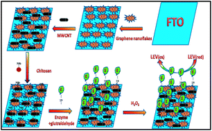
Current research work presents the detection of an antiepileptic drug i.e. leviteracetum by employing electrochemical impedance spectroscopy (EIS) using nanoflakes of graphene (GNF) and multiwalled carbon nanotube (MWCNT) decoratedfluorine-doped tin oxide (FTO) glass as sensing platform. This method also involved the covalent immobilized enzyme i.e. horse radish peroxidase (HRP) for LEV detection. Various stages of biosensor fabrication were characterized by TEM, XRD, SEM, EIS and CV. The sensing surface demonstrated a wide linear range (200 to 1000 μM) and a low detection limit 1 μM. The nano flakes-tubes composite based sensor showed good precision, analytical recovery and anti-interference ability which makes this sensing interface suitable for many pharmaceutical analyses. The applicability of the biosensor is to determine LEV levels in spiked serum samples.


1. Introduction

Nanomaterials have been broadly used in many research areas because of their exceptional physico-chemical properties.1 Nanostructured materials, including nanoflakes, microspheres composed of nanoflakes, microflowers, and nanowires have been used for various applications. Among these, nanoflakes have been extensively used because of their unique and distinct properties. Graphene nano flakes (GNFs) are graphene based materials with a size controllable energy band gap, which may be useful for different technological applications.2,3 Graphene nano-flakes are important due to their potential for fabrication of molecular devices, spintronics and quantum dot technology4 because of their excellent properties like very fast electron transport, the highest mechanical strength and greatest thermal conductivity yet measured.2,3 Hybrid nanocomposites are also of considerable interest because of their exceptional properties.5 Carbon nanotubes (CNTs), in combination with other nanoparticles have been used extensively for electrochemical sensing due to their unique properties such as high electronic conductivity, high surface/volume ratio and ability to promote electron transfer.6,7 CNT-based composites have been used to modify electrodes for fabrication of various biosensors in the determination of various compounds like ascorbic acid,8 glutathione9 hydrogen peroxide10 and choline.11 In the current work we employed multiwalled carbon nanotube wrapped nanoflakes of graphene for electrochemical sensing of an antiepileptic drug i.e. leviteracetum.

Epilepsy is one of the most common neurological disorders, affecting millions of people worldwide. An extremely debilitating condition if left untreated, epilepsy may lead to the loss of consciousness, physical injury, disorientation and loss of self confidence in some cases.12 Drug treatment is the mainstay of any epilepsy management approach, and over the past few years, many new antiepileptic drugs (AEDs) have been developed and licensed for clinical use. Levetiracetam (LEV) is the most recent of the AEDs to be introduced and is indicated as adjunctive therapy for the treatment of partial-onset seizures with or without secondary generalization.13 This drug also applied in the cure of many neurologic diseases like autism and Tourette syndrome.14

Levels of LEV concentration should be monitored to reduce its adverse methods of LEV determination.15 The objective of this study is to evaluate the levels of LEV in pharmaceuticals drugs for therapeutic drug monitoring. Various methods are accessible for determination of LEV among them are gas chromatography (GC),16 LC tandem mass,17 HPLC18 and capillary electrophoresis.19 Most of these methods are precise and suitable for many applications, however, they do not satisfy the requirements for a simple, fast, accurate and specific analysis, as these are complicated, require time-consuming sample pre-treatment, expensive instrumental set-up and skilled person to operate.20 Biosensors are considered a viable alternative to the earlier methods for LEV determination for on-site analysis. Besides being specific and sensitive, they are portable, less expensive and do not require tedious sample pretreatment.

In this report we used electrochemical sensing method like voltammerty and electrochemical impedance spectroscopy (EIS) technique for fast and sensitive detection of LEV. We describe herein preparation of multiwalled carbon nanotubes wrapped nanoflakes of graphene composite on fluorine-doped tin oxide (FTO) glass for sensitive biosensing of LEV. Finally, it is stressed here that our approach provides a promising method for the fabrication of biosensor as multiwalled carbon nanotubes wrapped nanoflakes composite provided large surface area, high electron transfer kinetics and it can also includes the advantages of low-cost when this fabrication strategy miniaturized into chip form.

2. Experimental

2.1 Materials

Leviteracetum was procured from vendors of Pharmaceautical Company. Graphite powder, Sodium dodecylbenzenesulfonate (SDBS), sodium hydroxide solution (NaOH) and chitosan were procured from Sigma. All other chemicals were of analytic reagent grade. Double distilled water (DW) was used throughout the experiments. Stock standard solutions of LEV (were prepared by dissolving the appropriate amount in water).

2.2 Apparatus and methods

Cyclic voltammetry (CV) and electrochemical impedance spectroscopy (EIS) measurements were performed on a potentiostat/galvanostat (Autolab, Eco Chemie, The Netherlands. Model: AUT83785) with a three electrode system consisting of a Pt wire as an auxiliary electrode, an Ag/AgCl electrode as reference electrode and modified fluorine-doped tin oxide (FTO) glass as a working electrode. All the electrochemical experiments were performed at an ambient temperature (25 °C). Scanning electron microscopy (SEM) measurements were carried out at Department of Amity institute of advanced research and studies, Amity University, Noida. Ultrasonication was performed on Misonix Ultrasonic Liquid Processors (mode XL-2000 series).

2.3 Preparation of graphene nanoflakes (GNFs)

Graphene nanoflakes were prepared after some modification in the protocol of literature.21 SDBS surfactant (20 mg ml−1) was prepared in distilled water with continuous stirring for 12 h. Graphite powder (10 mg) and SDBS (15 ml) were mixed together and then subjected to ultrasonication for 30 min. The resulting solution was centrifuged at 1000 rpm for 45 min. After centrifugation, supernatant was collected and sonicated for 20 min. Thus graphene nanoflakes were obtained.

2.4 Preparation of the nanoflakes composite and enzyme modified sensing electrode i.e. FTO

MWCNTs (MWCNTs) and graphene nanoflakes (GNF) were dispersed together in ratio of 3[thin space (1/6-em)]:[thin space (1/6-em)]1 in 2.0 ml distilled water with ultrasonication for half an hour. The multiwalled carbon nanotubes wrapped nanoflakes of graphene composite were decorated by dip coating the surfaces of fluorine-doped tin oxide (FTO) glass with 5% suspension of nano flake-tube composite. After composite deposition, chitosan was dropped onto modified FTO electrode and was kept overnight for physical adsorption. Modified electrode (MWCNTs–GNF/CHIT/FTO) was dipped into glutaraldehyde (2.5%; 1 ml) for cross-linking of enzyme. Electrode was dipped into enzyme solution (50 μl) and kept for 24 h at 4 °C. The electrode was finally washed with 0.1 M phosphate buffer (pH 8.5) to remove any unbound enzyme. Thus modified sensing electrode (HRP/MWCNTs–GNF/CHIT/FTO) was fabricated and kept at 4 °C, when not in use. This working electrode was characterized by SEM and CV at different stages of its fabrication (Scheme 1.).
image file: c4ra14716b-s1.tif
Scheme 1 Schematic representation of various stages of sensing electrode.

2.5 Characterization study of modified sensing electrode

X-ray diffraction (XRD) study of graphite nanoflakes was carried out at Physics Department G. J. University, Hisar, using X-ray diffractometer (Make: Rigaku Mini Flex II, Americas Corporation) and scanning electron microscopy (SEM) studies have been used to characterize nanoflakes composite modified sensing electrode. Electrochemical study was done using electrochemical impedance spectra (EIS) and cyclic voltammetry technique, cell composed of HRP/MWCNTs–GNF/CHIT/FTO as sensing electrode, Ag/AgCl as reference electrode and Pt wire as auxiliary electrode was recorded in PBS (50 mM, pH 6.5, 0.9% NaCl) containing 5 mM [Fe(CN)6]3−/4−. Frequency range: 0.01 Hz to 10 kHz. EIS and CV pattern of various stages of sensing electrode were taken. Various variable conditions like pH, incubation temperature and time were optimized. For evaluation of functioning of biosensor, parameters like precision and accuracy were also studied. Serum samples were acquired from University of Health Sciences (UHS), Rohtak. Various concentrations of LEV were spiked into serum samples. The measurements were performed after successive additions of spiked samples. After each addition, CV was recorded by cycling the potential between −1.0 and +1.0 V at a scan rate of 100 mV s−1.

3. Results and discussions

3.1 Characterization of various stages of fabrication of nanoflakes composite modified sensor

Results for confirmation of prepared graphene nanoflakes are shown below. Fig. 1A reveals the microscopic image of graphene nanoflakes by TEM. Image depicts that graphene nanoflakes were with size 30 nm.
image file: c4ra14716b-f1.tif
Fig. 1 (A) Transmission electron microscope (TEM) image of nanoflakes of graphene and (B). XRD spectra of (a) MWCNTs (b) FTO (c) GNF (d) MWCNTs–GNF.

Fig. 1B illustrates the X-ray diffraction (XRD) pattern of MWCNT, FTO, GNF, MWCNT/GNF. All peaks were consistent with the peaks of MWCNT, FTO, GNF, MWCNT/GNF (JCPDS card). In the XRD pattern of GNF, we can see a sharp diffraction peak at 10.9°, which can be assigned to GNF. In XRD pattern of MWCNT, we can see a sharp diffraction peak at 24.2°. Sharp diffraction peak at 24.2° can be assigned to MWCNT. In the XRD pattern of MWCNT/GNF, we can see a sharp diffraction peak at 10.9° and 24.2° proving that there was conjugation between GNF and MWCNT. In addition, no peaks were observed for other impurities. All results revealed that there is formation of all sensing platform materials.

Modification of electrode (HRP/MWCNTs–GNF/CHIT/FTO) was characterized by SEM, EIS and CV techniques.

Surface morphologies of the sensing electrode was studied by SEM, at an acceleration voltage of 13 kV. Various stages of electrode were characterized i.e. (a) bare FTO (b) MWCNTs–GNF/CHIT/FTO (c) and HRP/MWCNTs–GNF/CHIT/FTO. SEM image of the unmodified electrode showed rough surface which might be due to electrolyte thin film (Fig. 2a). SEM image of the MWCNTs–GNF modified electrode showed some flakes and tube like structures (Fig. 2b) these results confirmed that there was deposition of both nanofakes and nanotubes on the sensing surface while after bioconjugation of enzyme, electronic microstructures reveals that small spherical molecules other than flakes and tubes were also apparent on the surface. So these results confirmed that there is deposition of all sensing materials for detection of analyte (Fig. 2c).


image file: c4ra14716b-f2.tif
Fig. 2 SEM image of (a) bare FTO, (b) MWCNTs–GNF/CHIT/FTO and (c) HRP/MWCNTs–GNF/CHIT/FTO.

CV pattern of (a) HRP/MWCNTs/CHIT/FTO and (c) HRP/GNF/CHIT/FTO in a sodium phosphate buffer 0.05 M (pH 7.0) at a scan rate of 50 mV s−1 in the potential range of −1.0 to +1.0 V s−1 (Fig. 3A). For detection of difference in sensing signal of two differently modified electrode, electrode was scanned in the potential range of −1.0 to +1.0 V s−1. It was observed that there is amplification of current when MWCNT modified electrode was used as compared to graphene nanoflakes modified electrode. After that multiwalled carbon nanotubes wrapped nanoflakes of graphene composite was used to depict the synergistic effect of composite on electrode.


image file: c4ra14716b-f3.tif
Fig. 3 (A) CV pattern of (a) HRP/MWCNTs/CHIT/FTO and (c) HRP/GNF/CHIT/FTO in a sodium phosphate buffer 0.05 M (pH 7.0) at a scan rate of 50 mV s−1 in the presence of LEV. (B) CV pattern of (a) bare FTO, (b) MWCNTs–GNF/CHIT/FTO and (c) HRP/MWCNTs–GNF/CHIT/FTO in a sodium phosphate buffer 0.05 M (pH 7.0) at a scan rate of 50 mV s−1 in the presence of LEV. (C) EIS of (a) bare FTO, (b) MWCNTs–GNF/CHIT/FTO and (c) HRP/MWCNTs–GNF/CHIT/FTO containing 1 mM Fe(CN)63−/4− with 0.1 M KCl at 0.20 mV s−1 (frequency range of 0.01 Hz to 10 kHz) in the presence of LEV.

CV pattern of (a) bare FTO, (b) MWCNTs–GNF/CHIT/FTO and (c) HRP/MWCNTs–GNF/CHIT/FTO in a sodium phosphate buffer 0.05 M (pH 7.0) at a scan rate of 50 mV s−1 in the presence of LEV (Fig. 3B) in the presence of LEV. For detection in the difference of sensing signal, bare electrode (FTO) was scanned in the potential range of −1.0 to +1.0 V s−1 insignificant sensing signal was explicted, but there is increase in sensing signal when graphene nanoflakes and MWCNT get deposited (MWCNTs–GNF/CHIT/FTO) onto FTO electrode which proved that synergistic effect of both nanotubes and nanoflakes produces amplified signal (curve b). After introduction of enzymes on the sensing electrode (HRP/MWCNTs–GNF/CHIT/FTO), signal was greater than before as enzyme initiate and catalyzes redox reactions of LEV which generates more sensing signal (curve c). Additionally nano flakes-tube composite might provide large and effective surface for immobilization of enzyme which causes close proximity of enzyme and analyte and make the redox reactions fast.

Another evidence for modification of electrode was done by electrochemical impedance spectroscopy (EIS) technique. Electrochemical impedance spectroscopy (EIS) is an efficient way for analysis of the properties of a surface modified electrode. Semi-circle portion at higher frequencies corresponds to the electron transfer limited process and the linear portion at lower frequencies may attribute to diffusion process. Nyquist diameter (real axis value at lower frequency intercept) gives value of charge transfer resistance (RCT) i.e. hindrance provided by the electrode material to transfer of charge from solution to the electrode that can be correlated with the modification of surface. EIS was done at different phases of construction of sensing electrode. EIS of (a) bare FTO, (b) MWCNTs–GNF/CHIT/FTO and (c) HRP/MWCNTs–GNF/CHIT/FTO containing 1 mM Fe(CN)63−/4− with 0.1 M KCl at 0.20 mV s−1 (frequency range of 0.01 Hz to 10 kHz) in the presence of LEV. Nano flakes-tube composite based sensing electrode produce smaller resistance charge transfer (800 Ω) compared to unmodified FTO electrode (1000 Ω) while after introduction of enzyme on the surface of sensing electrode in the presence of LEV, the Rct get further decreased (700 Ω) (Fig. 3C). Decrease in Rct value can be explained that after introduction of enzyme catalysed redox reaction was occurred and reaction rate become faster. Principle behind electrochemical sensing of LEV drug is oxidation of HRP by H2O2 to an intermediate compound, which is subsequently reduced by a substrate donor or directly at the electrode, regenerating the native enzyme as LEV is a pyrrolidine derivative that can act as a substrate donor for the reduction of the HRP previously oxidized by H2O2.

Results proved that sensing electrode was modified with nano flakes-tubes composite and enzyme. Above illustrations also confirmed that nano flakes-tubes composite based sensing interface can prove to be excellent platform for electrochemical sensing of various pharmaceutical drugs.

3.2 Optimization & analytical performances of nano flakes-tubes composite modified sensor

Experimental variables were also studied for optimization so that maximum response can be achieved for electrochemical sensing of LEV.

Influence of the pH value was observed on the sensing signal of nano flakes-tubes composite modified sensor. Value of pH was varied between 4.0 and 8.0 in 0.05 M PBS. Optimum pH observed for sensing interface was pH 7.0 (Fig. 4a). Variation in sensing signal was also observed after change in temperature. Sensing signal observed maximum at 45 °C (Fig. 4b). Effects of serum interferents such as glucose, uric acid, urea and cholesterol were also studied in order to check the selectivity of the sensor. Serum interferants does not affect electrochemical sensing of LEV.


image file: c4ra14716b-f4.tif
Fig. 4 Effects of pH (a) and temperature (b) on the electrochemical response of fabricated nano flakes-tubes composite LEV biosensor in 0.1 M sodium phosphate buffer.

For evaluating the sensor, various parameters like analytic recovery, precision and accuracy of proposed method were also studied. Analytic recovery of known amount of added LEV was determined by the present biosensor. The mean analytic recoveries of added 200 and 400 μM LEV were 99.5 ± 1.4 and 98.9 ± 1.1 respectively. For checking the reproducibility and reliability of the present biosensor LEV content in ten spiked serum samples was estimated on single day (within batch) and five times again after storage at −20 °C (Table 1). The accuracy of the proposed method was determined by spiking serum samples with different concentrations of LEV. Accuracy of proposed method was 99%.

Table 1 Determination of LEV by nano flakes-tubes composite sensor in human spiked serum samples
S. no. Spiked serum samples (μM) Present method (μM)
1 10 15
2 20 22
3 40 40
4 60 62
5 80 81
6 100 100
7 200 202
8 300 310
9 400 405
10 500 500


3.3 Impediometric detection of antiepileptic drug i.e. LEV by nano flakes-tubes composite modified sensor

The RCT difference was observed after varying the concentration of LEV in the range of 200 to 1000 μM. EI spectra were recorded in 5 mM [Fe(CN)6]3−/[Fe(CN)6]4− containing 0.1 M KCl with incubation time of 2 s (Fig. 5). The incubation time was kept 2 s, after that analytical signal was produced. With increase in concentration value of Rct gets decreased as it is evident from Fig. 5. The results of experiments carried out in duplicate sets reveal reproducibility of the system within 1%. Limit of detection was found to be 1 μM. The electron transfer resistance decreased with the increasing concentrations of LEV, which gives rise to a linear-type detection response from 200 to 1000 μM, and the regression equation obtained with a correlation coefficient of R2 = 0.97. From these observations, we can conclude that the composite modified electrode provides highly conductive and expected as a good podium for sensing applications. Calibration was also performed using CV with increasing concentration of LEV. The peak current is linearly related to the various concentrations of LEV (Fig. 6a and b).
image file: c4ra14716b-f5.tif
Fig. 5 Impediometric response of nano flakes-tubes composite based sensor for LEV detection.

image file: c4ra14716b-f6.tif
Fig. 6 (a) Linear response of resistance versus concentration of LEV (substrate concentration/μM) (I/mA). (b) CVs response of modified electrode using different concentrations of substrate ranging between 200 and 1000 μM.

Stability of the sensing electrode was also observed. Value of Rct is increased to 4% after 1 week while Rct increases after 10 weeks resulting in about 40% loss in activity.

A comparison of analytic parameters of various nanoparticles based biosensors for detection of LEV with the present biosensor is summarized in Table 2.

Table 2 Comparison of the present method with other biosensing methodsa
Matrix/method Enzyme Response time Detection limit (μM) Linearity (μM) Stability References
a NR= not reported.
HPLC-UV/chromatography Non enzymatic NR 0.1 1–75 NR 19
Screen-printed carbon electrodes/cyclic voltammetery HRP NR 17.5 100 to 830 NR 20
HRP/GNF–MWCNT/FTO/impediometric and CV HRP 2 s 1 200 to 1000 1 month Present


4. Conclusions

In this we used MWCNT wrapped GNF composite on FTO glass as sensing interface for sensitive biosensing of leviteracetum (LEV). The fabricated technique provided wide linear range (200–1000 μM), good value of evaluation parameters (analytical recovery: 99.5 ± 1.4 and 98.9 ± 1.1 & accuracy 99%) and anti-interference ability. Fabrication of this sensing interface is easy as compared to other sensing matrix. Moreover our approach provides a promising method for the fabrication of biosensor as multiwalled carbon nanotubes wrapped nanoflakes composite provided large surface area and high electron transfer kinetics.

Acknowledgements

The present work was supported to one of the author (Jagriti Narang) by SERB, Department of Science and Technology (DST), India. Thanks to all scientists referenced throughout the paper whose valuable work has guided the way through to this research work.

References

  1. K. O. Varghese, D. Gong, M. Paulose, K. G. Ong and C. A. Grimes, Sens. Acuta. B: Chemical, 2003, 93, 338–344 CrossRef.
  2. D. S. L. Abergel, V. Apalkov, J. Berashevich, K. Zieler and T. Chakraborty, Adv. Phys., 2010, 59, 261–482 CrossRef CAS.
  3. A. H. C. Neto, F. Guinea, N. Peres, K. Novoselov and A. Geim, Rev. Mod. Phys., 2009, 81, 109 CrossRef.
  4. Y. W. Son, M. L. Cohen and S. G. Louie, Phys. Rev. Lett., 2006, 97, 216803 CrossRef.
  5. C. L. Sun, H. H. Lee, J. M. Yang and C. C. Wu, Biosens. Bioelectron., 2011, 22, 3450–3455 CrossRef PubMed.
  6. H. T. Zhao and H. X. Ju, Anal. Biochem., 2006, 350, 138–144 CrossRef CAS PubMed.
  7. B. Y. Weia, M. C. Hsub, P. G. Suc, H. M. Linb, R. J. Wud and H. J. Laia, Sens. Acuta. B: Chemical, 2004, 101, 81–89 CrossRef PubMed.
  8. N. Chauhan, J. Narang and C. S. Pundir, Analyst, 2011, 136, 1938–1945 RSC.
  9. J. Narang, N. Chauhan, P. Jain and C. S. Pundir, Int. J. Biol. Macromol., 2012, 50, 672–678 CrossRef CAS PubMed.
  10. J. Narang, N. Chauhan and C. S. Pundir, Analyst, 2011, 136, 4460–4466 RSC.
  11. S. Pundir, N. Chauhan, J. Narang and C. S. Pundir, Anal. Biochem., 2012, 427, 26–32 CrossRef CAS PubMed.
  12. B. S. Chang and D. H. Lowenstein, N. Engl. J. Med., 2003, 3, 1257–1266 CrossRef PubMed.
  13. UCB Pharmaceuticals Ltd. Keppra® (levetiracetam), Summary of product characteristics, Watford, UK, September 2004 Search PubMed.
  14. M. U. Farooq, A. Bhatt, A. Majid, R. Gupta, A. Khasnis and M. Y. Kassab, Am. J. Health-Syst. Pharm., 2009, 66, 541–561 CrossRef CAS PubMed.
  15. B. A. Khalil, Neuropsychiatr. Dis. Treat., 2008, 4, 507 CrossRef.
  16. N. Isoherranen, M. Roeder, S. Soback, B. Yagen and V. Bialer, J. Chromatogr. B: Biomed. Sci. Appl., 2000, 745, 325–332 CrossRef CAS.
  17. K. Matar, J. Pharm. Biomed. Anal., 2008, 48, 822–828 CrossRef CAS PubMed.
  18. J. Martens-Lobenhoffer and S. M. Bode-Boger, J. Chromatogr. B: Anal. Technol. Biomed. Life Sci., 2005, 819, 197–200 CrossRef CAS PubMed.
  19. Z. K. Shihabi, K. Oles and M. Hinsdale, J. Chromatogr. A, 2003, 1004, 9–12 CrossRef CAS.
  20. J. Narang and C. S. Pundir, Int. J. Biol. Macromol., 2011, 49, 707–715 CrossRef CAS PubMed.
  21. Y. C. Lai, D. Y. Wang, I. S. Huang, Y. T. Chen, Y. H. Hsu, T. Y. Lin, H. F. Meng, T. C. Chang, Y. J. Yang, C. C. Chen, F. C. Hsu and Y. F. Chen, J. Mater. Chem. C., 2013, 1, 2–559 RSC.

This journal is © The Royal Society of Chemistry 2015
Click here to see how this site uses Cookies. View our privacy policy here.