The electrocatalytic activity of IrO2–Ta2O5 anode materials and electrolyzed oxidizing water preparation and sterilization effect

Zhandong Ren*, Shanshan Quan, Jie Gao, Wenyang Li, Yuchan Zhu*, Ye Liu, Bo Chai and Yourong Wang
School of Chemical and Environmental Engineering, Wuhan Polytechnic University, Wuhan, 430023, China. E-mail: renzhandong@163.com; zhuyuchan@163.com; Tel: +86 27 83943956

Received 16th November 2014 , Accepted 18th December 2014

First published on 18th December 2014


Abstract

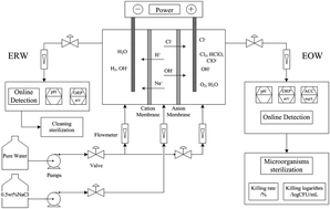
Ti/IrO2–Ta2O5 anode electrocatalysts with different contents and preparation temperatures were prepared by thermal decomposition in this work. The crystallite characterization and morphology were examined via XRD and SEM. The electrochemical properties were examined via cyclic voltammetry (CV) in 0.5 M H2SO4 and linear sweep voltammetry (LSV) in saturated sodium chloride. Through the study of different series of Ti/IrO2–Ta2O5 anodes, we find that the preparation conditions have a great impact on the electrode catalytic activity. Experimental results indicate that the electrochemically active surface area is determined by the content and morphology of the anode coating. When the IrO2 content and the preparation temperature are 70% and 500 °C, the surface of the electrode is aggregated with segregated crystallite flower-like particles, which brings about the best electrode catalytic activity. The current density in the chlorine evolution reaction of IrO2–Ta2O5 (70% and 500 °C) is 0.4 A cm−2 in saturated sodium chloride. The properties and sterilization effect of EO water are closely related to the electrode catalytic activity. The higher the current density is in chlorine evolution, the higher the available chlorine and HClO content. When the IrO2 content is 70% and the preparation temperature is 500 °C, the maximum values of the killing logarithm value and killing rate are 3.01–3.05 and 99.9023–99.9109%, respectively. In addition, when the Ti substrate undergoes 40 minutes of activation treatment, the Ti/IrO2–Ta2O5 anodes have the highest stability.


1. Introduction

Diseases caused by food are widespread throughout the world. Food-borne diseases are the most prominent health problems in the world and are directly related to people's lives, safety and health. Therefore, new food sterilization technology which has wide adaptability, high efficiency, broad spectrum, safety and no residues is very important for the food industry. Electrolyzed oxidizing water (EO water), with low pH (2.2–2.7), high oxidation–reduction potential (ORP, >1100 mV), and an available chlorine content of 30 to 80 mg L−1, has high bactericidal activity and wide adaptability against many food-borne pathogens.1 EO water has been designated as a food fungicide in Japan2 and has high sterilization ability for agricultural products, including lettuce3 (Escherichia coli); carrot4 (aerobic bacteria, molds, yeasts); fish5 (Listeria monocytogenes, Morganella morganii); shrimp6 (Vibrio parahaemolyticus); pork7–9 (Escherichia coli, Listeria monocytogenes); egg10 (Salmonella enteritidis); apple11 (Escherichia coli, Listeria innocua, Salmonella choleraesuis); peanut12 (aflatoxin B1); and cabbage2 (Escherichia coli). In addition, several studies have shown that EO water can be effective in killing fungi (Candida spp.; Aspergillus spp.), blood viruses (Hepatitis B virus; hepatitis C virus; human immunodeficiency virus) and toxins (Staphylococcal enterotoxin-A; Aspergillus parasiticus aflatoxin).1 Regarding the sterilization mechanism of EO water, there are two main theories: one is physical (high ORP and low pH),13–19 and the other is chemical (available chlorine concentration and active oxygen).20–25 Nevertheless, there is no definite conclusion about the disinfection targets of EO water for lack of authentic evidence at the molecular biological level.

EO water is generated by electrolysis of a dilute salt (NaCl) solution in an electrolysis chamber where anode and cathode electrodes are separated by a membrane. In 2005, Hsu first explored the influence of velocity, salinity and temperature on the preparation of EO water.26,27 However, little information concerning preparation of the electrode materials has been investigated until now.28 For example, commercial electrode materials were still dominated by platinum for a long time because of its electrolysis stability. However, the performance in chlorine evolution of platinum is very poor compared with that of ruthenium (see Table S1 and ref. 29). Ruthenium has good activity not only for chlorine evolution in saturated sodium chloride solution used in the chlor-alkali industry,29–35 but also for oxygen evolution in sulfuric acid or sodium sulfate solution.36–41 It should be noted that the chloride concentration (0.05 wt%) in EO water preparation is much lower than that in the chlor-alkali industry. Thus, a large quantity of oxygen would be produced by an evolution reaction in the electrolysis process of EO water preparation, which may result in low current efficiency. This would destroy the low-oxygen solid solution structure of the oxide electrode surface, resulting in a reduction in electrolysis efficiency and lifetime of the electrode.42–46 Iridium has a stable structure in acid medium and can effectively catalyze the oxygen evolution reaction.33,34,46–52 Although there are a few literature references about the chlorine evolution reaction of IrO2 electrodes,47,53,54 the activity of the chlorine evolution reaction on iridium is also poor in contrast to ruthenium (see Table S1). So the activity of the chlorine evolution reaction on iridium should be further enhanced and this is very important for EO water preparation. It would enhance the activity in the chlorine evolution reaction of the electrode through alloying with other metals.28

In this work, IrO2–Ta2O5 electrocatalysts with different contents and preparation temperatures were prepared by thermal decomposition. XRD, SEM and XRF characterizations were employed to study the performance of the IrO2–Ta2O5. The relationship between the physicochemical properties of the IrO2–Ta2O5 electrode and its electrochemical behavior was established through cyclic voltammograms (CV) and the chlorine evolution reaction (CER). Furthermore, the values of pH, ORP, ACC and the sterilization effect of EO water using a series of IrO2–Ta2O5 electrodes (different compositions and preparation temperatures) as anode materials has been systematically discussed in this article. In addition, the stabilities of IrO2–Ta2O5 electrodes with different activation treatment times of the Ti substrate were also investigated for their further industrial application.

2. Materials and methods

2.1 Electrode preparation

A titanium plate of grade TA1 (1 cm × 1 cm) was utilized as the electrode substrate, which was sandblasted, degreased in acetone and then in boiling 10% oxalic acid at 96 °C for 2 h to produce a gray surface with uniform roughness. The preparation of the titanium-based IrO2–Ta2O5-coated anode by the conventional method of thermal decomposition is described elsewhere in detail.33,47,55–57 The precursors H2IrCl6 and TaCl5 were dissolved in 1[thin space (1/6-em)]:[thin space (1/6-em)]1 volume ratio isopropanol and ethanol mixed solutions, in which the total metal concentration was kept at around 0.2 mol L−1. When the mole ratios of H2IrCl6 and TaCl5 were 1[thin space (1/6-em)]:[thin space (1/6-em)]18, 4[thin space (1/6-em)]:[thin space (1/6-em)]12, 6[thin space (1/6-em)]:[thin space (1/6-em)]8, 7[thin space (1/6-em)]:[thin space (1/6-em)]6 and 9[thin space (1/6-em)]:[thin space (1/6-em)]2, titanium-based IrO2–Ta2O5-coated anodes with nominal contents of 10%, 40%, 60%, 70% and 90% IrO2 would be obtained. The precursors were applied with a brush to the pretreated titanium substrate, which was dried at 100 °C for 10 min in an oven, and then sintered in a furnace at 500 °C for 10 min. The above steps were repeated 10 times, with a total oxides loading of about 1.5 mg cm−2, after which the samples were heated at the same annealing temperature for 1 h.

2.2 Material characterization

X-ray diffraction (XRD) was used to analyze the structure of the electrode coating. The inspection was carried out at room temperature on a D/Max-IIIA diffractometer (Rigaku, Japan), using Cu Kα radiation operating at 40 kV and 30 mA. The surface morphology of the oxide electrode was characterized by scanning electron microscopy (SEM: S-3000N, Hitachi Co., Japan). The analysis of the composition of the electrode was carried out by X-ray fluorescence (XRF: EDX-720, Shimadzu, Japan).

2.3 Electrochemical measurements

All the electrochemical measurements were carried out in a typical three-electrode electrochemical glass cell. A platinum plate was used as the counter electrode, and KCl saturated calomel electrode (SCE) as the reference. Cyclic voltammetry (CV) measurements were performed in 0.5 mol L−1 H2SO4 solution at 25 °C with a CHI760D potentiostat. Linear sweep voltammetry measurements were performed in saturated NaCl solution. Accelerated electrolysis life tests were carried out under conditions of 0.5 mol L−1 H2SO4 solution, 2 A cm−2 anodic current density, Ti plate as counter electrode and temperature 40 °C. For electrochemical tests, the surface of the specimen was covered with epoxy resin except for the working area (1 cm2) on one side.

2.4 Preparation and analysis of EO water

The generation of EO water involved electrolysis of NaCl in a three-compartment cell containing inert positively charged and negatively charged electrodes separated by an anion membrane and a cation membrane as shown in Fig. 1. The anode was IrO2–Ta2O5 and the cathode was titanium plate. A salt solution (0.5 wt% NaCl) was pumped into the EO water generator. The current densities were 40 mA cm−2 and 160 mA cm−2 and the electrode distance was 2 cm. Two types of water possessing different characteristics were generated: electrolyzed oxidizing water (pH < 2.6, ORP > 1100 mV) containing dilute hypochlorous acid (HOCl) was produced from the anode side with approximately 20–200 mg L−1 free chlorine, and electrolyzed reduced water (pH > 11, ORP < −795 mV) containing dilute NaOH was produced by the cathode.
image file: c4ra14671a-f1.tif
Fig. 1 Schematic representation of the electrolyzed oxidizing water preparation, detection and sterilization.

The values of pH and ORP were analyzed by a pH meter (3-Star, Thermo Orion). The concentration of total active chlorine dissolved in the solution was determined using the TMB (3,3′,5,5′-tetramethylbenzidine) colorimetric method. In this method, TMB was oxidized to form a yellow product and its concentration was analyzed immediately using a spectrophotometer (UV-2102PC, UNICO, US) at 450 nm. The content of HClO was measured directly by a spectrophotometer at 233 nm, where the absorption coefficient ε was 100.58 These were measured three times and the average values were obtained.

2.5 Bactericidal effect of EO water

Bacillus subtilis var. niger (ATCC 9372) (purchased from China General Microbiological Culture Collection Center) was employed as an indicator bacterium in all disinfection experiments. The initial population of B. subtilis for each disinfection experiment was approximately 7.53[thin space (1/6-em)]log[thin space (1/6-em)]CFU mL−1. During the experiments, 1.0 mL bacteria suspension was mixed with 9.0 mL EO water in a sterile tube and was immediately quenched with excess Na2S2O3 (10 mmol L−1) to eliminate residual disinfectants in the sample solution after 30 min.

The survival of B. subtilis was determined by the colony counting method using a tryptic soy agar (TSA) plate with serial dilutions in Butterfield's phosphate buffer (pH 7.2–7.4). To prepare the counting plate, 1 mL of each appropriate dilution of B. subtilis culture was mixed carefully with 15 mL TSA at 45–47 °C and then transferred into a Petri dish at ambient temperature for solidification. After complete solidification, an overlay adding 5 mL to 10 mL VRBL was poured onto the surface of the inoculated medium. The plates were incubated at 35 °C for 48 h and the colonies formed on the plates were counted. Significant differences between treatments were established at a significance level of p = 0.05.

3. Results and discussion

3.1 Composition analysis of IrO2–Ta2O5

The actual compositions of the oxide electrodes with different proportions and preparation temperatures were examined by XRF and the results are listed in Table S2. The majority of electrode actual compositions are consistent with the nominal compositions, such as 60% (450 °C), 70% (450 °C, 500 °C, 550 °C, 650 °C) and 90% (450 °C). However, for the 10% sample, there is a large deviation between the actual composition and the nominal composition. A possible reason is that when the content of TaCl5 is too high, hydrolysis of TaCl5 will easily occur due to its unstable nature and reduce the content of TaCl5 in a solid solution.

3.2 Effect of IrO2 content on surface morphology

Since the kinetics of the chlorine evolution reaction is very fast, the main active site is the outer surface. In order to improve the catalytic activity of a chlorine electrocatalyst it is necessary to specifically increase the outer surface area. So the surface morphology plays an important role in the chlorine gas-evolving catalysis reaction. As can be seen from Fig. 2, dried-mud cracks appear in all the samples as a result of the sintering process, which is quite typical for oxide electrodes. The reasons are the quick volatilization of organic solvents and the stress caused by different coefficients of thermal expansion between the substrate and the coatings. But the surface morphologies are different with varying IrO2 content in the coating. It can be seen that there are islands separated by wide and deep cracks and a few crystallite particles in Ti/IrO2–Ta2O5 anodes with 10% IrO2 in Fig. 2a. In Fig. 2b, it is found that the cracks become fewer and narrower in comparison with the sample in Fig. 2a, while there are some large IrO2 crystallite particles with average sizes about 20–30 nm. As for Ti/IrO2–Ta2O5 anodes with 60% and 70% IrO2 (Fig. 2c and d), the surfaces of the anodes are covered by more and more fine crystal particles in flower-like agglomerates which tend to connect to form networks without cracks. In addition, the film thickness is about 150 μm from a cross-section SEM of a 70% IrO2 anode in Fig. 2f and the oxide crystals are lined up tightly. However, it was found that the crystallite particles disappeared and dried-mud cracks appeared again in Ti/IrO2–Ta2O5 anodes with 90% IrO2 in Fig. 2e, which indicates that IrO2 crystallite agglomerates emerged when the content of IrO2 was too high. So the existence of proper quantities of Ta2O5 modification in the coating results in a typical morphology of the oxide solid solution, which features dried-mud cracks, networks and fine crystal particles.
image file: c4ra14671a-f2.tif
Fig. 2 SEM images for Ti/IrO2–Ta2O5 anodes with (a) 10% IrO2, (b) 40% IrO2, (c) 60% IrO2, (d) 70% IrO2, (e) 90% IrO2, (f) a cross-section of 70% IrO2.

This deduction has been confirmed by XRD curves as shown in Fig. 3. The resulting peak positions and intensities were compared with the JCPDS reference files for IrO2 (no. 15-870), Ta2O5 (no. 25-0922) and Ti (no. 44-1294). There is no evident diffraction peak of Ta2O5 in any XRD curve except 10% IrO2, which indicates that crystallization of Ta2O5 is restrained by the existence of IrO2. The obvious diffraction peaks of β-Ta2O5 (22.8°, 28.7°, 36.8°) only appear in the 10% IrO2 coating, although their intensities are not very strong. IrO2 rutile began to appear in 40% IrO2 films and the peaks of β-Ta2O5 had disappeared. As the IrO2 content reaches 60%, the crystallite phase in the mixture exists entirely as a rutile phase with 110 (27.9°) and 101 (34.6°) crystals. This result indicates that crystallization of Ta2O5 is affected by the IrO2 component. The intensity of the diffraction peaks of IrO2 appears to increase at high IrO2 concentrations (60%, 70% and 90%), which means that aggregation of IrO2 crystallites occurs with increasing content of IrO2 in the coating. In addition, diffraction peaks corresponding to the Ti support are also observed but no TiO2 is detected. Ti peaks may arise from the penetration of X-rays reaching the substrate in some thin areas and/or diffusion into the coating from the substrate during the thermal preparation.33 SEM observation, together with XRD results, indicates that the composition plays a vital role in determining the surface morphology and structure.


image file: c4ra14671a-f3.tif
Fig. 3 X-ray diffraction curves for Ti/IrO2–Ta2O5 anodes with (a) 10% IrO2, (b) 40% IrO2, (c) 60% IrO2, (d) 70% IrO2, (e) 90% IrO2.

3.3 Effect of IrO2 content on electrocatalytic activity

Cyclic voltammograms of Ti/IrO2–Ta2O5 anodes with different contents of IrO2 recorded at a potential scan rate of 20 mV s−1 in 0.5 mol L−1 H2SO4 solution are shown in Fig. 4. The anodes with different IrO2 contents exhibit voltammograms typical of oxide electrodes with pseudocapacitive behavior, while the electrochemical response is typical of a capacitor, exhibiting accurately rectangular mirror images and rapid reversals of the direction of the response current. The process of charge and discharge of IrO2 is as follows: IrOa(OH)b ↔ IrO(a+δ)(OH)(bδ) + δH+ + δe. When the potential was higher than 0.2 V, the process of OH adsorption and further oxidation with oxygen species began. In the cyclic voltammetric curves, the peak at 0.69 V (vs. SCE) corresponds to the conversion of Ir(III)/Ir(IV) and the conversion of Ir(IV)/Ir(V) or Ir(IV)/Ir(VI) is 1.0 V (vs. SCE).30,47,57,59 Regarding the Ir(III)/Ir(IV) transformation, a = 1, b = 1, δ = 1; for the Ir(IV)/Ir(V) transformation, a = 1, b = 2, δ = 1; for the Ir(IV)/Ir(VI) transformation, a = 1, b = 2, δ = 2. The pair of peaks at 0.69 V (vs. SCE) shows good reversibility, which is deduced from the symmetrical shape of the peaks. This indicates that the surface electrochemistry of the electrode is governed by the active component IrO2.
image file: c4ra14671a-f4.tif
Fig. 4 Cyclic voltammograms of Ti/IrO2–Ta2O5 anodes with different contents of IrO2 in 0.5 mol L−1 H2SO4 solution at a sweeping rate of 20 mV s−1.

As shown in Fig. 4, it is quite obvious that the voltammetric current of the Ti/IrO2–Ta2O5 anode with 70 mol% IrO2 content is greater than that of the other anodes. The voltammetric charge capacity (q*) obtained by integration of the cyclic voltammograms indicates the amount of protons exchanged with the solution. Therefore, the value of q* is expected to be proportional to the electrochemically active surface area and is thought to be able to represent the number of electrochemically active sites on the surface.30,47,57 Fig. S1 gives the value of q* at different contents of IrO2. The electrochemically active surface area increases gradually as the content of IrO2 increases in the beginning. When the content of IrO2 reaches 70%, the sample has the highest electrochemically active surface area because of its special morphology, then the electrochemically active surface area of the 90% IrO2 electrode decreases evidently because of the aggregation of IrO2 crystal particles. So the addition of Ta2O5 can improve the dispersion of iridium oxide grains and change the electrode morphology, and therefore increase the electrochemically active surface area. From the above discussion, it is evident that the electrochemically active surface area is determined not only by the content of the active component, but also by the structure and morphology of the anode coating.

In consideration of the fact that the electrolytic production of chlorine and hypochlorous acid from chloride could be used for food disinfection, the electrocatalytic activity was investigated by studying the model chlorine evolution reaction (CER). Linear sweep voltammetry curves of Ti/IrO2–Ta2O5 anodes with different contents of IrO2 are shown in Fig. 5. The results show that, accounting for the intrinsic catalytic activity and morphology,41,44,51,60,61 significant differences in current density still remain at a constant electrode overpotential. In agreement with the electrochemically active surface area, the current in the CER reaches a maximum at an IrO2 content of 70%. This result confirms that the electrochemically active surface area plays an important role in determining the chlorine evolution.


image file: c4ra14671a-f5.tif
Fig. 5 Linear sweep voltammetry curves of Ti/IrO2–Ta2O5 anodes with different contents of IrO2 in saturated sodium chloride solution at a sweeping rate of 20 mV s−1.

Furthermore, the mechanism of the CER was studied via a Tafel plot. In Fig. 6, the Tafel curve shows a deviation from linearity at high overpotentials, requiring correction for ohmic drop in order to permit an appropriate interpretation. A single linear segment is obtained showing the kinetics of the CER is independent of potential. The Tafel slope is 46.8 mV dec−1 for the 70% IrO2 electrode, indicating that desorption is the rate-determining step of the reaction (Volmer–Heyrovsky).34,47 The reaction process is divided into two steps; the second step is the reaction rate-controlling step (S represents the active site).

 
image file: c4ra14671a-t1.tif(1)
 
image file: c4ra14671a-t2.tif(2)


image file: c4ra14671a-f6.tif
Fig. 6 Tafel plot curve of Ti/IrO2–Ta2O5 with 70% IrO2 and preparation temperature 500 °C.

3.4 Effect of IrO2 content on EO water preparation

The properties of EO water prepared by Ti/IrO2–Ta2O5 anodes with different contents of IrO2 are shown in Table 1. The available chlorine content (ACC) and the content of HClO are very important characteristics in EO water preparation.47,62 From Table 1, it is clear that EO water has a maximum value of ACC at an IrO2 content of 70%, which confirms again that the Ti/IrO2–Ta2O5 anode with 70% IrO2 has the best electrocatalytic activity for chlorine evolution. It can also be seen from Table 1 that the higher the chlorine content is, the better the sterilization effect of EO water is. When the IrO2 content is 70%, the maximum values of the killing logarithm value and killing rate are 3.05 and 99.9109%, respectively. So this is the most suitable IrO2 content for the preparation of EO water. Among different forms of chlorine, hypochlorous acid is the main active substance in sterilization; its sterilization efficiency is 80 times that of sodium hypochlorite.62 From Fig. 7, the HClO content is low for 10% IrO2, so the killing logarithm value and killing rate are also lower than for the others. For 60%, 70% and 90% IrO2, the contents of HClO are similar, so the sterilization effects are also similar. However, we can see that the HClO content is the highest for 70% IrO2, so this has the best bactericidal activity.
Table 1 Sterilization effect and EO water physicochemical properties depending on the IrO2 content of Ti/IrO2–Ta2O5 anodes
Content/mol% Physicochemical property Sterilization effect
pH ORP/mV ACC/mg L−1 Killing logarithm valuea/log[thin space (1/6-em)]CFU mL−1 Killing ratea/%
a Positive control group (Bacillus subtilis var. niger): 7.53 log[thin space (1/6-em)]CFU mL−1, killing time: 30 min.
10 2.24 1146 31.64 2.30 99.4988
40 2.24 1149 69.83 2.76 99.8682
60 2.24 1152 85.66 2.92 99.8798
70 2.20 1150 90.17 3.05 99.9109
90 2.17 1150 83.56 2.90 99.8741


image file: c4ra14671a-f7.tif
Fig. 7 Absorbance of HClO from Ti/IrO2–Ta2O5 anodes with different contents of IrO2 at 233 nm.

3.5 Effect of preparation temperature on the surface morphology and electrocatalytic activity

Fig. 8 and 9 show SEM images and X-ray diffraction curves of Ti/IrO2–Ta2O5 anodes with 70% IrO2 prepared at different temperatures. From Fig. 8 and 9, it was observed that the morphology and surface composition of the electrodes are different when prepared at different temperatures. The obvious differences indicate that the temperature has a significant effect on the surface microstructure and the elemental distribution. At a sintering temperature of 450 °C, there is no obvious peak of the oxide component (IrO2) in Fig. 9. The amorphous phases of IrO2 were formed due to incomplete firing of the gel coating. When the sintering temperature was increased to 500 °C, the amount of IrO2 agglomerated particles increased quickly (Fig. 8). As can be seen from the XRD patterns, the diffraction peak of IrO2 is extremely broad and unsymmetrical, which suggests unsatisfactory crystallinity of IrO2. When the sintering temperature rose to 550 °C and 650 °C, the amount of IrO2 agglomerated particles decreased quickly and cracks began to increase and deepen, which greatly reduced the active area.
image file: c4ra14671a-f8.tif
Fig. 8 SEM images for Ti/IrO2–Ta2O5 anodes with 70% IrO2 prepared at different temperatures: (a) 450 °C, (b) 500 °C, (c) 550 °C, (d) 650 °C.

image file: c4ra14671a-f9.tif
Fig. 9 X-ray diffraction curves for Ti/IrO2–Ta2O5 anodes with 70% IrO2 prepared at different temperatures: (a) 450 °C, (b) 500 °C, (c) 550 °C, (d) 650 °C.

Cyclic voltammograms of Ti/IrO2–Ta2O5 anodes prepared at different temperatures tested in 0.5 mol L−1 H2SO4 solution at a sweeping rate of 20 mV s−1 are shown in Fig. 10. It can be seen that the sample prepared at 500 °C has the highest electrochemically active surface area in Fig. S2. Then the electrochemically active surface area decreases evidently with the increase in sintering temperature. The reduction in the active area should be attributed to the high temperature, which leads to grain growth and aggregation. Secondly, Ta enrichment occurs easily at high temperatures, reducing the distribution of the IrO2 active component. This phenomenon can lead to a decrease in the number of active surface sites. In addition, the Ti substrate tends to be oxidized at temperatures higher than 500 °C, leading to a decrease in electrode conductivity and a decline in apparent electrode activity. This result proves again that the electrochemically active surface area is determined by the structure and morphology of the anode coating.


image file: c4ra14671a-f10.tif
Fig. 10 Cyclic voltammograms of Ti/IrO2–Ta2O5 anodes with 70% IrO2 prepared at different temperatures tested in 0.5 mol L−1 H2SO4 solution at a sweeping rate of 20 mV s−1.

Linear sweep voltammetry curves of Ti/IrO2–Ta2O5 anodes prepared at different temperatures tested in saturated sodium chloride solution at a sweeping rate of 20 mV s−1 are shown in Fig. 11. It can be seen that the current during chlorine evolution also reaches a maximum for anodes prepared at 500 °C. This is in line with the above study as shown in Fig. 4 and 5.


image file: c4ra14671a-f11.tif
Fig. 11 Linear sweep voltammetry curves of Ti/IrO2–Ta2O5 anodes prepared at different temperatures tested in saturated sodium chloride solution at a sweeping rate of 20 mV s−1.

3.6 Effect of preparation temperature on EO water preparation

The properties of electrolyzed oxidizing water prepared by Ti/IrO2–Ta2O5 anodes at different preparation temperatures are shown in Table 2. It can be seen that EO water has maximum values of ACC and HClO content when prepared at 500 °C, which also proves again that Ti/IrO2–Ta2O5 anodes prepared at 500 °C have the best electrocatalytic activity for chlorine evolution. It can also be seen from Table 2 that when the preparation temperature is 500 °C, the maximum values of the killing logarithm value and killing rate are 3.01 and 99.9023%, respectively. Therefore, when the anode preparation temperature is 500 °C, this is most suitable for EO water preparation. In Fig. 12, the hypochlorous acid content first increased and then decreased with the increase in preparation temperature. We can see that the HClO content is the highest at 500 °C, which makes it have the best bactericidal activity.
Table 2 Sterilization effect and EO water physicochemical properties depending on the preparation temperature of Ti/IrO2–Ta2O5 anodes
Temperature/°C Physicochemical property Sterilization effect
pH ORP/mV ACC/mg L−1 Killing logarithm valuea/log[thin space (1/6-em)]CFU mL−1 Killing ratea/%
a Positive control group (Bacillus subtilis var. niger): 7.53 log[thin space (1/6-em)]CFU mL−1, killing time: 30 min.
450 2.24 1146 83.51 2.71 99.8050
500 2.29 1150 90.17 3.01 99.9023
550 2.30 1141 83.96 2.90 99.8741
650 2.27 1146 66.22 2.88 99.8682


image file: c4ra14671a-f12.tif
Fig. 12 Absorbance of HClO from Ti/IrO2–Ta2O5 anodes with different preparation temperatures at 233 nm.

3.7 The accelerated lifetime of Ti/IrO2–Ta2O5 anodes

An accelerated lifetime test (ALT) of Ti/IrO2–Ta2O5 anodes with different activation times under constant current electrolysis in 0.5 mol L−1 H2SO4 solution at current density 2 A cm−2 is shown in Fig. 13, showing the change of cell voltage with electrolysis time per unit loading during the ALT. The whole period can be divided into three stages: activation, stable and deactivation periods. In the beginning, the potential stays at the activation stage, when the electrolyte does not penetrate into the inner surface. After a certain time, it tends to stabilize; this period occupies the main portion of the whole electrolysis time. The potential starts to increase abruptly at the end and within a short time (less than 15 h) the anode is deactivated. As can be seen from Fig. 13, the IrO2–Ta2O5-coated Ti substrate without activating treatment has the shortest accelerated life (35 h). The stability of a Ti/IrO2–Ta2O5 anode can be improved significantly when the Ti substrate undergoes activating treatment. Therefore activating treatment for the Ti substrate is an effective method to improve the stability of Ti/IrO2–Ta2O5 anodes. On the other hand, the Ti/IrO2–Ta2O5 anodes differ greatly in terms of anode stability with different activation times. When the activation time increases from 0 min to 40 min, the anode stability also increases significantly. However, when the activation time reaches 60 min, the anode stability decreases. The change in ALT may be attributed to the different structure of the Ti substrate. So the surface morphologies of Ti substrates with different activation times were studied by SEM images as shown in Fig. S3. Fig. S3(a) shows that the Ti substrate has a smooth surface. When activating treatment is used, the surface morphologies become increasingly rough. With the increase in the activation time of the Ti substrate, the corrosion pits formed on the surface of the Ti substrate become deeper. However, if the activation time is too long, it would cause excessive corrosion of the substrate surface. Gas can easily enter electrochemically active sites within the oxide coating, so that it will fall off and electrochemical dissolution will occur, resulting in consumption of electrochemically active sites, and so the accelerated life will be reduced.
image file: c4ra14671a-f13.tif
Fig. 13 Accelerated lifetime test of Ti/IrO2–Ta2O5 anodes with different activation times under constant current electrolysis in 0.5 mol L−1 H2SO4 solution at 2 A cm−2.

4. Conclusions

Through the study of different series of Ti/IrO2–Ta2O5 anodes, it can be seen that the preparation conditions have a large impact on the electrode catalytic activity. The influences of IrO2 content, preparation temperature and activation time on the electrode catalytic activity for Ti/IrO2–Ta2O5 anodes were investigated by SEM, XRD, CV, LSV and accelerated lifetime test. The experimental results indicate that the electrochemically active surface area is determined by the structure and morphology of the anode coating. IrO2 aggregated as segregated crystallite particles at a content of 70% prepared at 500 °C produces the best electrode catalytic activity. The properties and sterilization effect of EO water are closely related to the electrode catalytic activity. When the IrO2 content is 70% and the preparation temperature is 500 °C, the maximum values of the killing logarithm value and killing rate are 3.01–3.05 and 99.9023–99.9109%, respectively. In addition, when the Ti substrate undergoes 40 minutes of activation treatment, the Ti/IrO2–Ta2O5 anodes have the best stability.

Acknowledgements

The authors would like to acknowledge financial support from The National Natural Science Foundation of China (31101370) and The Natural Science Foundation of Hubei Province (2012FFB04803).

References

  1. R. M. S. Thorn, S. W. H. Lee, G. M. Robinson, J. Greenman and D. M. Reynolds, Eur. J. Clin. Microbiol. Infect. Dis., 2012, 31, 641–653 CrossRef PubMed.
  2. H. C. Zhang, Y. C. Zhu, Z. D. Ren, D. Pan, Y. Liu, Y. R. Wang and B. Cai, Trans. Chin. Soc. Agric. Eng., 2013, 29, 277–283 Search PubMed.
  3. L. A. Keskinen, A. Burke and B. A. Annous, Int. J. Food Microbiol., 2009, 132, 134–140 CrossRef PubMed.
  4. S. Koide, D. Shitanda, M. Note and W. Cao, Food Control, 2011, 22, 452–456 CrossRef PubMed.
  5. S. McCarthy and W. Burkhardt III, Food Control, 2012, 24, 214–219 CrossRef PubMed.
  6. J. Xie, X. H. Sun, Y. J. Pan and Y. Zhao, Food Control, 2012, 23, 320–324 CrossRef PubMed.
  7. S. M. E. Rahman, J. Wang and D.-H. Oh, Food Control, 2013, 30, 176–183 CrossRef PubMed.
  8. Z. D. Ren, Y. C. Zhu, Y. Liu, Z. Y. Zhang and Q. Zhang, Trans. Chin. Soc. Agric. Mach., 2009, 12, 139–143 Search PubMed.
  9. Y. Liu, Z. D. Ren, Z. K. Liu and X. W. Zhang, Food Science, 2009, 14, 119–122 Search PubMed.
  10. W. Cao, Z. W. Zhu, Z. X. Shi, C. Y. Wang and B. M. Li, Int. J. Food Microbiol., 2009, 130, 88–93 CrossRef PubMed.
  11. A. Graca, M. Abadias, M. Salazar and C. Nunes, Postharvest Biol. Technol., 2011, 61, 172–177 CrossRef PubMed.
  12. K. Xiong, H. J. Liu and L. T. Li, J. Agric. Food Chem., 2012, 60, 9770–9778 CrossRef PubMed.
  13. C. Kim, Y. C. Hung and R. E. Brachett, J. Food Prot., 2000, 63, 19–24 Search PubMed.
  14. L. B. Liao, W. M. Chen and X. M. Xiao, J. Food Eng., 2007, 78, 1326–1332 CrossRef PubMed.
  15. S. Koseki and K. Itoh, J. Food Prot., 2001, 64, 1935–1942 Search PubMed.
  16. Z. D. Ren, Y. C. Zhu, Y. Liu, X. R. Zhou and Z. Y. Zhang, Chin. J. Prev. Med, 2008, 8, 578–581 Search PubMed.
  17. Z. D. Ren, Y. C. Zhu, Y. Liu, X. R. Zhou and Z. Y. Zhang, Chin. J. Hosp. Pharm., 2009, 17, 1448–1450 Search PubMed.
  18. Z. D. Ren, Y. C. Zhu, Y. Liu and Z. Y. Zhang, J. Chem. Ind. Eng., 2009, 10, 2583–2589 Search PubMed.
  19. J. X. Hao, S. Qiu, H. Y. Li, T. P. Chen, H. J. Liu and L. T. Li, Int. J. Food Microbiol., 2012, 155, 99–104 CrossRef CAS PubMed.
  20. H. Kiura, K. Sano, S. Morimatsu, T. Nakano, C. Morita, M. Yamaguchi, T. Maeda and Y. Katsuoka, J. Microbiol. Methods, 2002, 49, 285–293 CrossRef CAS.
  21. Y. R. Huang, Y. C. Hung, S. Y. Hsu, Y. W. Huang and D. F. Hwang, Food Control, 2008, 19, 329–345 CrossRef PubMed.
  22. J. Jeong, J. Y. Kim and J. Yoon, Environ. Sci. Technol., 2006, 40, 6117–6122 CrossRef CAS.
  23. J. X. Hao, S. Qiu, H. Y. Li, T. P. Chen, H. J. Liu and L. T. Li, Int. J. Food Microbiol., 2012, 155, 99–104 CrossRef CAS PubMed.
  24. S. D. Stan, J. S. Woods and M. A. Daeschel, J. Agric. Food Chem., 2005, 53, 4901–4905 CrossRef CAS PubMed.
  25. T. Mokudai, K. Nakamura, T. Kanno and Y. Niwano, PLoS One, 2012, 9, e46392 Search PubMed.
  26. S. Y. Hsu, J. Food Eng., 2003, 60, 469–473 CrossRef.
  27. S. Y. Hsu, J. Food Eng., 2005, 66, 171–173 CrossRef PubMed.
  28. X. P. Zeng, M. Zhang, X. D. Wang, X. Y. Chen, X. Z. Su and W. W. Tang, J. Electroanal. Chem., 2012, 677–680, 133–138 CrossRef CAS PubMed.
  29. S. Neodoa, D. Rosestolato, S. Ferro and A. D. Battisti, Electrochim. Acta, 2012, 80, 282–291 CrossRef PubMed.
  30. L. K. Xu, Y. L. Xin and J. T. Wang, Electrochim. Acta, 2009, 54, 1820–1825 CrossRef CAS PubMed.
  31. K. S. Exner, J. Anton, T. Jacob and H. Over, Angew. Chem., Int. Ed., 2014, 53, 1–5 CrossRef PubMed.
  32. N. Menzel, E. Ortel, K. Mette, R. Kraehnert and P. Strasser, ACS Catal., 2013, 3, 1324–1333 CrossRef CAS.
  33. Y. Zeng, K. N. Chen, W. Wu, J. R. Wang and S. Lee, Ceram. Int., 2007, 33, 1087–1091 CrossRef CAS PubMed.
  34. M. H. P. Santana and L. A. D. Faria, Electrochim. Acta, 2006, 51, 3578–3585 CrossRef CAS PubMed.
  35. A. R. Zeradjanin, N. Menzel, W. Schuhmann and P. Strasser, Phys. Chem. Chem. Phys., 2014, 16, 13741–13747 RSC.
  36. I. Katsounaros, S. Cherevko, A. R. Zeradjanin and K. J. J. Mayrhofer, Angew. Chem., Int. Ed., 2014, 53, 102–121 CrossRef CAS PubMed.
  37. A. R. Zeradjanin, A. A. Topalova, Q. V. Overmeereb, S. Cherevkoa, X. X. Chen, E. Ventosac, W. Schuhmannc and K. J. J. Mayrhofer, RSC Adv., 2014, 4, 9579 RSC.
  38. V. Petrykin, K. Macounova, O. A. Shlyakhtin and P. Krtil, Angew. Chem., 2010, 122, 4923–4925 CrossRef.
  39. V. Petrykin, K. Macounova, J. Franc, O. Shlyakhtin, M. Klementova, S. Mukerjee and P. Krti, Chem. Mater., 2011, 23, 200–207 CrossRef CAS.
  40. T. Reier, M. Oezaslan and P. Strasser, ACS Catal., 2012, 2, 1765–1772 CrossRef CAS.
  41. R. Y. Chen, V. Trieu, A. R. Zeradjanin, H. Natter, D. Teschner, J. Kintrup, A. Bulan, W. Schuhmann and R. Hempelmann, Phys. Chem. Chem. Phys., 2012, 14, 7392–7399 RSC.
  42. S. Y. Chen, Y. H. Zheng, S. W. Wang and X. M. Chen, Chem. Eng. J., 2011, 172, 47–51 CrossRef CAS PubMed.
  43. V. Petrykin, K. Macounová, M. Okube, S. Mukerjee and P. Krtil, Catal. Today, 2013, 22, 63–69 CrossRef PubMed.
  44. V. Trieu, B. Schley, H. Natter, J. Kintrup, A. Bulan and R. Hempelmann, Electrochim. Acta, 2012, 78, 188–194 CrossRef CAS PubMed.
  45. J. Jirkovský, H. Hoffmannová, M. Klementová and P. Krtil, J. Electrochem. Soc., 2006, 153, E111–E118 CrossRef PubMed.
  46. N. W. Fan, Z. K. Li, L. Zhao, N. M. Wu and T. Zhou, Chem. Eng. J., 2013, 214, 83–90 CrossRef CAS PubMed.
  47. X. L. Zhou, Z. G. Ye, X. Z. Hua, A. H. Zou and Y. H. Dong, J. Solid State Electrochem., 2010, 14, 1213–1219 CrossRef CAS.
  48. L. Vazquez-Gomez, S. Ferro and A. De Battisti, Appl. Catal., B, 2006, 67, 34–40 CrossRef CAS PubMed.
  49. P. Lakshmipathiraj, G. B. Raju, Y. Sakai, Y. Takuma, A. Yamasaki, S. Kato and T. Kojima, Chem. Eng. J., 2012, 198–199, 211–218 CrossRef CAS PubMed.
  50. B. Johnson, F. Girgsdies, G. Weinberg, D. Rosenthal, A. Knop-Gericke and R. Schlögl, J. Phys. Chem. C, 2013, 117, 25443–25450 CAS.
  51. E. Ortel, T. Reier, P. Strasser and R. Kraehnert, Chem. Mater., 2011, 23, 3201–3209 CrossRef CAS.
  52. R. Tolba, M. Tian, J. L. Wen, Z. H. Jiang and A. C. Chen, J. Electroanal. Chem., 2010, 649, 9–15 CrossRef CAS PubMed.
  53. K. C. Pillai, T. O. Kwon, B. B. Park and I. S. Moon, J. Hazard. Mater., 2009, 164, 812–819 CrossRef CAS PubMed.
  54. T. L. Luu, J. Y. Kim and J. Y. Yoon, J. Ind. Eng. Chem., 2015, 21, 400–404 CrossRef PubMed.
  55. L. K. Xu and J. D. Scantlebury, Corros. Sci., 2003, 45, 2729–2740 CrossRef CAS.
  56. J. M. Hu, H. M. Meng, J. Q. Zhang and C. N. Cao, Corros. Sci., 2002, 44, 1655–1668 CrossRef CAS.
  57. Z. W. Yan and H. M. Meng, Rare Met., 2011, 30(5), 439–446 CrossRef CAS.
  58. J. Carrell Morris, J. Phys. Chem., 1966, 70(12), 3798–3805 CrossRef.
  59. S. Ardizzone, C. L. Bianchi, G. Cappelletti, M. Ionita, A. Minguzzi, S. Rondinini and A. Vertova, J. Electroanal. Chem., 2006, 589, 160–166 CrossRef CAS PubMed.
  60. A. R. Zeradjanin, F. L. Mantiab, J. Masaa and W. Schuhmann, Electrochim. Acta, 2012, 82, 408–414 CrossRef CAS PubMed.
  61. H. Z. Cao, D. H. Lu, J. P. Lin, Q. Ye, J. J. Wu and G. Q. Zheng, Electrochim. Acta, 2013, 91, 234–239 CrossRef CAS PubMed.
  62. C. Kim, Y. C. Hung and R. E. Brackett, Int. J. Food Microbiol., 2000, 61, 199–207 CrossRef CAS.

Footnote

Electronic supplementary information (ESI) available. See DOI: 10.1039/c4ra14671a

This journal is © The Royal Society of Chemistry 2015
Click here to see how this site uses Cookies. View our privacy policy here.