Optical, electrochemical, third order nonlinear optical, and excited state dynamics studies of bis(3,5-trifluoromethyl)phenyl-zinc phthalocyanine

Narra Vamsi Krishna a, Varun Kumar Singh a, Debasis Swain b, Soma Venugopal Rao *b and Lingamallu Giribabu *a
aInorganic and Physical Chemistry Division, Indian Institute of Chemical Technology, Hyderabad 500 007, India. E-mail: giribabu@iict.res.in
bAdvanced Centre of Research in High Energy Materials (ACRHEM), University of Hyderabad, Prof. C.R. Rao Road, Hyderabad 500046, Andhra Pradesh, India. E-mail: svrsp@uohyd.ernet.in

Received 12th November 2014 , Accepted 4th February 2015

First published on 5th February 2015


Abstract

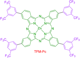
A zinc phthalocyanine with bis(3,5-trifluoromethyl)phenyl groups at peripheral positions has been synthesized and its optical, emission, electrochemical and third-order nonlinear optical properties were investigated. Both the Soret and Q bands were red-shifted and obeyed Beer–Lambert's law. Electrochemical properties indicated that both oxidation and reduction processes were ring centred. Emission spectra were recorded in different solvents and the fluorescence yields obtained were in the range of 0.29 to 0.10, while the time-resolved fluorescence data revealed lifetimes of typically a few ns. Nonlinear optical coefficients were retrieved from the Z-scan measurements using picosecond and femtosecond pulses. Excited state lifetime was deduced from the femtosecond degenerate pump–probe measurements. Our data suggests that this compound has potential for applications in the field of photonics.


Introduction

Nonlinear optical (NLO) materials play a pivotal role in the future evolution of photonics in general and their impact in technology and industrial applications in particular.1–3 The goal is to find and develop materials presenting large nonlinearities and satisfying at the same time all the technological requirements for applications such as wide transparency range, ultrafast response, high damage threshold nonetheless processability, adaptability and interfacing with other materials. NLO active materials displaying both second- and third-order NLO properties such as second harmonic generation, third harmonic generation and optical limiting.4–7 Among all the NLO applications, optical limiting is one of the most promising in practice, used in applications such as the protection of human eyes and optical sensors.8–11 Several mechanisms could lead to optical limiting behaviour, such as reverse saturable absorption, two photon absorption, nonlinear refraction and optically induced scattering.12–14 Organic NLO materials continue attracting attention because of their additional potential application in the fields of optical communications, optical storage, optical computing, harmonic generation, optical switching, etc. In the context of NLO active materials, organic dyes with long π-conjugated structures have attracted consideration owing to their interesting electronic and optical characteristics.15–17

Phthalocyanine type organic dyes with highly conjugated aromatic cyclic structures have thus emerged as exceptional candidates for NLO applications.18 Phthalocyanine derivatives are thermally, optically, and chemically stable.19 Moreover, their NLO properties have been actively studied by many researchers in this field including our group.20–28 Phthalocyanines are remarkable molecules with a characteristic sharp Q-band arising from the π–π* absorption in the 600–700 nm region (Q-bands) and a Soret band in the 300–450 nm region. The sharp Q-bands with high molar extinction coefficient in the far visible region is the source of inspiration for such molecules to be able to find applications in chemical sensors, electrochromic displays, liquid crystals, photodynamic therapy, dye sensitized solar cells and catalysis.29–32 What makes phthalocyanines even more suitable for NLO applications is the choice of modifications in the periphery and the possibility of accommodating more than 70 different elements in the core of the macrocycle directed towards tailoring of these molecules. But a drawback of these macrocycles is their lack of solubility in common organic solvents and their tendency to aggregate at higher concentrations. As a result, it is important to use specific functional groups at the periphery for optimal structural design to target particular applications. Recently, we have introduced sterically hindered phthalocyanine derivatives for applications in non-linear optical applications with strong NLO coefficients possibly because of the long-lived lifetimes.33 In our efforts towards the synthesis of an efficient phthalocyanine macrocycle capable of achieving strong NLO responses we have designed bis-trifluromethyl phenyl substituted phthalocyanines. The trifluromethyl group is strongly electron withdrawing and bulky with a van der Waals radius of 2.2 Å or more, and we therefore anticipated that the introduction of trifluromethyl groups onto the phthalocyanine periphery would impact on the electronic and steric properties of the macrocycle. Also, fluorine compounds are known to enhance the solubility of phthalocyanine in polar aprotic solvents as well as shift the redox processes towards positive potentials. Some groups have previously reported phthalocyanines with trifluromethyl groups.34–36 However, to the best of our knowledge no studies of NLO applications have so far been reported. We report here a new phthalocyanine molecule with conjugated 3,5-bis(trifluromethyl)phenyl substituents at the four peripheral positions as shown in Scheme 1. We have studied the aggregation properties, electrochemical, spectro-electrochemical, excited state dynamics and the NLO properties of the new zinc phthalocyanine.


image file: c4ra14390f-s1.tif
Scheme 1 Synthetic route of TFM-Pc.

Experimental section

Materials

4-Iodophthalonitrile, bis(3,5-trifluoromethyl)phenylboronic acid, 1-pentanol, 1,8-diazabicyclo[5.4.0] undec-7-ene and bis(triphenylphosphine)palladium(II) dichloride were purchased from Sigma Aldrich and were used as supplied. All the solvents viz., dichloromethane, tetrahydrofuran, dimethyl sulfoxide, N,N-dimethylformamide, toluene, chloroform, methanol were purchased from SD Fine chemicals limited, India and were dried before further use. Zinc(II) acetate and sodium carbonate were purchased from Qualigens Chemicals Ltd, India.

Synthesis

Bis(3,5-trifluoromethyl)phenylphthalonitrile (1). A sealed tube with a teflon cap was charged with 4-iodophthalonitrile (0.32 g, 1.26 mmol), 3,5-bis(trifluoromethyl)phenyl boronic acid (1.0 g, 3.88 mmol) and a catalytic amount of bis(triphenylphosphine)palladium(II) dichloride and then flushed with nitrogen for 15 minutes before the addition of tetrahydrofuran. To this 2 M sodium carbonate (aq.) was added and the sealed tube was capped under a nitrogen atmosphere. The reaction mixture was stirred at 70 °C for 6 h. After cooling to room temperature, the reaction mixture was washed twice with water. The combined organic layers were washed once with water, subsequently dried over anhydrous Na2SO4, filtered and concentrated using a rotary evaporator. The resultant residue was subjected to silica gel column chromatography and eluted with hexane–ethyl acetate (90/10% v/v) to obtain the desired product as a white powder. Elemental analysis of anal. calcd for C16H6F6N2% (340.23): C, 56.48; H, 1.78; N, 8.23. Found: C, 56.45; H, 1.79; N, 8.24. ESI-MS (m/z): C16H6F6N2 [340.23]: M+ 343. 1H NMR (300 MHz, CDCl3): δ (ppm) 8.05 (m, 1H), 8.02 (s, 3H), 7.98 (d, JHH = 1.22, 2H); FT-IR (KBr) νmax 2238 (CN) cm−1.
TFM-Pc . A mixture of anhydrous zinc acetate (1.90 g, 10 mmol), bis(3,5-trifluoromethyl)phenylphthalonitrile (1) (1.70 g, 5 mmol), DBU (catalytic amount) and dry 1-pentanol (5 ml) was refluxed at 150 °C for 16 h under a nitrogen atmosphere. After cooling, the solution was precipitated by addition of methanol, filtered and dried. The obtained solid material was subjected to silica gel column chromatography and eluted with dichloromethane. The green coloured band was collected and recrystallized from methanol, to get the desired compound in 70% yield. Elemental analysis of anal. calcd for C64H24F24N8Zn% (1426.29): C, 53.90; H, 1.70; N, 7.86. Found: C, 53.81; H, 1.74; N, 7.85. MALDI-TOF (m/z): C64H24F24N8Zn [1426.29]: 1426.15 (M+), FT-IR (KBr) νmax 2928, 2840, 1254 and 1024 cm−1. UV-vis: (in DCM, λmax, ε M−1 cm−1) 682 (99[thin space (1/6-em)]800), 616 (27[thin space (1/6-em)]000), 349 (51[thin space (1/6-em)]400).

Instrumentation

Absorption spectra were recorded with a Shimadzu UV-3600, UV-visible-NIR spectrophotometer. Steady-state fluorescence spectra were recorded using a Fluorolog-3 spectrofluorometer (Spex model, JobinYvon) for solutions with optical density at the wavelength of excitation (λex) ≈ 0.05. Fluorescence quantum yields (ϕ) were estimated by integrating the fluorescence bands with zinc tert-butyl phthalocyanine (ϕ = 0.37 in benzene).37 Fluorescence lifetime measurements were carried on a picosecond time-correlated single photon counting (TCSPC) setup (FluoroLog3-Triple Illuminator, IBH Horiba JobinYvon) employing a picosecond light emitting diode laser (NanoLED, λex = 670 nm) as excitation source. The decay curves were recorded by monitoring the fluorescence emission maxima of the phthalocyanine macrocycle (λem = 700 nm). Photomultiplier tube (R928P, Hamamatsu) was employed as the detector. The lamp profile was recorded by placing a scatterer (dilute solution of Ludox in water) in place of the sample. The width of the instrument function was limited by the full width at half maximum (FWHM) of the excitation source, ∼635 ps at 670 nm. Decay curves were analyzed by nonlinear least-squares iteration procedure using IBH DAS6 (version 2.3) decay analysis software. The quality of the fits was judged by the χ2 values and distribution of the residuals.

Electrochemical measurements were performed on a PC-controlled CH instruments model CHI 620C electrochemical analyzer. The experiments were performed in a 1 mM phthalocyanine solution in CH2Cl2 at scan rate of 100 mV s−1 using 0.1 M tetrabutyl ammonium perchlorate (TBAP) as supporting electrolyte. The working electrode was glassy carbon, a standard calomel electrode (SCE) was the reference electrode and platinum wire was an auxiliary electrode. After a cyclic voltammogram (CV) had been recorded, ferrocene was added, and a second voltammogram was measured. The optical thin layer electrochemical studies were carried on Maya 2000 Ocean Optics software using DT-MINI-2-GS, UV-vis-NIR LIGHTSOURCE. 1H NMR spectra were recorded in CDCl3 solutions on AVANCE 300 MHz spectrophotometer using TMS as standard. FT-IR spectra (KBr pellets) were recorded on a Perkin Elmer spectrophotometer. Mass spectra were acquired using ElectroSpray Ionization (ESI) method, operated in positive ion mode using m/z range 100–2000 and MALDI-TOF analysis.

NLO and pump–probe studies

The Z-scan38–41 measurements were performed using ∼2 picosecond (ps) pulses at 800 nm and ∼70 femtosecond (fs) pulses at 690 nm. The ps/fs experiments were performed on solutions (samples dissolved in THF) with typically 0.7 mM concentration. In the fs case beam waist at focus was estimated to be ∼22 µm with a corresponding Rayleigh range of 2.2 mm. In the ps case beam waist at focus was estimated to be ∼35 µm with a corresponding Rayleigh range of 4.5 mm. The degenerate pump–probe experiments were performed at 600 nm using ∼70 fs pulses from an optical parametric amplifier which was pumped by ∼40 fs pulses from an amplifier (Coherent). Pump–probe power ratios were typically 40–50. The probe diameter was <1 mm. The probe beam was aligned such that during the delay scan it was always located within the pump beam, whose diameter was ~3 mm. Data was acquired through the combination of a photo-diode and lock-in amplifier. Complete details can be found in some of our earlier work.38–41

Results and discussion

The synthetic route to new phthalocyanine TFM-Pc is shown in Scheme 1. This complex was prepared by the template cyclotetramerization of bis(3,5-trifluoromethyl)phenylphthalonitrile (1) with anhydrous zinc acetate in 1-pentanol at reflux temperature under nitrogen atmosphere in the presence of DBU as a non-nucleophilic base catalyst. The phthalocyanine was purified by silica gel column chromatography and characterized by elemental analysis, mass spectrometry, FT-IR, 1H NMR, UV-vis, and emission spectroscopy as well as electrochemical techniques. The elemental analysis of TFM-Pc as shown in the experimental section was found to be satisfactory. The MALDI-TOF mass spectrum of phthalocyanine consists of a molecular ion peak at m/z 1426.15 (M+), which confirms the presence of the phthalocyanine macrocycle (see ESI). The absence of a ‘–CN’ stretching group in the FT-IR spectrum of phthalocyanine further supports the macrocycle formation.

UV-visible absorption studies

Typical UV-visible absorption spectra of monomeric phthalocyanines show two featured absorption bands, namely the Q-band and B-band, which can be readily interpreted by using Gouterman's four orbital model.42 These intense band systems can be shifted or broadened depending upon peripheral substitution, metallation and aggregation of the molecules. The electronic absorption spectra of phthalocyanine in various solvents at a concentration of 1.0 × 10⁻⁵ M−1 cm−1 are shown in Fig. 1. At this concentration the absorption spectrum of the phthalocyanine molecule looks the same for all solvents except DCM and toluene where an increase in the absorption above 700 nm was observed. This difference in the absorption above 700 nm is anticipated to be due to J-aggregation. As the solvents THF, DMF and DMSO are coordinating solvents and therefore might coordinate with the central metal ion this would reduce the aggregation. In the UV-visible spectrum of TFM-Pc a characteristic Q-band was observed at 682 nm in DCM. The intense Q-band absorptions were also observed at 680 nm in THF, 685 nm in DMF, 684 nm in toluene and 685 nm in DMSO. The Q-band absorption in THF is slightly blue shifted compared to other solvents but shows the highest molar absorption coefficient, Table 1. Aggregation is usually depicted as coplanar association of phthalocyanine rings which is dependent on concentration, nature of solvent, nature of substituents, central metal ion and temperature. In this study, the aggregation behaviour of TFM-Pc was investigated in DCM. The concentration dependence of the absorption spectra of this Pc was assessed to show minimal aggregation. As the concentration was increased to 1.5 × 10⁻⁵ M and above, the absorbance change with concentration started deviating from Beer–Lambert law, Fig. 2.
image file: c4ra14390f-f1.tif
Fig. 1 Absorption spectra of TFM-Pc in different solvents at 1.0 × 10⁻⁵ M concentration.
Table 1 UV-visible, emission, excited state life-time data of TFM-Pc
Solvent λ max a, nm (log[thin space (1/6-em)]ε, M−1 cm−1) λ em, nm ϕ f b τ f (ns)
a Error limits: λmax, ±1 nm, log[thin space (1/6-em)]ε, ±10%. b Error limits: ϕ, ±10%.
Toluene 684 (4.83) 693 0.26 2.90
DCM 682 (5.27) 697 0.27 2.83
THF 680 (5.63) 693 0.26 3.02
DMF 684 (5.36) 698 0.09 2.39
DMSO 685 (5.12) 691 0.10 2.76


image file: c4ra14390f-f2.tif
Fig. 2 Absorption spectra of TFM-Pc in DCM at different concentrations.

To show that the increase in absorbance values above 700 nm in DCM and toluene is due to J-aggregation, a study of the absorption of the monoprotonated species of TFM-Pc was carried out to rule out the possibility that this increase in absorbance is not due to the protonated species.4,38–46 For protonation, trifluoroacetic was added to the TFM-Pc solution of DCM. With an increase in the trifluoroacetic acid concentration the absorption at 682 nm was found to decrease and a new band was formed around 700 nm. The Q-band was found to degrade at even higher concentration of acid. Comparison of the absorption spectrum of TFM-Pc in DCM at higher concentration and the protonated TFM-Pc species spectrum suggests that the absorbance above 700 nm in DCM is not due to the protonation. From Fig. 2, it can be said that with an increase in concentration above 1 × 10⁻⁵ M the molar extinction coefficient at 682 nm becomes smaller and the intensity of the band above 700 nm increased. These concentration dependent spectral changes of TFM-Pc are typical of J-aggregates.

Electrochemical studies

The phthalocyanine unit is an 18 π-electron aromatic system that, in its common oxidation state, carries two negative charges. This unit is capable of oxidation, by losing one to three electrons, and reduction by gaining one or two electrons. The relative positions of the HOMO and LUMO levels can be shifted via changes in the electron density of the molecule brought about by electron-donating or electron-withdrawing substituents. To investigate the redox properties of TFM-Pc, we adopted cyclic voltammetry and differential pulse voltammetry techniques. Fig. 3 shows the cyclic voltammogram of TFM-Pc in DCM and 0.1 M tetrabutyl ammonium perchlorate (TBAP) using ferrocene as an external standard.
image file: c4ra14390f-f3.tif
Fig. 3 Cyclic voltammogram of TFM-Pc in DCM.

Three one-electron oxidations and two one-electron reductions are observed for this complex when studied in DCM solvent. All the redox processes are of either reversible or quasi-reversible nature. The HOMO–LUMO gap as calculated from the difference between first oxidation and first reduction potential was found to be 1.64 V. The HOMO–LUMO value was found to be within the range that has been recorded previously for other metallophthalocyanines.47,48 Based on well-known electrochemical behaviour of phthalocyanine complexes, all redox couples for TFM-Pc are assigned to the phthalocyanine ring. The nature of redox couples will also be confirmed using spectro-electrochemical measurements.

Spectroelectrochemical studies

Spectro-electrochemical studies were performed to monitor changes during redox reactions of the newly synthesized phthalocyanine. Fig. 4a–c illustrate the spectral changes of TFM-Pc under applied potential. Fig. 4a shows the spectra changes during the controlled potential oxidation of TFM-Pc at an applied potential of 0.90 V. While the Q-band decreases in intensity, new bands at 510 and 839 nm are recorded during the oxidation process. At the same time the B-band at 349 nm reduces its intensity. The shoulder present at 616 nm, which generally represents the aggregation in phthalocyanines, was observed to vanish completely during the first oxidation. Clear isosbestic points were observed at 336, 416, 595 and 714 nm, which confirms the generation of new species. These changes are typical of a ring based oxidation and were assigned to the [ZnIIPc2−]/[ZnIIPc1−]1+ process.49–51 Spectroscopic changes during the controlled potential application at 1.60 V support the further oxidation of the monocationic species confirming the CV assignment of the couple to [ZnIIPc1−]1+/[ZnIIPc0]2+, as shown in Fig. 4b. The band at 510 nm has decreased in intensity. While the Q band further decreased in intensity, the generation of a new band at 738 nm was seen, which is known to be characteristic absorption band of Pc cation.
image file: c4ra14390f-f4.tif
Fig. 4 In situ UV-visible absorption changes of TFM-Pc at an applied potential of (a) 0.9 V (b) 1.6 V (c) 1.9 V.

Clear isosbestic points were recorded at 416, 555, 588 nm. Almost the same changes were observed during a third oxidation of the phthalocyanine macrocycle where the band at 738 nm further increased in intensity while the Q band along with a decrease in the intensity also shifted to 706 nm from 685 nm. These changes in the absorption spectrum were assigned to oxidation of species from [ZnIIPc]2+ to [ZnIIPc]3+. Fig. 5a shows the spectral changes during the reduction process at a controlled potential of −1.20 V. The intensity of Q band absorption at 683 nm decreased without shift, while new bands at 404 and 723 nm appeared with increasing intensity. The B band at 348 nm and shoulder at 628 nm decrease in intensity without shift. Clear isosbestic points were recorded at 416 and 707 nm. These changes are typical of the ring based reduction and are assigned to the [ZnIIPc2−]/[ZnIIPc3−]1− process (Fig. 5).49–51 Spectroscopic changes were observed during the controlled potential application at −1.50 V and further decrease the intensity of Q-band (Fig. 5b).


image file: c4ra14390f-f5.tif
Fig. 5 In situ UV-visible absorption changes of TFM-Pc at an applied potential of (a) −1.20 V (b) −1.50 V.

Emission studies

Qualitative evaluation of emission, including quantitative analysis of the fluorescence spectra and determination of the quantum yields (φf), was performed for TFM-Pc in order to understand the effect of peripheral substitution on the phthalocyanine macrocycle. The emission spectra were recorded in different solvents by exciting the phthalocyanine at 680–685 nm. The steady state spectrum of the complex was found to be similar except the different emission maxima when studied in different solvents. Fluorescence quantum yields were determined by a comparative method according to
image file: c4ra14390f-t1.tif
where ϕsample is the quantum yield of the sample, ϕstandard is the quantum yield of the standard, Fsample is the fluorescence intensity of the sample, Fstandard is the fluorescence intensity of the standard, Abssample is the absorbance of the sample at excitation wavelength, Absstandard is the absorbance of the standard at the excitation wavelength.

From the data presented in Fig. 6 it is evident that the emission spectrum of TFM-Pc lies within the limits of stokes rule and the rule of mirror symmetry between the absorption and fluorescence bands. From the data presented in Table 1, it is clear that as the polarity of the solvent increases, the quantum yield is reduced which might be due to the increase of aggregation in polar solvents which reduces the possibility of radiative deactivation i.e., fluorescence through dissipation of energy by the aggregates and this has direct influence on the excited state life time of the complex. The excited state life-time decay signal of TFM-Pc in different solvents is shown in Fig. 7 and the corresponding data is summarized in Table 1. The complex showed mono-exponential decay in all the solvents studied and a maximum lifetime of 3 ns was found in THF, which may be an indication of the slow degradation of this complex in this solvent following excitation.


image file: c4ra14390f-f6.tif
Fig. 6 Steady state fluorescence spectrum of TFM-Pc in different solvents.

image file: c4ra14390f-f7.tif
Fig. 7 Excited state lifetime spectra of TFM-Pc in different solvents.

NLO results and discussion

Fig. 8a and b depict the fs open aperture and closed aperture data of TFM-Pc, respectively. It is evident that saturable absorption (SA) was the dominant mechanism in the open aperture case. Since the excitation wavelength is 690 nm, where the molecule has strong absorption, it is expected to demonstrate SA. The saturation intensity (Is = 5.0 × 10¹¹ W cm−2) was lower than the peak intensity used (I00 = 6.6 × 10¹¹ W cm−2). The signature of the closed aperture data indicates a peak-valley structure suggesting negative nonlinearity. The magnitude of the NLO coefficient n2 was 6.8 × 10⁻¹⁷ cm2 W−1. Since the pulses are too short (<100 fs duration with true 1 kHz repetition rate) the value of the NLO coefficient clearly represents a pure electronic contribution.
image file: c4ra14390f-f8.tif
Fig. 8 (a) Open aperture and (b) closed aperture data of TFM-Pc recorded with ∼70 fs, 1 kHz pulses at 690 nm.

Fig. 9a and b depict the ps open aperture and closed aperture data of TFM-Pc, respectively. The open aperture data clearly suggest the presence of reverse saturable absorption (RSA) type behaviour recorded at a peak intensity of 82 GW cm−2. In this case we fitted the data for an effective two-photon absorption (2PA) coefficient. There is a possibility of excited state absorption contribution to the observed RSA. Further detailed intensity dependent studies are required to decouple the contribution of excited state absorption in the present case. The value of 2PA (βeff) coefficient obtained from the fit to experimental data was 5.9 × 10⁻¹² cm W−1. The closed aperture data again suggested negative nonlinearity with a magnitude of 1.64 × 10⁻¹⁵ cm2 W−1. The huge magnitude of n2 obtained in this case when compared to the fs case indicates the contribution of other processes to the overall optical nonlinearity apart from purely electronic contribution. The large n2 value obtained in the ps case clearly suggests the possible involvement of excited state population to the overall nonlinearity. The NLO coefficients obtained for this molecule are on par with some of the recently reported molecules from our group.15,16,22,33


image file: c4ra14390f-f9.tif
Fig. 9 (a) Open aperture and (b) closed aperture data of TFM-Pc recorded with ∼2 ps, 1 kHz pulses at 800 nm.

Fig. 10 depicts the fs pump–probe data obtained for TFM-Pc at 690 nm. The data clearly indicate bleaching of ground state population. The data was fitted to a single exponential decay constant of ∼205 ps. The radiative lifetime in this compound is in the ns regime as confirmed from the radiative lifetime measurements. The decay constant obtained indicates the presence of non-radiative decay processes (through release of energy to the solvent) from the first excited singlet state to the ground state and/or triplet states. Our future studies will focus on doping this molecule in a suitable polymer and evaluate the enhanced NLO coefficients and explore the possible applications in the field of optical limiting and all-optical switching.


image file: c4ra14390f-f10.tif
Fig. 10 Degenerate pump probe data of TFM-Pc recorded with ∼70 fs pulses at a wavelength of 690 nm.

Conclusions

A novel zinc phthalocyanine has been synthesized and its optical, emission, electrochemical and third-order NLO properties were investigated. Electrochemical properties indicated that both oxidation and reduction processes were ring centred. The emission spectra were recorded in different solvents and the fluorescence yields obtained were in the range of 0.29 to 0.10. The time-resolved fluorescence data revealed lifetimes of typically a few ns. Z-scan measurements using ps and fs pulses revealed strong NLO coefficients suggesting the potential of this molecule for applications in photonics. Excited state lifetime of ∼205 ps was deduced from the fs degenerate pump–probe measurements and attributed to the non-radiative decay from first excited state.

Acknowledgements

L.G. thanks CSIR-XII FYP ‘Intel Coat’ project CSC-0114. V.K.S. and N.V.K. thank CSIR for research fellowships. S.V.R. and D.S. thank DRDO for continued financial support.

References

  1. D. Wohrle, G. Schnurpfeil, S. G. Makarov, A. Kazarin and O. N. Suvoroa, Macroheterocycles, 2012, 5, 191 CrossRef.
  2. T. Nyokong, Pure Appl. Chem., 2011, 83, 1763 CrossRef CAS.
  3. G. de la Torre, P. Vazquez, F. Agullo-Lopez and T. Torres, Chem. Rev., 2004, 104, 3723 CrossRef CAS PubMed.
  4. A. Colombo, F. Nisic, C. Dragonetti, D. Marinotto, I. P. Oliveri, S. Righetto, M. G. Lobello and F. De Angelis, Chem. Commun., 2014, 50, 7986 RSC.
  5. S. Suresh, A. Ramanand, D. Jayaraman and P. Mani, Rev. Adv. Mater. Sci., 2012, 30, 175 CAS.
  6. E. D. D'silva, G. K. Podigatipalli, S. Venugopal Rao and S. M. Dharmaprakash, Opt. Laser Technol., 2012, 44, 1689 CrossRef PubMed.
  7. H. Liu, C. Chen, F. Xi, P. Wang, S. Zhang, P. Wu and C. Ye, J. Nonlinear Opt. Phys. Mater., 2001, 10, 423 CrossRef CAS.
  8. N. Venkatram, L. Giribabu, D. Narayana Rao and S. Venugopal Rao, Appl. Phys. B, 2008, 91, 149 CrossRef CAS.
  9. W. Sun, G. Wang, Y. Li, M. J. F. Calvete, D. Dini and M. Hanack, J. Phys. Chem. A, 2007, 111, 3263–3270 CrossRef CAS PubMed.
  10. M. Hanack, D. Dini, M. Barthel and S. Vagin, Chem. Rec., 2002, 2, 129 CrossRef CAS PubMed.
  11. Y. Chen, S. O'Flaherty, M. Fujitsuka, M. Hanack, L. R. Subramanian, O. Ito and W. J. Blau, Chem. Mater., 2002, 14, 5163 CrossRef CAS.
  12. N. Venkatram, L. Giribabu, D. Narayana Rao and S. Venugopal Rao, Chem. Phys. Lett., 2008, 464, 211 CrossRef CAS PubMed.
  13. S. J. Mathews, S. Chaitanya Kumar, L. Giribabu and S. Venugopal Rao, Opt. Commun., 2007, 280, 206 CrossRef CAS PubMed.
  14. G. S. He, G. C. Xu, P. N. Prasad, B. A. Reinhardt, J. C. Bhatt and A. G. Dillard, Opt. Lett., 1995, 20, 435 CrossRef CAS.
  15. D. Swain, A. Rana, P. K. Panda and S. Venugopal Rao, Chem. Phys. Lett., 2014, 610–611, 310 CrossRef CAS PubMed.
  16. D. Swain, T. Sarma, P. K. Panda and S. Venugopal Rao, Chem. Phys. Lett., 2013, 580, 73 CrossRef CAS PubMed.
  17. T. Sarma, P. T. Anusha, A. Pabbathi, S. Venugopal Rao and P. K. Panda, Chem.–Eur. J., 2014, 20, 15561 CrossRef CAS PubMed.
  18. S. Venugopal Rao, Proc. SPIE, 2010, 7728, 77281N CrossRef PubMed.
  19. Phthalocyanines Properties and Applications, ed. C. C. Leznoff and A. B. P. Lever, Wiley VCH publishers, New York, 1993 Search PubMed.
  20. S. J. Mathews, S. Chaitanya Kumar, L. Giribabu and S. Venugopal Rao, Mater. Lett., 2007, 61, 4426 CrossRef CAS PubMed.
  21. D. Swain, V. K. Singh, N. V. Krishna, L. Giribabu and S. Venugopal Rao, J. Porphyrins Phthalocyanines, 2014, 18, 305 CrossRef CAS.
  22. S. Hamad, S. P. Tewari, L. Giribabu and S. Venugopal Rao, J. Porphyrins Phthalocyanines, 2012, 16, 140 CrossRef CAS.
  23. F. Dumoulin, M. Durmuş, V. Ahsen and T. Nyokong, Coord. Chem. Rev., 2010, 254, 2792 CrossRef CAS PubMed.
  24. H. Bertagnolli, W. J. Blau, Y. Chen, D. Dini, M. P. Feth, S. M. O'Flaherty, M. Hanack and V. Krishnan, J. Mater. Chem., 2005, 15, 683 RSC.
  25. A. Santhi, V. V. Naboodiri, P. Radhakrishnan and V. P. N. Nampoori, J. Appl. Phys., 2006, 100, 053109 CrossRef PubMed.
  26. K. P. Unnikrishnan, J. Thomas, V. P. N. Nampoori and C. P. G. Vallabhan, Appl. Phys. B, 2002, 75, 871 CrossRef CAS PubMed.
  27. F. Z. Henari, J. Opt. A: Pure Appl. Opt., 2001, 3, 188 CrossRef CAS.
  28. G. Torre, P. Vazquez, F. Aqullo-Lopez and T. Torres, J. Mater. Chem., 1998, 8, 1671 RSC.
  29. A. B. Sorokin, Chem. Rev., 2013, 113, 8152 CrossRef CAS PubMed.
  30. M. E. Ragoussi, M. Ince and T. Torres, Eur. J. Inorg. Chem., 2013, 6475 CrossRef CAS.
  31. V. K. Singh, R. K. Kanaparthi and L. Giribabu, RSC Adv., 2014, 4, 6970 RSC.
  32. K. Ishii, N. Kobayashi, K. M. Kadish, K. M. Smith and R. Guilard, The Porphyrin Handbook, Academic Press/Elsevier, New York, 2003, vol. 16, ch. 102, pp. 1–40 Search PubMed.
  33. D. Swain, V. K. Singh, N. V. Krishna, L. Giribabu and S. Venugopal Rao, J. Mater. Chem. C, 2014, 2, 1711 RSC.
  34. N. Lida, E. Tokunga, N. Saito and N. Shibata, J. Fluorine Chem., 2014, 168, 93 CrossRef PubMed.
  35. S. Vagin and M. Hanack, Eur. J. Org. Chem., 2004, 600 CrossRef CAS.
  36. L. Gao, X. Qian, L. Zhang and Y. Zhang, J. Photochem. Photobiol., B, 2001, 65, 35 CrossRef CAS.
  37. D. S. Lawrence and D. G. Whitten, Photochem. Photobiol., 1996, 64, 923 CrossRef CAS PubMed.
  38. P. T. Anusha, P. S. Reeta, L. Giribabu, S. P. Tewari and S. Venugopal Rao, Mater. Lett., 2010, 6, 1915 CrossRef PubMed.
  39. K. V. Saravanan, K. C. James Raju, M. G. Krishna, S. P. Tewari and S. Venugopal Rao, Appl. Phys. Lett., 2010, 96, 232905 CrossRef PubMed.
  40. G. K. Podagatlapalli, S. Hamad, S. Sreedhar, S. P. Tewari and S. Venugopal Rao, Chem. Phys. Lett., 2012, 530, 93 CrossRef CAS PubMed.
  41. S. Venugopal Rao, J. Mod. Opt., 2011, 58, 1024 CrossRef.
  42. A. Ceulemans, W. Oldenhof, C. Gorller-Walrand and L. G. Vanquickenborne, J. Am. Chem. Soc., 1986, 108, 1155 CrossRef CAS.
  43. G. Bottari, A. Kahnt, D. M. Guldi and T. Torres, ECS J. Solid State Sci. Technol., 2013, 2, M3145 CrossRef CAS PubMed.
  44. K. Palewska, J. Sworakowski and J. Lipinski, Opt. Mater., 2012, 34, 1717 CrossRef CAS PubMed.
  45. E. Onal, F. Dumoulin and C. Hirel, J. Porphyrins Phthalocyanines, 2009, 13, 702 CrossRef CAS.
  46. L. Jianbo, Z. Yu, Z. Fuqun, Z. Fushi, T. Yingwu, S. Xinqi and C. Foo-Tiem, Acta Phys.-Chim. Sin., 1996, 12, 202 Search PubMed.
  47. K. Sakamoto and E. Ohno-Okumura, Materials, 2009, 2, 1127 CrossRef CAS PubMed.
  48. K. M. Kadish, G. Moninot, Y. Hu, D. Dubois, A. Ibnlfassi, J.-M. Barbe and R. Guilard, J. Am. Chem. Soc., 1993, 115, 8513 CrossRef.
  49. B. Simicglavaski, S. Zecevic and E. Yeager, J. Electrochem. Soc., 1987, 134, C130 Search PubMed.
  50. B. Agboola, K. I. Ozoemena and T. Nyokong, Electrochim. Acta, 2006, 51, 4379 CrossRef CAS PubMed.
  51. Z. Ou, Z. Jiang, N. Chen, J. Huang, J. Shen and K. M. Kadish, J. Porphyrins Phthalocyanines, 2008, 12, 1123 CrossRef CAS.

Footnote

Electronic supplementary information (ESI) available: ESI-MS, IR, 1H NMR, MALDI-TOF-MS spectra. See DOI: 10.1039/c4ra14390f

This journal is © The Royal Society of Chemistry 2015