Synergetic effect of sodium polystyrene sulfonate and guanidine hydrochloride on the surface properties of lysozyme solutions

Olga Yu Milyaevaa, Richard A. Campbellc, Shi-Yow Linb, Giuseppe Loglioe, Reinhard Millerf, Michail M. Tihonova, Imre Vargad, Anna V. Volkovaa and Boris A. Noskov*a
aDepartment of Colloid Chemistry, St. Petersburg State University, Universitetsky pr. 26, 198504 St. Petersburg, Russia. E-mail: borisanno@rambler.ru
bNational Taiwan University of Science and Technology, Chemical Engineering Department, 43 Keelung Road, Section 4, Taipei 106, Taiwan
cInstitut Laue-Langevin, 71 avenue des Martyrs, CS 20156, 38042 Grenoble, Cedex 9, France
dInstitute of Chemistry, Eötvös Lorand University, P.O. Box 32, Budapest 112, H-1518, Hungary
eDipartimento di Chimica Organica, Universita degli Studi di Firenze, Via della Lastruccia 13, 50019 Sesto Fiorentino, Firenze, Italy
fMPI für Kolloid-und Grenzflächenforschung, Wissenschaftspark Golm, D-14424 Golm, Germany

Received 11th November 2014 , Accepted 19th December 2014

First published on 19th December 2014


Abstract

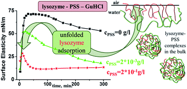
A study of the dilational surface viscoelastic properties of mixed solutions of lysozyme and denaturing agents with different chemical natures allows us to characterize the changes of protein tertiary structure in the surface layer upon adsorption at the liquid–gas interface. We show that guanidine hydrochloride (GuHCl) and urea influence the dynamic surface properties of lysozyme solutions less than the properties of previously studied solutions of bovine serum albumin and β-lactoglobulin. Although the addition of sodium polystyrene sulfonate (PSS) changes the kinetic dependencies of the surface properties and leads to the formation of large aggregates in the bulk phase, the dependencies of the dynamic surface elasticity on the surface pressure almost coincide with the results for pure lysozyme solutions thereby indicating the preservation of the adsorption layer structure. At the same time, the simultaneous addition of PSS and GuHCl to lysozyme solutions results in a strong synergistic effect: the kinetic dependency of the dynamic surface elasticity becomes non-monotonical and similar to that for solutions of amphiphilic polymers and non-globular proteins. The local maximum of this dependency indicates the destruction of the protein globular structure and formation of the distal region of the surface layer. The simultaneous addition of PSS and urea to lysozyme solutions does not lead to a similar effect. These results confirm different mechanisms of protein denaturation under the influence of urea and GuHCl, and the important role of electrostatic interactions in the latter case.

Introduction

Aqueous solutions of protein–polyelectrolyte mixtures have been widely studied in recent years.1–4 The interest in these systems stems from their applications in biocatalysis, biosensoring, protein separation and purification, and drug delivery.1 An important step in most of these processes is the adsorption of bound or free protein molecules at liquid–fluid interfaces. At the same time, the main focus of studies of complex formation between proteins and polyelectrolytes is the bulk properties of concentrated solutions and the formation of coacervates.1–5 Information on the mechanisms of formation of primary complexes in dilute solutions and especially on their surface properties is quite scarce.6 Indeed the current understanding of the surface properties of protein–polyelectrolyte solutions is often limited by the general difficulties in the investigation of protein adsorption layers at liquid–fluid interfaces.7–9 Although the structure of protein spread and adsorbed layers at the liquid–gas interface has been discussed for about ninety years,10–14 progress in this field proved to be rather slow and reliable information on the changes of protein tertiary structure in the course of adsorption is still extremely limited. For example, different authors came to the opposite conclusions on the preservation12,15 or destruction7,13 of the globular structure of lysozyme at the water–air interface.

It has been shown some time ago that the destruction of the protein tertiary structure results in strong changes of the kinetic dependencies of the dilational dynamic surface elasticity.9,16,17 They become non-monotonical and close to equilibrium the surface elasticity approaches the values characteristic for solutions of amphiphilic polymers. Measurements of the dynamic surface elasticity allowed registration of the unfolding of bovine serum albumin (BSA) and β-lactoglobulin (BLG) in the surface layer under the influence of strong denaturants. The aim of the present work is to characterize the influence of denaturants and especially sodium polystyrene sulfonate (PSS) on the dynamic surface properties of lysozyme solutions. This protein is characterized by high internal cohesion18 and higher stability of the tertiary structure at liquid–liquid interfaces as compared with many other proteins.19 PSS is not a typical denaturant but it can also destroy the lysozyme tertiary structure, for example, in concentrated solutions if its concentration exceeds that of the protein.4 To the best of our knowledge the dynamic surface properties of lysozyme–PSS solutions have not previously been studied.

Lysozyme interacts strongly with PSS in aqueous solutions and the complex formation in these systems has been studied in detail during the last two decades.4 The primary lysozyme–PSS complexes are capable of further aggregation into large particles.4,20–24 Depending on the ratio of the components and their concentrations the solutions are turbid, transparent or one can observe precipitation. In clear solutions components coexist in the form of charged flexible chains. In this case PSS acts like a surfactant causing unfolding of the protein due to specific interactions with the polyelectrolyte. In other concentration ranges lysozyme molecules can keep their globular structure and the interaction with the polyelectrolyte leads to the formation of large aggregates. The formation of primary protein–polyelectrolyte complexes occurs at lower bulk polyelectrolyte concentrations that those which result in the formation of large aggregates. Indeed primary complexes are formed, even when both the components are similarly charged due to the presence of positively charged patches on the globule surface. The total charge of lysozyme globule at the beginning of complex formation is about −2.6 and the formation of large aggregates begins at the overall protein charge of more than 0.7.24 It has been shown recently that one can also use dilational surface rheology to estimate the conformational changes in the surface layer of mixed polyelectrolyte–protein solutions.25,26 The complex formation between BSA and oppositely charged polyelectrolytes leads to non-monotonical kinetic dependencies of the dynamic surface elasticity. Although the effect on the protein tertiary structure of polyelectrolyte is less than that of guanidine hydrochloride (GuHCl) or urea, the elasticity maximum corresponds to the same surface pressure (12 mN m−1) and also indicates the distortion of the protein tertiary structure in the surface layer. If polyelectrolytes and BSA were similarly charged, the effect depended on the chemical nature of polyelectrolyte. For example, at pH = 2.3 the addition of positively charged poly(diallyldimethylammonium chloride) (PDADMAC) to solutions of positively charged BSA did not lead to any changes of the dynamic surface properties but PSS at pH = 7 interacted with the negatively charged BSA and influenced strongly the kinetic dependencies of the dynamic surface elasticity. This difference is probably caused by the higher flexibility of PPS chains allowing its interaction with a relatively large number of positively charged patches on the surface of negatively charged BSA globules.20

In this work, surface dilational rheology and ellipsometry are applied to lysozyme–PSS solutions with the aim to elucidate conformation changes of the adsorbed protein molecules in the surface layer under the influence of polyelectrolyte. The detailed information on the corresponding changes in the bulk phase makes this task easier.4 Another aim consists in the investigation of the mutual effect of PSS and GuHCl on the dynamic surface properties of lysozyme solutions. Although the impact of GuHCl on the structure of lysozyme adsorption layer was investigated in ref. 15, to the best of our knowledge no information on the dynamic surface properties of the mixed lysozyme–GuHCl and lysozyme–PSS–GuHCl solutions has been published until now.

Experimental

Lysozyme (Sigma-Aldrich), molecular weight 14.3 kDa, was used as received. The lysozyme solutions in phosphate buffer at pH 7 were prepared by dilution of stock solution with concentration 1.256 g l−1, which had been stored in a refrigerator at a temperature lower than 10 °C for no longer than two days. All the measurements in this work were carried out at lysozyme concentrations of 3.5 × 10⁻⁶ M.

PSS (Sigma-Aldrich) with a molecular weight of 70 kDa was used as received. The PSS solutions were prepared by dilution of stock solutions with concentrations of 2 g l−1 and 0.02 g l−1. GuHCl (molecular weight 95.53 Da) (Sigma-Aldrich) was used as received. In the most cases GuHCl concentration was 3 M (286.6 g l−1). The pH of solutions was regulated by the addition of phosphate buffer (NaH2PO4 and Na2HPO4, Sigma-Aldrich). The ionic strength of lysozyme solutions without GuHCl was 0.002 M. Triple-distilled water was used for the preparation of all the solutions.

The surface tension of solutions was measured by the Wilhelmy plate method using a roughened glass plate attached to an electronic balance. The complex dynamic surface elasticity was measured by the oscillating barrier method at a fixed frequency of 0.1 Hz. The corresponding experimental procedure has been described in detail elsewhere.27 The oscillations of the surface area of the solution in a polytetrafluoroethylene (PTFE) Langmuir trough were produced by a movable PTFE barrier sliding along polished brims of the trough. A mechanical generator transformed the rotation of an electric motor into the translational motion with reversion and allowed the control of the oscillation amplitude and frequency. The moving part of the generator was connected to the barrier by a steel rod. The barrier glided back and forth along the Langmuir trough and produced oscillations of the liquid surface area A with a relative amplitude of 3%. The corresponding oscillations of the surface tension γ were measured by the Wilhelmy plate method. The complex dynamic surface elasticity E was determined from the oscillations of the surface tension γ and surface area according to the following relation

E(ω) = Er + iEi = δγ[thin space (1/6-em)]ln[thin space (1/6-em)]A
where E, δγ, δA – are complex quantities.

In the case of a pure elastic system E becomes a real quantity and coincides with Er (storage modulus). In the more general case E depends not only on the amplitudes of the surface tension and the surface area oscillations but also on the phase shift between these two quantities and becomes a complex quantity where the imaginary part Ei (loss modulus) describes the viscous contribution.

The imaginary part of the complex dynamic surface elasticity of the solutions under investigation proved to be much less than the real part. Therefore, only the results for the real part are discussed below.

A null-ellipsometer Multiskop (Optrel GBR, Germany) at a single wavelength of 632.8 nm was used to estimate the adsorbed amount using a fixed compensator (±45°) and a 2-zone averaging nulling scheme. The scheme of this apparatus has been described in detail elsewhere.28 All the ellipsometric measurements were performed at an incidence angle of 50° close to the Brewster angle because this condition ensured the highest sensitivity of the ellipsometric angles to the properties of the adsorption layer.

Elliptically polarized light consists of two components with the electric vectors oscillating parallel and perpendicular to the plane of incidence. The reflection of light at an interface results in different changes of the phase and amplitude of these two components. These changes depend on the optical properties of the interface and can be characterized by two ellipsometric angles, an amplitude change Ψ and a phase shift Δ, which are related to the Fresnel reflectivity coefficients of the parallel and perpendicular components, rp and rs, respectively28

image file: c4ra14330b-t1.tif

The difference Δsurf between the ellipsometric angle Δ for the investigated solution and that of pure water Δ0 can be approximated as a rough measure of the interfacial excess as the value is proportional to the adsorbed amount Γ for thin, transparent, isotropic layers of uniform density at the liquid–gas interface.29,30 Ψ is very insensitive to the optical properties of a thin film at the liquid–air interface. Therefore we present below only the data on the ellipsometric angle Δ to characterize the adsorption kinetics.

The size distribution of the globules and aggregates in the bulk solution were determined by dynamic light scattering (DLS) using a Zetasizer ZS Nano analyzer (Malvern Instruments, United Kingdom). The measurements were carried out at a scattering angle of 173°. The solutions studied using DLS were previously filtered by microporous membrane film made of the mixture of cellulose acetates with a pore size of 2 micron and a total porosity of 80–85%.

All the measurements were performed at room temperature (20 °C ± 1 °C).

Results and discussion

The dynamic surface properties of mixed solutions of lysozyme and various denaturants were measured as a function of surface age and denaturant concentration at a constant protein concentration of 3.5 × 10⁻⁶ M and at pH = 7. The imaginary part of the dilational dynamic surface elasticity of all the investigated solutions was much less than the real one and only the latter quantity is under discussion below.

The kinetic dependencies of the surface properties for solutions of pure lysozyme are characterized by an induction period of about one hour when no changes of the surface properties take place (Fig. 1a). This effect has been already discussed in the literature and is thought to be connected with the peculiarities of the equation of state for the adsorption layer of this protein.31,32 The addition of GuHCl to lysozyme solutions results in the acceleration of the changes of surface properties with the surface age as well as decreases in both the induction period and the static surface tension. These observations may be attributed to the increase of the solution ionic strength. If the denaturant concentration exceeds about 1.5 M, the kinetic dependencies of the dynamic surface elasticity change and become non-monotonical (Fig. 1a). A similar effect has been observed recently for solutions of other globular proteins when the denaturant concentration was higher than a certain critical value.9,16,17 The local maximum of the dynamic surface elasticity can be connected with the globule unfolding at the interface leading to the formation of the distal region of the surface layer. A similar conclusion on the changes of the adsorption layer structure with the increase of GuHCl concentration follows also from the neutron reflectivity data.15 In this case the surface stresses can be relaxed at the expense of the exchange of some parts of the macromolecules between different regions of the layer, and the dynamic surface elasticity starts to decrease after a local maximum.


image file: c4ra14330b-f1.tif
Fig. 1 Kinetic dependences of the dynamic surface elasticity of lysozyme solutions mixed with (a) GuHCl at various GuHCl concentrations: 0 M (black squares), 1 M (red circles), 3 M (green triangles), and 4 M (dark blue diamonds); (b) PSS at various polyelectrolyte concentrations: 0 g l−1 (black squares), 2 × 10⁻⁵ g l−1 (red circles), 2 × 10⁻⁴ g l−1 (green diamonds), 2 × 10⁻³ g l−1 (dark blue triangles), 2 × 10⁻² g l−1 (blue stars), 5 × 10⁻² g l−1 (orange pentagons), and 1 × 10⁻¹ g l−1 (purple snowflakes); (c) PSS and GuHCl at various concentrations of polyelectrolyte: 0 g l−1 (black squares), 2 × 10⁻⁴ g l−1 (red circles), 2 × 10⁻³ g l−1 (green triangles), 2 × 10⁻² g l−1 (blue pentagons), and 2 × 10⁻¹ g l−1 (dark blue stars) and at the concentration of GuHCl 3 M; (d) PSS and urea solutions at constant concentration of polyelectrolyte (0) and at various concentrations of urea: 5 M (green triangles), and 8 M (red circles), and at a constant concentration of polyelectrolyte (2 × 10⁻² g l−1) and at various concentrations of urea: 5 M (black triangles), and 8 M (blue circles).

The corresponding peak of the kinetic curves of the dynamic surface elasticity of lysozyme solutions is much smoother than that for solutions of other globular proteins. The maximal value of the surface elasticity in the solution of 4 M GuHCl is 47 mN m−1 while the plateau value close to equilibrium is 37 mN m−1. In the case, for example, of BSA solutions in 4 M of GuHCl the corresponding values are, respectively, 49 and 13 mN m−1.17 This distinction between the properties of the two proteins is caused by the stronger stability of lysozyme globules at liquid–fluid interfaces.19 Further, the data indicate that the lysozyme tertiary structure is not entirely destroyed in the surface layer of even 4 and 6 M GuHCl solutions and the distal region of the layer is not developed to a sufficient extent.

Similar results were also obtained for lysozyme solutions in urea (see ESI).

In this case one can observe the non-monotonical kinetic dependencies of the dynamic surface elasticity only at higher concentrations of the denaturant (>2 M) due to the weaker denaturing activity of urea as compared with GuHCl. The plateau value close to equilibrium is also rather high in this case (∼42 mN m−1) even when the urea concentration is 8 M for the same reason.

PSS influences noticeably the dynamic surface properties of lysozyme solutions even at very low concentrations.

The changes of the surface properties are decelerated strongly and the induction period increases by almost a factor of two even at the PSS concentration of 2 × 10⁻⁵ g l−1 (Fig. 1b and 2).


image file: c4ra14330b-f2.tif
Fig. 2 Kinetic dependences of the surface tension of mixed lysozyme–PSS solutions at various polyelectrolyte concentrations: 0 g l−1 (black squares), 2 × 10⁻⁵ g l−1 (red circles), 2 × 10⁻⁴ g l−1 (green diamonds), 2 × 10⁻² g l−1 (dark blue stars), and 1 × 10⁻¹ g l−1 (purple snowflakes).

Although in this case the adsorption on the walls of the trough can influence the real bulk concentration, the results are reproducible and indicate strong interactions between the solutes in the surface layer. A further increase of the PSS concentration leads to the decrease of the induction period and it disappears finally at concentrations higher than 2 × 10⁻² g l−1. The rate of change of the surface properties after the induction period or immediately after the surface formation decreases noticeably with increasing polyelectrolyte concentration. The representation of the dynamic surface elasticity as a function of surface pressure can give additional information on the adsorption layer structure. For example, although PSS influences significantly the kinetic dependencies of the dynamic surface properties of BSA solutions at pH = 7, all the graphs of the surface elasticity vs. surface pressure at different PSS concentrations coincide with the corresponding results for pure BSA solutions, which indicates that the adsorption layer structure is determined by the adsorbed protein.26 In the case of lysozyme–PSS solutions all the dependencies of the dynamic surface elasticity on the surface pressure are similar only if the surface elasticity is relatively high (≥10 mN m−1) (Fig. 3).


image file: c4ra14330b-f3.tif
Fig. 3 Dependences of the dilatational surface elasticity on the surface pressure of mixed lysozyme–PSS solutions at various concentrations of polyelectrolyte: 0 g l−1 (black squares), 2 × 10⁻⁵ g l−1 (red circles), 2 × 10⁻⁴ g l−1 (green triangles), 2 × 10⁻² g l−1 (dark blue stars), and 2 × 10⁻¹ g l−1 (purple snowflakes).

At the beginning of adsorption the obtained dependencies differ from those for pure lysozyme solutions if the PSS concentration is higher than about 2 × 10⁻⁵ g l−1. This result indicates that PSS can penetrate into the proximal region of the adsorption layer of lysozyme–PSS solutions and contribute to the surface pressure at the initial adsorption step when the surface elasticity is low. The further adsorption of lysozyme–PSS complexes is accompanied by the displacement of polyelectrolyte segments from the proximal region and the shape of corresponding curves of the dynamic surface elasticity becomes similar to those of pure lysozyme solutions.

The ellipsometric data confirm the deceleration of protein adsorption at the increase of PSS concentration. The difference of the ellipsometric angle Δsurf, which we take as a rough measure of the total adsorbed amount, changes slower than for pure protein solution during at least the first 16 hours after the surface formation (Fig. 4). This effect becomes more pronounced at PSS concentrations close to 2 × 10⁻² g l−1.


image file: c4ra14330b-f4.tif
Fig. 4 Kinetic dependences of the ellipsometric angle Δsurf of mixed lysozyme–PSS solutions at various concentrations of polyelectrolyte: 0 g l−1 (black squares), 2 × 10⁻⁵ g l−1 (red circles), 2 × 10⁻⁴ g l−1 (green diamonds), 2 × 10⁻³ g l−1 (blue triangles), and 1 × 10⁻¹ g l−1 (orange pentagons).

The changes of the kinetic dependencies of the dynamic surface properties with changing PSS concentration can be connected with aggregate formation in the solution bulk.

The solutions become opalescent at the concentrations exceeding about 1 × 10⁻² g l−1. The results from dynamic light scattering support these observations. The particle diameters were calculated from the obtained diffusion coefficients by the Stokes–Einstein relation and Fig. 5 shows some examples of the dependencies of the scattered light intensity on the particle size.


image file: c4ra14330b-f5.tif
Fig. 5 The dependency of the scattering intensity on the particle size for mixed lysozyme–PSS solutions at various concentrations of polyelectrolyte: 0 g l−1 (black line), 2 × 10⁻² g l−1 (dark blue line), and 1 × 10⁻¹ g l−1 (red line).

The solution of pure lysozyme is characterized by a particle diameter of about 4 nm coinciding approximately with the size of lysozyme globule.4 The addition of PSS to lysozyme solutions even at the concentration of 2 × 10⁻⁵ g l−1 results in new peaks of the scattered intensity beyond 100 nm but the system is characterized by the high polydispersity and poor reproducibility of the results up to the concentration of 2 × 10⁻³ g l−1. This behavior can be attributed to the formation of large loose lysozyme–PSS aggregates with a large number of interconnected protein and polyelectrolyte macromolecules. The poor reproducibility of the particle size characterization is likely to be related to the nonequilibrium nature of the aggregation process between oppositely charged mixtures involving charged macromolecules.33 Another reason of the poor reproducibility can consist in the polydispersity of aggregates. This means that the DLS data indicate only the formation of large aggregates and do not allow reliable estimation of their size. Nevertheless, the peak corresponding to large aggregates becomes more reproducible at the concentrations of 2 × 10⁻² g l−1 and higher. At the same time, a new peak appears close to 20 nm, which can be attributed to free PSS molecules in the solution. Simultaneously the peak of globules disappears and consequently most of the protein molecules at high PSS concentrations are present in the form of large loose lysozyme–PSS aggregates.

The electrostatic interactions between protein and polyelectrolyte molecules are the main driving force of the formation of primary complexes, which are capable to further aggregation (Fig. 6a).1,4 If the ionic strength of solution increases due to the addition of an inert electrolyte, for example, NaCl, the surface properties of mixed lysozyme–PSS solutions became closer to the properties of pure protein solutions (see ESI) confirming the main role of relatively weak electrostatic interactions between the components in the complex formation. The flexibility of PSS molecules and hydrophobic interactions between the polyelectrolyte polystyrene chains and hydrophobic groups of lysozyme can also contribute to the complex formation.4,34


image file: c4ra14330b-f6.tif
Fig. 6 A scheme of protein–polyelectrolyte interactions in lysozyme–PSS (a) and lysozyme–PSS–GuHCl (b) solutions and at the liquid–gas interface.

The formation of aggregates in the bulk phase can explain some of the kinetic dependencies in Fig. 1b and 2. The concentration of the aggregates increases and the concentration of free protein molecules decreases with increasing polyelectrolyte concentration. The diffusion coefficients of the large loose aggregates are much lower than those of protein molecules and thereby the adsorption rate decreases with increasing PSS concentration leading to the increase of the induction period at low polyelectrolyte concentrations (Fig. 1b). Such interfacial depletion effects have recently been related to bulk aggregation in solutions of the mixture of synthetic and biological polyelectrolytes.35 PSS does not exhibit any surface activity at low concentrations36 and the adsorption of lysozyme–PSS complexes is possible only due to the amphiphilic protein component. However, hydrophilic polyelectrolyte segments connected with lysozyme molecules can also penetrate the proximal region of the surface layer. The overall surface concentration increases and the lysozyme–PSS complexes start to interact at the interface even when the surface concentration of protein molecules alone is insufficient to create any finite surface pressure. It follows that this synergistic interaction is related to the decrease of induction period (Fig. 1b and 2) and the corresponding changes of the dependency of the dynamic surface elasticity on the surface pressure at PSS concentrations higher than 2 × 10⁻⁴ g l−1 (Fig. 3). The further increase of surface concentration in the course of adsorption results in gradual displacement of PSS segments from the proximal region of the surface layer and the graphs of the dynamic surface elasticity vs. surface pressure become similar to those of pure protein solutions. The deceleration of the changes of surface properties with the surface age at high PSS concentrations (>2 × 10⁻² g l−1), when all the aggregates are negatively charged, can be also caused by the increase of the electrostatic barrier due to the repulsion between the adsorbing complexes and similarly charged interface. The ellipsometric results on the protein adsorbed amount agree with the proposed interpretation of the kinetic dependencies of the dynamic surface elasticity (Fig. 4).

It is noteworthy that all the dependencies of the dynamic surface elasticity of lysozyme–PSS solutions on the surface pressure and surface age are monotonical and do not indicate noticeable changes of the protein tertiary structure in the surface layer.

The corresponding dependencies for the mixed solutions of lysozyme and GuHCl indicate some changes of the globular structure but this effect is less than for the mixed solutions of GuHCl with other globular proteins.16,17 However, if lysozyme solutions contain both PSS and GuHCl, the kinetic dependencies of the dynamic surface elasticity change strongly (Fig. 1c).

The decrease of the surface elasticity beyond the maximum for the mixed solution of lysozyme and GuHCl at the concentrations of 3.5 × 10⁻⁶ M and 3 M correspondingly becomes steeper even at the PSS concentration of 2 × 10⁻⁴ g l−1. If the PSS concentration exceeds 2 × 10⁻³ g l−1, the surface elasticity close to equilibrium drops to about 11 mN m−1. This value is typical for solutions of non-globular proteins and only a little higher than for most solutions of amphiphilic linear polymers.9,27 The further increase of PSS concentration results in a decrease of the maximum value and a faster decrease of the surface elasticity after the maximum.

All the kinetic dependencies before the maximum of the dynamic surface elasticity almost coincide (Fig. 1c). In this case the lysozyme molecules are enclosed in the protein–polyelectrolyte aggregates, the high ionic strength of the solution at the expense of high GuHCl concentration leads to the disappearance of the kinetic adsorption barrier, and the adsorption rate does not depend on PSS concentration. Beyond the maximum, the decrease of the dynamic surface elasticity indicates the destruction of the protein tertiary structure and the rate of this process depends strongly on polyelectrolyte concentration. The globule unfolding leads to the changes of the adsorption layer structure. Although the disulfide bonds in lysozyme molecule between the 6th and 127th and also between the 30th and 115th amino acid residues limit its flexibility and do not allow formation of long loops and tails in the surface layer,37 some parts of the adsorbed unfolded macromolecules can protrude into the aqueous phase and ensure effective relaxation of surface stresses due to the exchange of matter between different regions of the adsorption layer.

Note that the changes of the lysozyme tertiary structure are inevitably connected with the alterations of the secondary structure. For example, it is known that GuHCl induces the unfolding of α-helix structures.38 Unfortunately, the surface rheology and ellipsometry do not allow elucidation of any details of these changes in the surface layer.

It is certain only that the flexibility of macromolecules increases strongly under the combined action of GuHCl and PSS and they start to form loops and tails. The best way to estimate the corresponding alterations of the secondary structure is probably the molecular dynamic simulations39 and this study is in progress now.

The reorganization of the adsorption layer under the influence of PSS does not lead to any noticeable changes of the kinetic dependencies of the surface tension (Fig. 7).


image file: c4ra14330b-f7.tif
Fig. 7 Kinetic dependencies of the surface tension of mixed lysozyme–PSS–GuHCl solutions at various concentrations of polyelectrolyte: 0 g l−1 (black squares), 2 × 10⁻⁴ g l−1 (red circles), 2 × 10⁻³ g l−1 (green triangles), and 2 × 10⁻² g l−1 (blue pentagons).

The surface tension of the solutions of flexible macromolecules is determined by the local segment concentration in the proximal region of the surface layer and consequently the data in Fig. 7 and 8 show that almost all the changes in the layer structure occur in its distal region. The proximal region contains approximately the same concentrations of relatively hydrophobic amino acid residues at all PSS concentrations independently of the degree of the globule unfolding. On the other hand, the data in Fig. 7 and 8 confirm the high sensitivity of the dilational dynamic surface elasticity to the conformation of macromolecules at the interface.27 The sensitivity of the surface tension proves to be much lower.


image file: c4ra14330b-f8.tif
Fig. 8 Dependences of the dilatational surface elasticity on the surface pressure of mixed lysozyme–PSS–GuHCl solutions at various concentrations of polyelectrolyte: 0 g l−1 (black squares), 2 × 10⁻⁴ g l−1 (red circles), 2 × 10⁻³ g l−1 (green triangles), 2 × 10⁻² g l−1 (blue pentagons), and 2 × 10⁻¹ g l−1 (dark blue stars).

The dependencies of the dynamic surface elasticity on the surface pressure of mixed lysozyme–PSS–GuHCl solutions show a consistent decrease of the maximum elasticity with increasing PSS concentrations (Fig. 8). The surface pressure corresponding to the maximum at low PSS concentrations coincides with that of pure lysozyme solutions and is about 15 mN m−1. Unlike the corresponding results for solutions without GuHCl, these data corroborate strong changes of the adsorption layer structure due to the displacement of some parts of unfolded protein molecules from the proximal region of the surface layer in the vicinity of the gas phase. In this case one can use an analogy with the adsorption layer of flexible amphiphilic polymers where the relaxation of surface stresses occurs mainly due to the segment exchange between the proximal and distal regions of the surface layer.27

The ellipsometric data also indicate a strong difference between the adsorption layer structures for lysozyme–PSS solutions with and without GuHCl. The data in Fig. 9 show faster adsorption for lysozyme–PSS–GuHCl solutions, which may be attributed to the higher ionic strength.


image file: c4ra14330b-f9.tif
Fig. 9 Kinetic dependencies of the ellipsometric angle Δsurf of mixed lysozyme–PSS–GuHCl solutions at various concentrations of polyelectrolyte: 0 g l−1 (black squares), 2 × 10⁻⁴ g l−1 (blue snowflakes), 2 × 10⁻³ g l−1 (green triangles), and 2 × 10⁻² g l−1 (grey stars).

A more important difference is that Δsurf and thereby the adsorbed amount increase with PSS concentration, which is unlike the case of lysozyme–PSS solutions (Fig. 4). This result corroborates the stronger unfolding of lysozyme under the mutual influence of both PSS and GuHCl leading to the formation of the distal region of the surface layer and the increase of the overall adsorbed amount. It is possible that some PSS segments interacting with the unfolded protein also penetrate the surface layer and contribute to the increase of Δsurf.

Although the surface properties differ strongly for lysozyme–PSS–GuHCl and lysozyme–PSS solutions, the aggregation in the bulk has similar characteristics and causes the main changes in the dynamic light scattering data upon the addition of PSS to lysozyme and lysozyme–GuHCl solutions (see ESI). The addition of GuHCl to pure lysozyme solution results in an increase of the size of the globules due to their partial unfolding. The distortion of the protein tertiary structure leads to the formation of large aggregates with increasing PSS concentration, and the peak in the data from the globules and the dependency of the scattered light intensity on the particle size both disappear even at PSS concentration of 2 × 10⁻⁵ g l−1. Unlike the case of lysozyme–PSS solutions, the peak of free PSS molecules does not appear at high polyelectrolyte concentrations probably due to the stronger aggregation in the solution under the influence of GuHCl. Another peculiarity of the dynamic light scattering data from lysozyme–PSS–GuHCl solutions is the formation of small particles with the mean diameter of 5–7 nm at PSS concentrations higher than about 2 × 10⁻³ g l−1. It follows that high PSS concentrations result in the refolding of some protein molecules but the most of them remain unfolded and connected with PSS inside the large aggregates.

The very strong influence of PSS on the structure of the lysozyme adsorption layer is specific to samples that contain GuHCl. If GuHCl is substituted by another strong denaturant, urea, the addition of PSS does not lead to strong changes of the kinetic dependencies of the dynamic surface elasticity. Moreover, they become monotonical even at high urea concentrations (Fig. 1d).

This distinction is obviously connected with the difference in the unfolding mechanisms under the influence of GuHCl and urea. In the former case the denaturant is charged in aqueous solutions and is bound mainly to hydrophilic regions on the surface of protein globules.38 The interaction of PSS with the globules reduces their charge and thereby facilitates their interaction with GuHCl and the subsequent unfolding. On the contrary, urea interacts mainly with the neutral hydrophobic regions on the surface of globules and PSS does not noticeably influence this process. The electrostatic interactions between the protein and denaturant are not important in the latter case. The differences in the unfolding mechanisms under the influence of PSS and urea have been widely discussed in the literature.38,40,41 These measurements of the surface dilational rheological properties of protein solutions in the presence of polyelectrolytes with and without stronger denaturants has given us the have allowed us to substantiate some proposed hypothesis on the peculiarities of protein unfolding.

Conclusions

Our application of dilational surface rheology has allowed the elucidation of the adsorption layer structure in lysozyme–PSS aqueous solutions with and without additions of GuHCl and urea. Strong mutual influence of PSS and GuHCl on the dynamic surface elasticity of lysozyme solutions was discovered and connected with the complete destruction of the protein globular structure. Lysozyme and PSS interact strongly in aqueous solutions with the formation of large loose aggregates. The aggregates diffuse slower to the surface than free protein molecules and the kinetic dependencies of the dynamic surface elasticity and surface tension change strongly with increasing PSS concentration. At the same time, the surface layer structure changes only at the initial adsorption steps when some PSS segments penetrate the adsorption layer. They are displaced from the surface as a result of further adsorption of material and the surface properties are determined by the adsorbed protein molecules preserving mainly their native form regardless of the PSS concentration.

Almost the same extent of bulk aggregation occurs in lysozyme–PSS–GuHCl solutions. However, in this case the mutual influence of GuHCl and PSS destroys the protein tertiary structure at the liquid–gas interface to a greater extent. The corresponding kinetic dependencies of the dynamic surface elasticity differ strongly from the kinetic dependencies for solutions of mixtures of lysozyme with GuHCl in the absence of PSS. One can observe a local maximum of the surface elasticity for lysozyme–PSS–GuHCl solutions. This feature in the data reveals the formation of the distal region of the surface layer as a result of the displacement of some parts of the unfolded protein molecule from the proximal region with the possible formation of loops and tails. The substitution of GuHCl by urea changes strongly the surface viscoelastic behavior but does not indicate significant changes in the protein tertiary structure in the surface layer. This distinction between the two systems is caused by different mechanisms of the lysozyme unfolding under the influence of GuHCl and urea and in particular the degree of electrostatic interactions involved.

Acknowledgements

This work was supported by the Russian Foundation for Basic Research (project no. 14-03-00670_a), the National Science Council of Taiwan (joint project RFFI_NSC no. 12_03_92004_NNS_a), and the grant of St. Petersburg state university no. 12.38.241.2014.

Notes and references

  1. C. L. Cooper, P. L. Dubin, A. B. Kayitmazer and S. Turksen, Polyelectrolyte-protein complexes, Curr. Opin. Colloid Interface Sci., 2005, 10, 52 CrossRef CAS PubMed.
  2. C. Schmitt and S. L. Turgeon, Protein/polysaccharide complexes and coacervates in food systems, Adv. Colloid Interface Sci., 2011, 167, 63 CrossRef CAS PubMed.
  3. E. Kizilay, A. B. Kayitmazer and P. L. Dubin, Complexation and coacervation of polyelectrolytes with oppositely charged colloids, Adv. Colloid Interface Sci., 2011, 167, 24 CrossRef CAS PubMed.
  4. F. Cousin, J. Gummel, S. Combet and F. Boue, The model Lysozyme PSSNa system for electrostatic complexation: Similarities and differences with complex coacervation, Adv. Colloid Interface Sci., 2011, 167, 71 CrossRef CAS PubMed.
  5. S. Llamas, E. Guzmán, F. Ortega, N. Baghdadli, C. Cazeneuve, R. G. Rubio and G. S. Luengo, Adsorption of polyelectrolytes and polyelectrolytes-surfactant mixtures at surfaces: a physico-chemical approach to a cosmetic challenge, Adv. Colloid Interface Sci. DOI:10.1016/j.cis.2014.05.007..
  6. R. A. Ganzevles, R. Fokkink, M. A. Cohen Stuart, T. van Vliet and H. J. Jongh, Structure of mixed beta-lactoglobulin/pectin adsorbed layers at air/water interfaces; a spectroscopy study, J. Colloid Interface Sci., 2008, 317, 137 CrossRef CAS PubMed.
  7. Y. F. Yano, Kinetics of protein unfolding at interfaces, J. Phys.: Condens. Matter, 2012, 24, 503101 CrossRef PubMed.
  8. V. Mitropoulos, A. Mütze and P. Fischer, Mechanical properties of protein adsorption layers at the air/water and oil/water interface: A comparison in light of the thermodynamical stability of proteins, Adv. Colloid Interface Sci., 2014, 206, 195 CrossRef CAS PubMed.
  9. B. A. Noskov, Protein conformational transitions at the liquid-gas interface as studied by dilational surface rheology, Adv. Colloid Interface Sci., 2014, 206, 222 CrossRef CAS PubMed.
  10. E. Gorter and F. Grendel, On the spreading of proteins, Trans. Faraday Soc., 1926, 22, 477 RSC.
  11. I. Langmuir and V. J. Schaefer, Properties and structure of protein monolayers, Chem. Rev., 1939, 24, 181–202 CrossRef CAS.
  12. D. J. F. Taylor, R. K. Thomas and J. Penfold, Polymer/surfactant interactions at the air/water interface, Adv. Colloid Interface Sci., 2007, 132, 69 CrossRef CAS PubMed.
  13. T. Furuno, Atomic force microscopy study on the unfolding of globular proteins in the Langmuir films, Thin Solid Films, 2014, 552, 170 CrossRef CAS PubMed.
  14. S. Vaidya and A. R. Narvaez, Understanding interactions between immunoassay excipient proteins and surfactants at air-aqueous interface, Colloids Surf., B, 2014, 113, 285 CrossRef CAS PubMed.
  15. A. W. Perriman, M. J. Henderson, C. R. Evenhuis, D. J. McGillivray and J. W. White, Effect of the air-water interface on the structure of lysozyme in the presence of guanidinium chloride, J. Phys. Chem. B, 2008, 112, 9532 CrossRef CAS PubMed.
  16. B. A. Noskov, D. O. Grigoriev, A. V. Latnikova, S.-Y. Lin, G. Loglio and R. Miller, Impact of Globule Unfolding on Dilational Viscoelasticity of beta-Lactoglobulin Adsorption Layers, J. Phys. Chem. B, 2009, 113, 13398 CrossRef CAS PubMed.
  17. B. A. Noskov, A. A. Mikhailovskaya, S.-Y. Lin, G. Loglio and R. Miller, Bovine Serum Albumin Unfolding at the Air/Water Interface as Studied by Dilational Surface Rheology, Langmuir, 2010, 26, 17225 CrossRef CAS PubMed.
  18. L. G. Smith, M. J. Sutcliffe, C. Redfield and C. M. Dobson, Structure of hen lysozyme in solution, J. Mol. Biol., 1993, 229, 930 CrossRef CAS PubMed.
  19. J. Zhai, L. Day, M.-I. Aguilar and T. J. Wooster, Protein folding at emulsion oil/water interfaces, Curr. Opin. Colloid Interface Sci., 2013, 18, 257 CrossRef CAS PubMed.
  20. J. M. Park, B. B. Muhoberac, P. L. Dubin and J. Xia, Effects of protein charge heterogeneity in protein-polyelectrolyte complexation, Macromolecules, 1992, 25, 290 CrossRef CAS.
  21. J. Xia, P. L. Dubin, Y. Kim, B. B. Muhoberac and V. J. Klimkowski, Electrophoretic and quasi-elastic light-scattering of soluble protein-polyelectrolyte complexes, J. Phys. Chem., 1993, 97, 4528 CrossRef CAS.
  22. O. N. Ivinova, V. A. Izumrudov, V. I. Muronetz, I. Y. Galaev and B. Mattiasson, Influence of complexing polyanions on the thermostability of basic proteins, Macromol. Biosci., 2003, 3, 210 CrossRef CAS.
  23. F. Cousin, J. Gummel, D. Ung and F. Boue, Polyelectrolyte-protein complexes: Structure and conformation of each specie revealed by SANS, Langmuir, 2005, 21, 9675 CrossRef CAS PubMed.
  24. J. Gummel, F. Boue, B. Deme and F. Cousin, Charge stoichiometry inside polyelectrolyte-protein complexes: A direct SANS measurement for the PSSNa-lysozyme system, J. Phys. Chem. B, 2006, 110, 24837 CrossRef CAS PubMed.
  25. O. Y. Milyaeva, B. A. Noskov, S.-Y. Lin, G. Loglio and R. Miller, Influence of polyelectrolyte on dynamic surface properties of BSA solutions, Colloids Surf., A, 2014, 442, 63 CrossRef CAS PubMed.
  26. O. Y. Milyaeva, S.-Y. Lin and B. A. Noskov, Influence of sodium polystyrene sulfonate on dynamic surface properties of bovine serum albumin solutions, Colloid J., 2014, 76, 459 CrossRef CAS.
  27. B. A. Noskov, A. V. Akentiev, A. Y. Bilibin and I. M. Zorin, Dilational surface viscoelasticity of polymer solutions, Adv. Colloid Interface Sci., 2003, 104, 245 CrossRef CAS.
  28. R. M. A. Azzam and N. M. Bashara, Ellipsometry and polarized light, North-Holland, Amsterdam, 1977 Search PubMed.
  29. C. Monteux, C. E. Williams, J. Meunier, O. Anthony and V. Bergeron, Adsorption of oppositely charged polyelectrolyte/surfactant complexes at the air/water interface: formation of interfacial gels, Langmuir, 2004, 20, 57 CrossRef CAS.
  30. E. Tyrode, C. M. Johnson, M. W. Rutland, J. P. R. Day and C. D. Bain, A study of the adsorption of ammonium perfluorononanoate at the air-liquid interface by vibrational sum-frequency spectroscopy, J. Phys. Chem. C, 2007, 111, 316 CAS.
  31. S. Sundaram, J. K. Ferri, D. Vollhardt and K. J. Stebe, Surface phase behavior and surface tension evolution for lysozyme adsorption onto clean interfaces and into DPPC monolayers: Theory and experiment, Langmuir, 1998, 14, 1208 CrossRef CAS.
  32. V. S. Alahverdjieva, D. O. Grigoriev, J. K. Ferri, V. B. Fainerman, E. V. Aksenenko, M. E. Leser, M. Michel and R. Miller, Adsorption behaviour of hen egg-white lysozyme at the air/water interface, Colloids Surf., A, 2008, 323, 167 CrossRef CAS PubMed.
  33. A. Naderi, P. M. Claesson, M. Bergstrom and A. Dedinaite, Trapped non-equilibrium states in aqueous solutions of oppositely charged polyelectrolytes and surfactants: effects of mixing protocol and salt concentration, Colloids Surf., A, 2005, 253, 83 CrossRef CAS PubMed.
  34. E. Sedlak, D. Fedunova, V. Vesela, D. Sedlakova and M. Antalk, Polyanion Hydrophobicity and Protein Basicity Affect Protein Stability in Protein-Polyanion Complexes, Biomacromolecules, 2009, 10, 2533 CrossRef CAS PubMed.
  35. Á. Ábraham, R. A. Campbell and I. Varga, New method to predict the surface tension of complex synthetic and biological polyelectrolyte/surfactant mixtures, Langmuir, 2013, 29, 11554 CrossRef PubMed.
  36. D. J. F. Taylor, R. K. Thomas and J. Penfold, The adsorption of oppositely charged polyelectrolyte/surfactant mixtures: neutron reflection from dodecyl trimethylammonium bromide and sodium poly(styrene sulfonate) at the air/water Interface, Langmuir, 2002, 18, 4748 CrossRef CAS.
  37. J.-Y. Chang and L. Li, The unfolding mechanism and the disulfide structures of denatured lysozyme, FEBS Lett., 2002, 511, 73 CrossRef CAS.
  38. A. Hedoux, S. Krenzlin, L. Paccou, Y. Guinet, M.-P. Flament and J. Siepmann, Influence of urea and guanidine hydrochloride on lysozyme stability and thermal denaturation; a correlation between activity, protein dynamics and conformational changes, Phys. Chem. Chem. Phys., 2010, 12, 13189 RSC.
  39. G. M. Seddon and R. P. Bywater, Inactivation and reactivation of ribonuclease A studied by computer simulation, Open Biol., 2012, 2, 120088 CrossRef PubMed.
  40. C. E. Dempsey, T. J. Piggot and P. E. Mason, Dissecting contributions to the denaturant sensitivities of proteins, Biochemistry, 2005, 44, 775 CrossRef CAS PubMed.
  41. S. Chodankar, V. K. Aswal, J. Kohlbrecher, R. Vavrin and A. G. Wagh, Structural evolution during protein denaturation as induced by different methods, Phys. Rev. E: Stat., Nonlinear, Soft Matter Phys., 2008, 77, 031901 CrossRef CAS.

Footnote

Electronic supplementary information (ESI) available: Additional information about the surface elasticity of mixed lysozyme–PSS–NaCl and lysozyme–urea solutions and additional DLS data. See DOI: 10.1039/c4ra14330b

This journal is © The Royal Society of Chemistry 2015
Click here to see how this site uses Cookies. View our privacy policy here.