Screening of antidote sensitivity using an acetylcholinesterase biosensor based on a graphene–Au nanocomposite

Linjuan Longa, Yanan Luoa, Bingwen Liud, Dan Du*abc and Yuehe Lin*bc
aKey Laboratory of Pesticide and Chemical Biology of Ministry of Education, College of Chemistry, Central China Normal University, Wuhan, 430079, P. R. China. E-mail: dan.du@mail.ccnu.edu.cn
bSchool of Mechanical and Materials Engineering, Washington State University, Pullman, WA 99164, USA. E-mail: yuehe.lin@wsu.edu
cPaul G. Allen School for Global Animal Health, Washington State University, PO Box 647090, Pullman, WA 99164, USA
dVoiland School of Chemical Engineering and Bioengineering, Washington State University, Pullman, WA 99164-6515, USA

Received 8th November 2014 , Accepted 9th December 2014

First published on 9th December 2014


Abstract

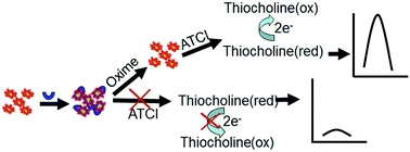
In this paper, we developed a new technique for in vitro screening of the therapeutic effects of three oxime compounds: obidoxime, 2-pralidoxime methiodide (2-PAM-I) and pyridine-2-aldoxime methochloride (2-PAM-Cl). Acetylcholinesterase (AChE) is first immobilized in a thin nanocomposite film composed of graphene and gold nanoparticles on a glassy carbon electrode by a self-assembly approach. The immobilized AChE in the nanocomposite is first exposed to an organophosphate agent, paraoxon-ethyl, and the enzyme activity is inhibited. The therapeutic effect is based on the reactivation capability of the oximes. AChE activity is measured by an electrochemical method. Our study indicated that oximes' reactivation capacity is correlated to their molecular structures. Obidoxime, having dual oxime groups, showed higher reactivation capacity than 2-PAM compounds with a single oxime group. Due to the similar molecular structures of 2-PAM-I and 2-PAM-Cl, a slight difference between their reactivation capacities was found, and this difference is attributed to the stronger electron withdrawing ability of iodine ions than that of chloride ions. The proposed electrochemical method thus provides a new simple tool for new drug screening.


1. Introduction

Organophosphates (OPs) represent a diverse group of highly toxic compounds used in insecticides, herbicides, and nerve agents. OPs attack serine residues of acetylcholinesterase (AChE), forming a phosphorylated adduct (OP-AChE) and thus inhibiting the function of this enzyme. The inhibition of AChE activity by OPs blocks the breakdown of the neurotransmitter, choline, which results in serious health problems such as paralysis, respiratory failure or even death.1–4 The wide use of OPs in agriculture causes tens of thousands of victims worldwide per year. Recently, a number of new drugs have been developed to treat OPs intoxication, such as nucleophiles,5–9 fluoride ions,10–13 and atropine.14 The detoxification principle is based on their reactivation of OP-AChE to gain restored enzyme activity. The therapeutic effect of antidotes can be determined by measuring reactivated AChE activity. Numerous methods have been developed to measure cholinesterase activity, including Ellman colorimetric assay,15 chemiluminescence assay,16,17 fluorescence assay,18,19 and radioactive assay.20 However, these methods need complicated pretreatment procedures, trained operators, and expensive instruments. Here, an electrochemical method was proposed for assessment of the therapeutic effect of different antidotes.

Graphene is an ideal material for electrochemistry because of its large surface area, excellent electrical conductivity, and low cost.21,22 More recently, many kinds of metal nanoparticles have been introduced onto graphene, such as Au,23,24 TiO2,25,26 and SnO2,27 to strengthen catalyst ability. A graphene-based nanocomposite has been developed as an enhanced sensing platform for biosensors28,29 because these kinds of nanocomposite films may generate synergistic effects to enhance the sensitivity.

Here, we developed a new enzyme electrode based on the self-assembly of AChE on graphene–Au nanocomposite to investigate the reactivation capacity of antidotes for drug screening. As shown in Scheme 1, AChE hydrolyzes the substrate acetylthiocholine (ATCl) to generate an electro-active product thiocholine which produces a detectable oxidation peak.30 Paraoxon-ethyl as a model OP compound reduces the enzymatic activity of AChE, and therefore decreases the response current. In this paper, different kinds of AChE reactivators, oximes, were used to treat OP poisoning by restoring the inhibited enzyme activity. The regenerated AChE activity was monitored by measuring the oxidation current, which served as an indicator of antidote effects.


image file: c4ra14085k-s1.tif
Scheme 1 Illustration of the principle of antidote reactivation.

2. Experimental

2.1. Reagents

Acetylthiocholine chloride (ATCl), AChE (Type C3389, 500 U mg−1 from the electric eel), 2-pralidoxime methiodide (2-PAM-I), pyridine-2-aldoxime methochloride (2-PAM-Cl), obidoxime chloride, poly(diallyldimethylammonium chloride) (PDDA 20%, w/w in water, MW: 200[thin space (1/6-em)]000–350[thin space (1/6-em)]000), phosphate buffer saline (PBS) were purchased from Sigma-Aldrich (St. Louis, USA). Paraoxon-ethyl was obtained from Dr Ehrenstorfer GmbH (Augsburg, Germany). Graphite powder, HAuCl4·4H2O, hydrazine solution (50% w/w), poly(N-vinyl-2-pyrrolidone) (PVP), acetic acid, trisodium citrate, potassium chloride, chitosan (95% deacetylation) were purchased from Shanghai Chemical Reagent Co. (Shanghai, China).

2.2. Apparatus

Electrochemical measurements were performed on a CHI 660C Electrochemical Workstation (Shanghai Chenhua Instrument Co., China) with a conventional three-electrode system comprising platinum wire as an auxiliary electrode, saturated calomel electrode (SCE) as a reference electrode, and AChE assembling graphene–Au nanocomposite modified screen printed carbon electrode (AChE/graphene–Au/SPCE) as a working electrode. Transmission electron microscopy (TEM) images were taken using JEM-100SX (JEOL, Japan). X-ray photoelectron spectroscopy (XPS) measurements were recorded with a thermo electron instrument MultiLab 2000 (Thermo Fisher, USA).

2.3. Immobilization of AChE on graphene–Au nanocomposite

2.3.1. Synthesis of Au nanoparticles. Au nanoparticles were synthesized using a method reported in literature.31 Briefly, 100 mL of 1 mM HAuCl4·4H2O aqueous solution was stirred and heated to boiling in which 10 mL of 38.8 mM trisodium citrate solution was quickly added. When the solution color changed from pale yellow to deep red, the solution was heated for an additional 15 min, resulting in the final Au nanoparticles. The final solution was cooled to room temperature and stored in a dark place. The size of AuNPs was ∼13 nm based on TEM analysis.
2.3.2. Synthesis of graphene–Au nanocomposite. Graphite oxides (GO) (0.5 mg mL−1) were synthesized from graphite by the modified Hummers method.32,33 GO exfoliation was carried out by sonicating graphite for 1 h. Polyelectrolyte-functionalized graphene was synthesized according to a previously reported method. Briefly, the mixture of 400 mg of PVP and 100 mL of homogeneous GO dispersion (0.5 mg mL−1) was stirred for 12 h. Then, 35 μL of hydrazine solution (50% w/w) and 400 μL of ammonia solution (25% w/w) were added into the resulting dispersion solution with stirring for 1 h at 95 °C. The resulting black aqueous solution (PVP/graphene) was centrifuged and dissolved in 25 mL of water (1 mg mL−1). Then 12 mL of aqueous solution containing 0.625 M potassium chloride and 1.25 mg mL−1 PDDA were mixed with 3 mL portion of 1 mg mL−1 PVP/graphene. The mixture was sonicated for 3 h and the excess PDDA was removed by centrifugation. Finally, the concentration of PDDA/graphene was adjusted to 0.5 mg mL−1 by dispersing in 6 mL of water.

A 160 μL portion of 0.5 mg mL−1 PDDA/graphene was added into 4 mL of Au nanoparticle solution. The precipitate was collected by centrifugation and washed with water several times. Then the graphene–Au precipitate was re-dispersed in 1 mL distilled water. A homogenous graphene–Au nanocomposite was prepared by mixing 300 μL graphene–Au and 600 μL 0.5 mg mL−1 chitosan.

2.3.3. Preparation of AChE enzyme electrode. 5.0 μL of graphene–Au was first coated on a SPCE. After drying for 2 h, 5.0 μL 100 mU AChE was dropped on the modified electrode and then incubated at 25 °C for 30 min to obtain the enzyme electrode (AChE/graphene–Au/SPCE).

2.4. Measurement procedure

The AChE/graphene–Au/SPCE was first exposed to 100 μL PBS solution containing 0.5 μg paraoxon-ethyl for 10 min and then transferred to different concentrations of antidotes dispersed in 100 μL PBS. After the electrode was washed with PBS, it was transferred to an electrochemical cell containing 1.0 mL PBS with 3 mM ATCl to measure the electrochemical response. The reactivation efficiency (IR) was defined as follows:
 
image file: c4ra14085k-t1.tif(1)
where i0 and iop are the peak currents of ATCL before and after Paraoxon-ethyl inhibition, ir is the peak currents of ATCl after antidote reactivation.

3. Results and discussion

3.1. Characterization of the graphene–Au nanocomposite

The typical TEM images of as-prepared graphene–Au hybrids were displayed in Fig. 1. It was observed that Au nanoparticles were distributed uniformly on the surface of graphene (Fig. 1A). The average size of AuNPs was ∼13 nm (Fig. 1B). XPS spectra of GO further showed that there were three different C groups in GO (Fig. 1C) including (1) C–C at 284.8 eV, (2) C–O at 286.8 eV, and (3) C[double bond, length as m-dash]O at 287.6 eV. Compared with GO, the peaks of three different oxygen-containing components in reduced GO displayed an obvious decrease (Fig. 1D), indicating that GO has been successfully reduced in chemical synthesis. Generally, the fully scanned spectra of GO (Fig. 1E) demonstrated the existence of C and O elements without Au, while in the XPS spectrum of graphene–Au nanocomposite (Fig. 1F), one can see that Au 4f7/2 peak and Au 4f5/2 peak respectively appeared at a binding energy of 83.8 eV and 87.5 eV, confirming the formation of graphene–Au nanocomposite.34
image file: c4ra14085k-f1.tif
Fig. 1 Typical TEM images of graphene–Au nanocomposite at different magnifications (A and B). XPS spectra of different C groups in GO (C) and (D) graphene–Au. XPS spectrum of GO (E) and Au element in graphene–Au nanocomposite (F).

3.2. Electrochemical behavior of the enzyme electrode

As shown in Fig. 2A, AChE can hydrolyze ATCl to produce thiocholine, which shows an irreversible oxidation peak at 685 mV (curve a). When the AChE/graphene–Au/SPCE was incubated in 5 μg mL−1 paraoxon-ethyl for 10 min, the current greatly decreased (curve b). However, this peak was regained after the biosensor was immersed in 0.5 mM obidoxime chloride for 25 min (curve c), indicating that the inhibited AChE could be greatly reactivated by obidoxime chloride. Other reactivators displayed similar performances.
image file: c4ra14085k-f2.tif
Fig. 2 Cyclic voltammograms of AChE/graphene–Au/GCE in PBS containing 3.0 mM ATCl (A). (a) Control, (b) incubation with 5 μg mL−1 paraoxon-ethyl for 10 min, and (c) reactivation with 0.5 mM obidoxime for 25 min. Structures of oximes (B). Effect of reactivation time (C) and reactivation concentration (D) on reactivation efficiency.

3.3. Effect of different reactivators on reactivation of AChE

Three reactivators: obidoxime, 2-PAM-I, and 2-PAM-Cl were chosen to study their reactivation activity. Their molecular structures are shown in Fig. 2B.

Fig. 2C shows the effect of reactivation time on reactivation efficiency (IR). It can be seen that the reactivation efficiency of the three reactivators gradually increased with increasing reactivation time and reached a maximum level at 10–15 min. Obidoxime, 2-PAM-I, and 2-PAM-Cl achieved their maximum IR of 90%, 70%, and 65%, respectively. We observed that obidoxime took the shortest time and achieved the highest reactivation efficiency. Moreover, the IR of the three reactivators also depends on the concentrations, and they reached the maximum reactivation at the concentration of 0.7 mol L−1 (Fig. 2D). The IR of obidoxime, 2-PAM-I and 2-PAM-Cl was 98%, 72% and 68%, respectively.

All three reactivators belong to oxime compounds, which work as nucleophilic reagents that can competitively bind to the phosphorus atom in OP-AChE adduct by forming an oxime-phosphonate so that the inhibited AChE can be released from OP-AChE and restore its activity. Results in Fig. 2 show that obidoxime possesses much higher detoxification than pralidoximes (2-PAM-Cl and 2-PAM-I). The different reactivation capacity of antidotes is attributed to their different molecular structures. Pralidoxime iodide (2-PAM-I) and pralidoxime chloride (2-PAM-Cl) belongs to single oxime, containing an oxime group in each molecule, while obidoxime belongs to the double oxime containing two oxime groups. Therefore the nucleophilicity of obidoxime is much higher than that of 2-PAM-I and 2-PAM-Cl. In addition, obidoxime as a bis-quaternary compound can improve its reactivation ability because the second cation interacts with the peripheral binding site of AChE and increases binding affinity. Similar to the structure of pralidoxime chloride and pralidoxime iodide it also shows slightly different reactivation capacity, which is attributed to different electron withdrawing ability of iodide ions and chloride ions. Since iodide ions own stronger electron withdrawing ability than chloride ions, the nucleophilicity of pralidoxime iodide is larger than that of pralidoxime chloride, and pralidoxime iodide possesses higher reactivation capacity than pralidoxime chloride. Therefore, the reactivation efficiency of antidotes is related to their spatial structure and electric charge. At the same time, we also noticed that the maximum efficiency of three reactivators cannot reach 100%, which was likely attributed to the binding equilibrium between antidotes and binding sites in enzyme.

4. Conclusions

We have successfully constructed an AChE biosensor based on graphene–Au nanocomposite for the investigation of reactivation ability of oxime antidotes. It is found that obidoxime shows better reactivation capacity than that of 2-PAM-I and 2-PAM-Cl. Their reactivation ability was explained based on their molecular structures. The proposed electrochemical approach for antidote sensitivity test is a sensitive and low-cost technique, which provides a new tool for screening of a large number of new drugs.

Acknowledgements

This work was supported by the National Natural Science Foundation of China (21275062) and the Program for New Century Excellent Talents in University (NCET-12-0871). YL acknowledges the financial support from a WSU start-up grant.

References

  1. T. Xu, J. Wang, X. Wang, R. Slawecki, F. Rubio, J. Li and Q. Li, Food Control, 2012, 27, 94–99 CrossRef CAS PubMed.
  2. A. Watson, D. Opresko, R. Young and V. Hauschild, J. Toxicol. Environ. Health, Part B, 2006, 9, 173–263 CAS.
  3. D. Quinn, Chem. Rev., 1987, 87, 955–979 CrossRef CAS.
  4. J. Järv, Bioorg. Chem., 1984, 12, 259–278 CrossRef.
  5. M. Pohanka, D. Jun and K. Kuca, Talanta, 2008, 77, 451–454 CrossRef CAS PubMed.
  6. K. A. Oh, N. J. Parka, N. S. Parka, K. Kuca, D. Jun and Y. Jung, Chem.-Biol. Interact., 2008, 175, 365–367 CrossRef CAS PubMed.
  7. D. Du, X. Huang, J. Cai and A. Zhang, Sens. Actuators, B, 2007, 127, 531–535 CrossRef CAS PubMed.
  8. D. Du, X. Huang, J. Cai and A. Zhang, Biosens. Bioelectron., 2007, 23, 285–289 CrossRef CAS PubMed.
  9. D. Jun, L. Musilova, K. Kuca, J. Kassa and J. Bajgar, Chem.-Biol. Interact., 2008, 175, 421–424 CrossRef CAS PubMed.
  10. L. P. A. de Jong and C. V. Dijk, Biochem. Pharmacol., 1984, 33, 663–669 CrossRef CAS.
  11. M. Polhuijs, J. P. Langenberg and H. P. Benschop, Toxicol. Appl. Pharmacol., 1997, 146, 156–161 CrossRef CAS PubMed.
  12. D. Noort, H. P. Benschop and L. P. A. de Jong, Vojen. Zdrav. Listy, 2001, 70, 14–17 Search PubMed.
  13. D. Noort, H. P. Benschop and R. M. Black, Toxicol. Appl. Pharmacol., 2002, 184, 116–126 CrossRef CAS.
  14. S. Wiener and R. Hoffman, J. Intensive Care Med., 2004, 19, 22–37 CrossRef PubMed.
  15. G. Ellman, K. Courtney, V. Andres and R. Featherstone, Biochem. Pharmacol., 1961, 7, 88–95 CrossRef CAS.
  16. S. Godoy, B. Leca-Bouvier, P. Boullanger, L. Blum and A. Girard-Egrot, Sens. Actuators, B, 2005, 107, 82–87 CrossRef CAS PubMed.
  17. S. Sabelle, P. Renard and C. Mioskowski, J. Am. Chem. Soc., 2002, 124, 4874–4880 CrossRef CAS PubMed.
  18. V. Vamvakaki, D. Fournier and N. Chaniotakis, Biosens. Bioelectron., 2005, 21, 384–388 CrossRef CAS PubMed.
  19. H. Maeda, H. Matsuno, M. Ushida, K. Katayama, K. Saeki and N. Itoh, Angew. Chem., Int. Ed., 2005, 44, 2922–2925 CrossRef CAS PubMed.
  20. B. Henn, S. McMaster and S. Padilla, J. Toxicol. Environ. Health, Part A, 2006, 69, 1805–1818 CrossRef CAS PubMed.
  21. K. Novoselov, A. Geim, S. Morozov, D. Jiang, M. Katsnelson, I. Grigorieva, S. Dubonos and A. Firsov, Nature, 2005, 438, 197–200 CrossRef CAS PubMed.
  22. Y. Zhang, Y. Tan, H. Stormer and P. Kim, Nature, 2005, 438, 201–204 CrossRef CAS PubMed.
  23. Y. Fang, S. Guo, C. Zhu, Y. Zhai and E. Wang, Langmuir, 2010, 26, 11277–11282 CrossRef CAS PubMed.
  24. S. Zhang, Y. Shao, H. Liao, J. Liu, I. Aksay, G. Yin and Y. Lin, Chem. Mater., 2011, 23, 1079–1081 CrossRef CAS.
  25. Y. Qiu, K. Yan, S. Yang, L. Jin, H. Deng and W. Li, ACS Nano, 2010, 4, 6515–6526 CrossRef CAS PubMed.
  26. O. Akhavan, M. Abdolahad, A. Esfandiar and M. Mohatashamifar, J. Phys. Chem. C, 2010, 114, 12955–12959 CAS.
  27. S. Paek, E. Yoo and I. Honma, Nano Lett., 2009, 9, 72–75 CrossRef CAS PubMed.
  28. Y. Yang, D. Du and Y. Lin, MRS Bull., 2012, 37, 1290–1296 CrossRef.
  29. Y. Yang, A. M. Asiri, Z. Tang, D. Du and Y. Lin, Mater. Today, 2013, 16, 365–373 CrossRef CAS PubMed.
  30. B. Barsoum, W. Watson, I. Mahdi and E. Khalid, J. Electroanal. Chem., 2004, 567, 277–281 CrossRef CAS PubMed.
  31. D. Du, S. Chen, J. Cai and A. Zhang, Biosens. Bioelectron., 2007, 23, 130–134 CrossRef CAS PubMed.
  32. W. Hummers and R. Offeman, J. Am. Chem. Soc., 1985, 80, 1339 CrossRef.
  33. N. Kovtyukhova, P. Ollivier, B. Martin, T. Mallouk, S. Chizhik, E. Buzaneva and A. Gorchinskiy, Chem. Mater., 1999, 11, 771–778 CrossRef CAS.
  34. T. Jaramillo, S. Baeck, B. Cuenya and E. McFarland, J. Am. Chem. Soc., 2003, 125, 7148–7149 CrossRef CAS PubMed.

This journal is © The Royal Society of Chemistry 2015
Click here to see how this site uses Cookies. View our privacy policy here.