Carbon quantum dots as a macromolecular crowder

Somnath Mondalab, Ravula Thirupathiabc and Hanudatta S. Atreya*ab
aNMR Research Centre, Indian Institute of Science, Bangalore-560012, India. E-mail: hsatreya@sif.iisc.ernet.in
bSolid State and Structural Chemistry Unit, Indian Institute of Science, Bangalore-560012, India
cInstitute Nanoscience Initiative, Indian Institute of Science, Bangalore-560012, India

Received 7th November 2014 , Accepted 8th December 2014

First published on 8th December 2014


Abstract

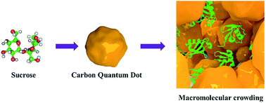
Fluorescent carbon quantum dots (CQD) induce macromolecular crowding making them suitable for probing the structure, function and dynamics of both hydrophilic and hydrophobic peptides/proteins under near in-cell conditions.


Carbon quantum dots (CQD) are nanomaterials with size less than ∼10 nm, first obtained in 2004 during purification of single-walled carbon-nanotubes.1 Since then CQDs have been used in a wide range of applications due to their low cost of preparation2,3 and favourable properties such as chemical inertness, biocompatibility, non-toxicity and solubility in aqueous medium. One of the applications of CQDs has been their use for imaging and tracking proteins inside cells, based on their intrinsic fluorescence.3,4 This has been demonstrated for a wide range of systems.5 Further, quantum dots exhibit concentration dependent aggregation6 while retaining their solubility. Considering these aspects, we show here that carbon quantum dots act as a macromolecular crowder confining proteins to a restricted space. This makes them suitable for structural and functional studies of proteins in near in-cell conditions (Fig. 1). Macromolecular crowding, which occurs primarily via the excluded volume effect,7 has been shown to mimic conditions inside living cells effecting the structural, dynamic and functional behaviour of proteins.8 Unlike conventionally used crowding agents the CQDs proposed here can be adapted for both hydrophilic as well as hydrophobic (e.g., membrane) proteins by a simple modification in the method of preparation. Further, their intrinsic fluorescence can be used for monitoring the state of aggregation to tune the degree of crowding. This is demonstrated for three proteins: a globular structured protein (ubiquitin), an intrinsically disordered protein: the linker domain of insulin like growth factor binding protein-2 (L-hIGFBP2; 12 kDa)9 and a 17-residue peptide comprising the hydrophobic region of the prion Protein in its N-terminal domain (residues 112–128).10
image file: c4ra14019b-f1.tif
Fig. 1 Schematic representation of carbon quantum dots as macromolecular crowders.

The CQDs were prepared by refluxing sucrose in water at high pH (∼12) for hydrophilic and with octadecyl-amine/octadecene for hydrophobic CQD2a (the complete details of sample preparation and characterization are provided in the ESI Sections S1–S4). CQDs thus prepared were characterized extensively using transmission electron microscopy (TEM), scanning electron microscopy (SEM), dynamic light scattering (DLS), fluorescence, infra-red (IR) and nuclear magnetic resonance (NMR) spectroscopy. The particle size of CQDs were determined to be ∼2–10 nm using TEM (Fig. 2A and D) for both hydrophilic and hydrophobic CQDs. The fluorescence spectra were measured using the excitation wavelength 346 nm (Fig. 2B and E) at different concentration in water and methanol, respectively. The intrinsic fluorescence CQDs was used for obtaining fluorescence microscope image also (Fig. S1). The composition the CQDs were characterized by IR spectroscopy, NMR and XPS (Fig. S2). The IR spectrum reveals the presence of OH, C[double bond, length as m-dash]O, COOH, C–O (ether) and NH2 (in hydrophobic CQD) functional groups (Fig. S3). The presence of sp2 and sp3 carbons including the functional groups were verified using XPS spectra.


image file: c4ra14019b-f2.tif
Fig. 2 Characterization of CQDs: the two sets A–C and D–F represents TEM image (at <0.1 mg ml−1), fluorescence emission spectra (with excitation at 346 nm), DLS spectra for hydrophilic and hydrophobic CQDs, respectively, at high concentration. The inset in A and C shows a magnified view of the CQDs. The fluorescence emission spectra of the CQDs at different concentrations indicates aggregation at higher concentration.

The CQDs aggregate as the concentration is increased (Fig. 2C and F). This was validated using SEM (Fig. S1A) and DLS (Fig. S4a). The size of the aggregated particles increase to 8454 nm at 100 mg ml−1 for hydrophilic CQD and 6930 nm at maximum saturated conc. for hydrophobic CQD. This is also followed by increase in the viscosity of the medium (Fig. S5) as measured by rheometer. This rapid increase in viscosity is one of the properties that CQDs share with other crowding agents such as Ficoll (Fig. S5A). The aggregation of CQDs results in solvent exclusion (discussed below), a property important for the crowding effect.7,11

Zeta-potential measurements indicate a charge of 0.0002 femto-coulombs and a zeta-potential value 0.8 mV for hydrophilic CQD, whereas the corresponding values for hydrophobic CQDs are ∼0 and 0.7 mV, respectively. Thus both the CQDs can be considered to be electrically neutral, a property useful for avoiding any electrostatic interaction with the protein being studied. These CQDs also do not photo-bleach under the exposure of light,4a and are thus chemically inert and stable.

The two properties above: self-aggregation and charge neutrality implies that the CQDs can act as molecular crowders through an excluded volume effect. Such an effect was tested on three proteins: ubiquitin, an intrinsically disordered protein: the linker domain of insulin like growth factor binding protein-2 (L-hIGFBP2)9 and a 17 residue peptide the hydrophobic region of the prion protein comprising residues 112–128.10 The first two proteins were probed using the hydrophilic CQD and the membrane protein was studied using the hydrophobic CQD.

Fig. 3A and B shows the two dimensional (2D) [15N–1H] heteronuclear single quantum (HSQC) spectra of ubiquitin in the absence and presence of hydrophilic CQDs. The structure of ubiquitin is not altered as evident from the absence of any change in the chemical shifts of the cross peaks. In order to verify if the protein is not aggregating and/or interacting with the CQDs, the rotational correlation time (τc) of the protein was measured using 15N R2 and 15N R1 relaxation times.12 The τc of the protein increases from 4.5 ns (in absence of CQD) to 6.5 ns (in presence of 100 mg ml−1 CQD). Note that for the same concentration, the viscosity of the medium changes significantly (Fig. S5). This implies that the increase in τc is primarily happening due to the crowding effect, wherein the local effective concentration of the protein goes up. On the contrary, a direct interaction of the protein with CQDs or an effect of the viscosity of the medium on the protein would have resulted in considerable broadening of NMR signals and a large measured τc, both of which are not observed. A concentration of 10–25 mg ml−1 of CQD resulting in τc of 4.8–5.25 ns comes closest to the in-cell conditions where a τc ∼ 4.9 ns is observed for ubiquitin in intact cells by NMR (Fig. S6A).


image file: c4ra14019b-f3.tif
Fig. 3 Effect of molecular crowding by CQDs studied using NMR spectroscopy: 2D [15N–1H] HSQC spectra of (A) ubiquitin and (B) its overlay with the spectra acquired for the protein in 25 mg ml−1 of hydrophilic CQDs. (C) 2D [15N–1H] HSQC spectra of L-hIGFBP2 and (D) its overlay with the spectra acquired for the protein in 25 mg ml−1 of hydrophilic CQD.

Molecular crowding in presence CQDs is also evident from its effect on the intrinsically disordered protein: L-hIGFBP2 (12 kDa). While the effect of macromolecular crowding on the structure and dynamics of intrinsically disordered proteins (IDPs) is less explored, it has been observed that IDPs behave differently from structured proteins.13,14 For instance, the flexibility of IDPs counteracts the effect of viscosity on their NMR spectra. Fig. 3C and D shows the spectra of L-hIGFBP2 in absence/presence of hydrophilic CQDs. As in the case of ubiquitin, no significant change is the chemical shifts are observed. However, a decrease in τc was observed in the presence of hydrophilic CQDs from 3.2 ns (in absence of CQDs) to ∼2.5 ns (in presence of 25 mg ml−1 of CQDs). The decrease in rotational correlation time could be due to an increase in compactness of the protein reducing its anisotropic motions. We found a similar reduction in the presence of the conventionally used crowding agent, Ficoll, (2.5 ns in 100 mg ml−1 of Ficoll, Fig. S7).

Using the hydrophobic CQDs we studied the effect of crowding on the 17 residue peptide comprising residues 112–128 (MAGAAAAGAVVGGLGGY) of Prion Protein (PrP) with NMR spectroscopy. The 17 residue peptide is part of the conserved hydrophobic region (CHR)10 and is responsible for the conversion of recombinant PrPc (normal cellular form) to PrPsc (amyloidic form) in the presence of phospholipid bicelles.15,16 We wanted to study the amylogenic properties of this peptide in solution in absence of the membrane. To understand the importance of the role of hydrophobic interactions in its amyloidogenesis, the peptide was dissolved in methanol, which is known to weaken hydrophobic interactions.17 The peptide adopts a β-sheet conformation as evident from circular dichroism (Fig. S8). Fig. 4 shows the 1D 1H spectra of the 1 mM of peptide in methanol. The solution was kept standing for several hours and no precipitate or aggregation effects were noticeable in the NMR spectrum. To test if molecular crowding results in the formation of the fibrils, hydrophilic and hydrophobic CQDs were added (Fig. S4 and S9). The 1D 1H NMR spectrum at two concentrations of hydrophobic CQDs is shown in Fig. 4C and D. Even in the presence of both the macromolecular crowders, which are known to accelerate the formation of aggregates/amyloid fibres,18 no visible precipitate or aggregates were observed up to several days. This indicates that the lipid environment is important for the formation of the PrP amyloid and reinforces results of the previous studies which have shown that binding to or interaction with the membrane is important for formation of the amyloids10,16,19 and non-membrane bound forms of PrP may be non-pathogenic.20


image file: c4ra14019b-f4.tif
Fig. 4 Effect of hydrophobic CQD on peptide (PrP): one dimensional 1H-NMR spectra of peptide (PrP) with two different concentration of hydrophobic CQD in methanol. The spectra in D shows peaks from both CQDs and the peptide. Absence of significant broadening of the peptide peaks supports the evidence that fibrils are not formed.

In summary, we have shown for the first time that carbon quantum dots induce molecular crowding. The CQDs have several advantages over other popularly used crowding agents due to: (1) the ability to prepare both hydrophilic and hydrophobic CQDs, (2) their high intrinsic fluorescence enabling the tuning of their concentration and (3) the ease and inexpensive method of preparation. To our knowledge this represents the first report of a macromolecular crowding agent for hydrophobic proteins. Taken together, this opens up new avenues for studying the structure, dynamics and function of biomolecules in near in-cell conditions.

Acknowledgements

The work was supported by research grants from Department of atomic energy (DAE), India. Support for NMR facility at IISc is gratefully acknowledged. We acknowledge the support from IISc Nanocentre for different experimental characterizations.

Notes and references

  1. H. T. Li, Z. H. Kang, Y. Liu and S. T. Lee, J. Mater. Chem., 2012, 22, 24230 Search PubMed.
  2. (a) S. K. Bhunia, A. Saha, A. R. Maity, S. C. Ray and N. R. Jana, Sci. Rep., 2013, 3, 1473 Search PubMed; (b) Q. Liang, W. Ma, Y. Shi, Z. Li and X. Yang, Carbon, 2013, 60, 421 CrossRef CAS PubMed.
  3. (a) Z. J. Zhu, Y. C. Yeh, R. Tang, B. Yan, J. Tamayo, R. W. Vachet and V. M. Rotello, Nat. Chem., 2011, 3, 963 CrossRef CAS PubMed; (b) S. Sahu, B. Behera, T. K. Maiti and S. Mohapatra, Chem. Commun., 2012, 48, 8835 RSC.
  4. (a) L. Wang, S. J. Zhu, H. Y. Wang, S. N. Qu, Y. L. Zhang, J. H. Zhang, Q. D. Chen, H. L. Xu, W. Han, B. Yang and H. B. Sun, ACS Nano, 2014, 8, 2541 CrossRef CAS PubMed; (b) A. Zhu, Q. Qu, X. Shao, B. Kong and Y. Tian, Angew. Chem., Int. Ed. Engl., 2012, 51, 7185 CrossRef CAS PubMed.
  5. (a) Q. Wang, X. Huang, Y. Long, X. Wang, H. Zhang, R. Zhu, L. Liang, P. Teng and H. Zheng, Carbon, 2013, 59, 192 CrossRef CAS PubMed; (b) H. Ding, L.-W. Cheng, Y.-Y. Ma, J.-L. Kong and H.-M. Xiong, New J. Chem., 2013, 37, 2515 RSC.
  6. M. Noh, T. Kim, H. Lee, C. K. Kim, S. W. Joo and K. Lee, Colloids Surf., A, 2010, 359, 39 CrossRef CAS PubMed.
  7. (a) H. Damien and P. M. Allen, Biochim. Biophys. Acta, Proteins Proteomics, 2003, 1649, 127139 Search PubMed; (b) Z. Huan-Xiang, R. Germán and P. M. Allen, Biophysics, 2008, 37, 375 Search PubMed.
  8. S. B. Zimmerman and A. P. Minton, Annu. Rev. Biophys. Biomol. Struct., 1993, 22, 27 CrossRef CAS PubMed.
  9. M. Swain, M. G. Slomiany, S. A. Rosenzweig and H. S. Atreya, Arch. Biochem. Biophys., 2010, 501, 195 CrossRef CAS PubMed.
  10. S. Sauve, D. Buijs, G. Gingras and Y. Aubin, J. Biol. Chem., 2012, 287, 1915 CrossRef CAS PubMed.
  11. A. P. Minton, G. C. Colclasure and J. C. Parker, Proc. Natl. Acad. Sci. U. S. A., 1992, 89, 10504 CrossRef CAS.
  12. A. G. Palmer III, Annu. Rev. Biophys. Biomol. Struct., 2001, 30, 129 CrossRef PubMed.
  13. C. Li, L. M. Charlton, A. Lakkavaram, C. Seagle, G. Wang, G. B. Young, J. M. Macdonald and G. J. Pielak, J. Am. Chem. Soc., 2008, 130, 6310 CrossRef CAS PubMed.
  14. E. A. Cino, M. Karttunen and W. Y. Choy, PLoS One, 2012, 7, e49876 CAS.
  15. F. Ceciliani and P. Pergami, Curr. Protein Pept. Sci., 2001, 2(3), 191–204 CrossRef CAS.
  16. T. Lührs, R. Zahn and K. Wüthrich, J. Mol. Biol., 2006, 357, 833 CrossRef PubMed.
  17. S. Hwang, Q. Shao, H. Williams, C. Hilty and Y. Q. Gao, J. Phys. Chem. B, 2011, 115, 6653 CrossRef CAS PubMed.
  18. (a) D. M. Hatters, A. P. Minton and G. J. Howlett, J. Biol. Chem., 2002, 277, 7824 CrossRef CAS PubMed; (b) J. Martin, J. Mol. Recognit., 2004, 17, 465 CrossRef CAS PubMed.
  19. (a) D. A. Kocisko, J. H. Come, S. A. Priola, B. Chesebro, G. J. Raymond, P. T. Lansbury and B. Caughey, Nature, 1994, 370, 471 CrossRef CAS PubMed; (b) V. A. Lawson, S. A. Priola, K. Wehrly and B. Chesebro, J. Biol. Chem., 2001, 276, 35265 CrossRef CAS PubMed.
  20. B. Chesebro, M. Trifilo, R. Race, K. Meade-White, C. Teng, R. LaCasse, L. Raymond, C. Favara, G. Baron, S. Priola, B. Caughey, E. Masliah and M. Oldstone, Science, 2005, 308, 1435 CrossRef CAS PubMed.

Footnote

Electronic supplementary information (ESI) available: Experimental details, other characterization and biophysical data. See DOI: 10.1039/c4ra14019b

This journal is © The Royal Society of Chemistry 2015
Click here to see how this site uses Cookies. View our privacy policy here.