Roozbeh Javad Kalbasi*a and
Ali Zirakbashb
aFaculty of Chemistry, Kharazmi University, Tehran, Iran. E-mail: rkalbasi@gmail.com; Fax: +98 21 88820992; Tel: +98 21 88848949
bDepartment of Chemistry, Shahreza Branch, Islamic Azad University, 311-86145, Isfahan, Iran
First published on 15th January 2015
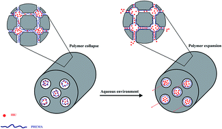
Poly(2-hydroxyethyl methacrylate)/KIT-5 as an ordered mesoporous polymer–silica nanocomposite was synthesized through in situ polymerization. The chemical and physical properties of the nanocomposite were analyzed using a series of different techniques, including XRD, BET, FT-IR, SEM and TEM analysis. Afterward, this innovative ordered mesoporous nanocomposite was utilized for the delivery and controlled release of the model drug ibuprofen. In addition, the in vitro cytotoxicity of the nanocomposite was assessed by MTT assay. Overall, the results were indicative of the potential of the nanocomposite as an excellent carrier system.
Versatile drug nanocarriers, including liposomes, dendrimers and polymeric nanoparticles have been utilized to approach the mentioned goals.3–5 Actually, liposome-based therapeutics have been on the market for more than a decade, but use of liposomes in clinics poses several challenges, including the high production cost. Whilst commercial drug delivery systems (DDSs) based on polymers have been very successful systems that cover several fields, such as implantable, oral, transdermal and injectable drug reservoirs, one of the main and most specific problems of such systems is the loss of activity of several drugs before reaching the target tissue as a result of premature degradation of the active agent.6,7
As an alternative to nanoscale drug carriers constructed from organic materials, innovative inorganic material-based systems have been greatly advanced. Ever since the discovery of MCM-41-type ordered mesoporous silica by Mobil Corporation scientists in the early 1990s, there has been large amount of research conducted on the controlled syntheses and applications of mesoporous silica.8,9 In particular, biomedical application research on mesoporous silica nanoparticles (MSNs) has received great attention over the last few years.10,11 For instant, MSNs have been intensively suggested for use in controlled drug/gene release and as delivery carriers due to the following unique properties:12–18
1. Tunable particle size. The particle size of MSNs can be tuned from 50 to 300 nm permitting a facile endocytosis by living animal and plant cell without any significant cytotoxicity.
2. Stable and rigid framework. In contrast to other polymer-based drug carriers, MSNs are more resistant to heat, pH, mechanical stress and hydrolysis-induced degradation.
3. Uniform and tunable pore size. The pore size distribution of MSNs is very narrow and pore diameter can be tailored between 2 and 20 nm. These features enable one to adjust the loading of various drug molecules and study the kinetics of drug release with high precision.
4. High surface area and large pore volume. The total surface area and pore volume of mesoporous silicas are very large, which allow high loadings of drug molecules.
However, conventional pure MSNs have little control over drug release mainly because of weak interactions between drug molecules and unmodified surfaces. To overcome this problem, the surface is usually functionalized with chemical groups that are able to link to the drug molecules through stronger bonds.19–23 Extensive efforts toward the functionalization of MSNs have been carried out using different approaches, such as post-synthesis grafting, co-condensation, and post-polymerization.24 In the post-polymerization method, which has been less studied on DDSs, mesoporous polymer–inorganic hybrid materials are synthesized using in situ free radical polymerization of vinyl-based monomers confined at the mesopore surface. After polymerization of the monomers, a thin layer of polymer is formed on the inner surface of the MSNs, while the mesopores remain fully accessible. This method makes it possible to exploit the desired physical properties of the MSNs such as large pore diameter and ordered mesostructure, as well as the additional properties of the introduced functional groups.25–27
Herein, and in continuing our previous works to develop new organic–inorganic hybrid materials, we will introduce a simple, yet efficient hybrid organic–silica carrier system based on a cage-type mesoporous silica with three-dimensional (3D) porous network (KIT-5) and 2-hydroxyethyl methacrylate as a biocompatible hydrogel.28–30 The potential of the nanocomposite to load drug molecules was investigated using ibuprofen (IBU) as a model drug and the release pattern was studied in simulated body fluid (SBF). To evaluate the efficiency of the carrier system, a comparison was made with pure KIT-5 as a vehicle, and as anticipated, the nanocomposite showed a more controllable release pattern.
In a typical synthesis method, 2.5 g (0.198 mmol) of F-127 was dissolved in 120 g (6.60 mol) of distilled water and 5.25 g (0.05 mol) of concentrated hydrochloric acid (35 wt% HCl). To this mixture, 12 g (0.057 mol) of TEOS was added. The mixture was stirred at 45 °C for 24 h for the formation of the mesostructured product. Subsequently, the reaction mixture was heated for 24 h at 95 °C under static conditions for hydrothermal treatment. The solid product was then filtered, washed with deionized water and dried at 80 °C. Another sample was prepared by varying the hydrothermal treatment temperature from 95 to 150 °C. The samples were designated KIT-5(T) where T denotes the hydrothermal treatment temperature. Finally, the samples were calcined at 550 °C for 6 h in order to remove the template.
Viability% = 100 − toxicity% |
![]() | ||
Fig. 1 The powder XRD patterns of KIT-5(150), PHEMA/KIT-5(150)1/2, KIT-5(95), PHEMA/KIT-5(95) and IBU/KIT-5(150). |
The length of the cubic cell a0 and the diameter of the cage Dm are calculated using the formulae and Dm = a0(6εme/πv)1/3 (εme is the volume fraction of a regular cavity and v is the number of cavities present in the unit cell which is equal to 4 for Fm3m space group) respectively33,34 (Table 1). The greater cubic cell length, cage diameter and other related parameters of KIT-5(150) compare to KIT-5(95) which are also suggested by the slight shift of its characteristic reflection peak to a lower angle, arise from the higher hydrothermal treatment temperature.33,34 Furthermore, the decrease of d111 spacing (or the pore center distance a0) in polymeric composites results from decreasing of the cage size after hybridization process which may be attributed to polymer chains distribution in pores of the KIT-5 (Table 1).
Sample | d111 (nm) | a0 (nm) | Dm (nm) | 2θ (°) |
---|---|---|---|---|
KIT-5(95) | 9.57 | 16.6 | 7.32 | 0.922 |
KIT-5(150) | 12.02 | 20.8 | 9.18 | 0.734 |
PHEMA/KIT-5(95) | 9.40 | 16.28 | — | 0.938 |
PHEMA/KIT-5(150)1/2 | 11.3 | 19.57 | — | 0.78 |
Nitrogen adsorption–desorption measurements were used to further characterize the mesoporous silicas and the nanocomposites as shown in Fig. 2 and 3. The BET specific surface areas and pore sizes of the host KIT-5(150), IBU/KIT-5(150) and PHEMA/KIT-5(150) were calculated using Brunauer–Emmentt–Teller (BET) and Barrett–Joyner–Halenda (BJH) methods, respectively. Both samples show a type IV adsorption–desorption isotherm with an H2 hysteresis loop, signifying the large uniform cage-type pores (Fig. 2). Besides, the corresponding BJH adsorption pore size distributions shown in Fig. 3 demonstrate the high structural ordering with a narrow pore size distribution of the samples.
![]() | ||
Fig. 2 N2 adsorption–desorption isotherms of mesoporous silica KIT-5(150), PHEMA/KIT-5(150)1/2 and IBU/KIT-5(150). |
BET surface area, pore size and pore volume of KIT-5(95), KIT-5(150), PHEMA/KIT-5(95) and PHEMA/KIT-5(150)1/2 samples have been summarized in Table 2. A comparison between the two pure mesoporous silicas reveals that KIT-5(150) possesses larger pore volume and pore size but smaller BET surface area relative to KIT-5(95). This observation, arisen from variation in hydrothermal treatment temperature, is not only in agreement with the XRD results but also with other studies.34 It is also noticeable that in comparison to KIT-5(150), PHEMA/KIT-5(150)1/2 features less BET surface area and pore volume which are acceptable consequences of immobilization the polymeric chains inside the pores. Despite the reduction of surface area and pore volume of PHEMA/KIT-5(150)1/2, there seems to be no significant change in the pore size of the resultant composite. This fact could be attributed to the occurrence of two simultaneous but opposing incidents. To state more precisely, the presence of PHEMA chains inside the channels, reduces the pore size of mesoporous material as expected. In the meanwhile, the physical pressure on the channels wall induced by polymeric strands can cause an increase in the pore diameter. However, it is wise to advert that the exact similarity of the numbers here is accidental (the same trends could be obviously seen for KIT-5(95) and PHEMA/KIT-5(95) from Table 2).28,29
Sample | BET surface area (m2 g−1) | Pore volume (cm3 g−1) | Pore diameter (nm) |
---|---|---|---|
KIT-5(95) | 1090 | 0.71 | 2.62 |
KIT-5(150) | 517 | 0.79 | 4.03 |
PHEMA/KIT-5(95) | 596 | 0.40 | 2.59 |
PHEMA/KIT-5(150)1/2 | 393 | 0.64 | 4.03 |
Fig. 4 presents the FT-IR spectra of KIT-5(150) and PHEMA/KIT-5(150)1/2 (spectra a and b). The spectra of pure KIT-5 (150) and the hybrid sample exhibit the typical vibrations of asymmetric and symmetric stretching in addition to the rocking of Si–O–Si at approximately around 1090, 820 and 470 cm−1 which are characteristic of mesoporous silica. Moreover, the intense band at 1727 cm−1 clearly shows the presence of PHEMA in the nanocomposite sample (Fig. 4b). This new band at the hybrid sample spectrum is attributed to the carbonyl bond of PHEMA.35 Furthermore, the aliphatic C–H stretching peaks at around 2800–3000 emphasize the existence of PHEMA in the hybrid sample. It is worthy to note that all the mentioned evidences signify the successful synthesis of the PHEMA/KIT-5(150) composite.
![]() | ||
Fig. 4 FT-IR spectra of (a) mesoporous silica KIT-5(150), (b) PHEMA/KIT-5(150)1/2, (c) IBU and (d) IBU/KIT-5(150). |
In order to measure the amount of polymer loaded inside the KIT-5(150), as well as thermal stability of the nanocomposite, thermo gravimetric analysis under N2 atmosphere was conducted. Fig. 5 shows the TGA curve of PHEMA/KIT-5(150)1/2. The mass loss at temperature below 100 °C (approximately 3%, w/w) is attributed to the thermo desorption of physically adsorbed water on the surface of the sample. The second weight loss in the range of 250 to 500 °C which is around 22% (w/w), results from the degradation of the polymer. By comparing the PHEMA/KIT-5(150)1/2 and pure PHEMA TG curves,35 it becomes clear that the weight loss of the confined PHEMA occurs at a higher temperature, which means that PHEMA/KIT-5(150)1/2 has a higher thermal stability and a slower degradation rate than the naked PHEMA. Therefore once again, it could be concluded that the polymeric strands are mainly present inside the pores.
The morphologies of the synthesized products i.e. KIT-5 host, PHEMA/KIT-5(150)1/2 and PHEMA/KIT-5(95) were inspected by means of SEM technique (Fig. 6). All samples are agglomerates of small irregular particles. It can be observed that there is negligible difference in particle surface morphology between the KIT-5 host and the PHEMA/KIT-5 composites, implying that the polymerization takes place more in the channels and less on the outer surface of the matrix.
![]() | ||
Fig. 6 Scanning electron microscopy (SEM) photographs of (a) mesoporous silica KIT-5, (b) PHEMA/KIT-5(150)1/2 and (c) PHEMA/KIT-5(95). |
In Fig. 7, characteristic TEM images of PHEAM/KIT-5(150)1/2 and PHEMA/KIT-5(95) are shown. It should be noted that despite the appearance of some darker patches on the images, which are attributed to the encapsulation of PHEMA chains inside the mesochannels, the ordered cubic Fm3m mesostructure of KIT-5 is obviously retained and no damage in the periodic structure of the silicate framework is observed.
Weight ratio of ibuprofen to mesoporous matrix (w/w) | 1![]() ![]() |
2![]() ![]() |
3![]() ![]() |
4![]() ![]() |
5![]() ![]() |
---|---|---|---|---|---|
Amount of loaded drug in KIT-5(95) (w/w) | 27% | 30% | 32% | 40% | 38% |
Sample | d111 (nm) | a0 (nm) | BET surface area (m2 g−1) | Pore volume (cm3 g−1) | Pore diameter (nm) |
---|---|---|---|---|---|
IBU/KIT-5(150) | 11.31 | 19.59 | 351 | 0.58 | 3.53 |
To further examine whether IBU molecules are mainly encapsulated into the mesostructure, nitrogen adsorption–desorption analysis was performed. Fig. 2 also shows the N2 adsorption–desorption isotherm of KIT-5(150) loaded with IBU molecules. Apparently, this isotherm is in accordance with the type IV isotherm with an H2 hysteresis loop that signifies the mesoporous structure of IBU/KIT-5(150). In other words, this means that the mesoporous structure of KIT-5(150) is preserved even after the confinement of IBU molecules inside the pores. Additionally, as shown in Table 4, the BET surface area, pore volume and diameter reduction of IBU/KIT-5(150) relative to KIT-5(150) (Table 2) could be further used to conclude that ibuprofen molecules are mainly placed on the internal surface of KIT-5(150).36 Moreover, the narrow pore size distribution of IBU/KIT-5(150) illustrated in Fig. 3, strongly suggest the even dispersion of drug molecules inside the channels.
FT-IR spectra of IBU (c) and IBU/KIT-5(150) (d) are shown in Fig. 4. The FT-IR spectrum of IBU/KIT-5(150) clearly shows the carbonyl and C–H stretching vibrations at 1712 and 2800–3000 cm−1 respectively. It is worthy to note that the slight shift of the carbonyl stretching adsorption is attributed to the hydrogen bonding present between ibuprofen carbonyl groups and hydroxyl groups of KIT-5. Nevertheless, these stretching absorbances which are obviously in consonance with the IBU spectrum could be used to further confirm the presence of IBU molecules in the mesoporous matrix.
Fig. 8 illustrates the scanning electron microscopic images of KIT-5(150) loaded with IBU molecules by 6000 (a) and 15000 (b) times of magnification. Making a comparison of these images with the one related to the pure KIT-5 (Fig. 6a), reveals the slight changes made in the particles morphology after loading with drug molecules. This observation suggests the presence of some IBU molecules on the outer surface of the matrix. It should be noted that this conclusion is in accordance with other studies.37
Sample | KIT-5 (95) | KIT-5 (150) | PHEMA/KIT-5 (95) | PHEMA/KIT-5 (95)1/2 | PHEMA/KIT-5 (150) | PHEMA/KIT-5 (150)1/2 |
---|---|---|---|---|---|---|
Amount of loaded ibuprofen (w/w) | 40% | 37% | 16% | 19% | 25% | 38% |
In the case of polymeric composites, PHEMA/KIT-5(95) contains relatively lower amounts of IBU molecules. This observation arises from the occupation of the composite pore volume with polymeric chains. To approach this problem, PHEMA/KIT-5(150)1/2 with greater free pore volume was utilized. It is wise to note that in this case, although some of the pore volume is still occupied with polymer strands, the stronger interaction between IBU molecules and PHEMA (mostly hydrogen bonds) results in a good loading capacity.
As mentioned previously, spectrophotometric analysis of the SBF supernatant of different matrices yielded information about the release profile of different mesoporous carriers. As illustrated in Fig. 9, cumulative release pattern of all samples, show an initial burst of drug release which could be due to the adsorbed IBU on the outer surface of the mesoporous matrices that is easily released in the first step to the delivery medium. Further release analysis of IBU from KIT-5(95), reveals a very slow and incomplete release of drug molecules which is acceptable due to its cage-type structure and narrow pore diameter. Because of the poor results obtained from KIT-5(95) based carriers, either in loading or release processes (16% drug loading and an incomplete and unrepeatable drug release for PHEMA/KIT-5(95)), the next experiments were mainly focused on KIT-5(150) based carriers. Studies on KIT-5(150) release profile reveals a more complete delivery of IBU compare to KIT-5(95) thanks to its greater pore diameter. On the other hand, IBU release from PHEMA/KIT-5(150)1/2 shows a more controlled release pattern which could be very profitable in some cases.
![]() | ||
Fig. 9 IBU release profile from different matrices (a) KIT-5(150), (b) KIT-5(95), and (c) PHEMA/KIT-5(150)1/2. |
As it can be deduced from Fig. 10, IBU molecules contain hydroxyl and carbonyl groups through which, they can be adsorbed to the silanol groups of KIT-5 by hydrogen bonds. However, these interactions are relatively week due to the hydrophobic characteristics of IBU (benzene ring and alkyl groups) which is in contrast to hydrophilic characteristics of KIT-5.
In the case of composite (PHEMA/KIT-5), although HEMA polymeric chains are adsorbed to the silanol groups by the same hydrogen bonds, this adsorption is much stronger than the previous one and polymeric chains are not leachable since there are much more hydrogen bonds as a consequence of more functional groups. It is important to note that in this case, not all the functional groups of PHEMA are adsorbed on the silica surface, therefore, there are still free functional groups to interact to IBU molecules through hydrogen bonds (these hydrogen bonds are also stronger than the hydrogen bonds between IBU and KIT-5 since both IBU and the polymer have hydrophobic characteristics). In addition, dipol–dipol interactions between polar parts as well as numerous van der Waals forces between nonpolar chains of polymers and nonpolar parts of ibuprofen molecules, make the adsorption of IBU on the composite much greater than its adsorption on the unmodified KIT-5 (in the latter, there is definitely no van der Waals force).
In the release process of the IBU/unmodified KIT-5 in water, it is in fact the stronger hydrogen bonds between IBU and water molecules which bring about the release process and the only decelerating factor might be the pore size of KIT-5.
However, the release process from the nanocomposite (PHEMA/KIT-5) is a different story. Since HEMA is a hydrogel, it collapses in non-aqueous environment as a result of intramolecular hydrogen bonds. Consequently, IBU molecules are trapped inside the chains and have difficulty releasing to the medium. However, in an aqueous environment, these intramolecular hydrogen bonds are replaced by intermolecular hydrogen bonds (with water molecules) and result in polymer expansion, allowing the IBU molecules to release. This release process is slower and more controllable compare to the release process from KIT-5 thanks to the mentioned stronger interactions.
It is also wise to mention that it is in fact the hydrophilic characteristics of polymer chains which bring about the release of drug molecules into the delivery medium. In other words, on the one hand, the expansion of internal hydrophilic polymer chains in aqueous environments would commence the release process, but on the other hand the limited expansion of internal chains, due to the rigid structure of KIT-5, makes the release process slow and controllable (Fig. 11).
![]() | ||
Fig. 11 Schematic release process of IBU molecules from PHEMA/KIT-5(150)1/2 in an aqueous environment. |
![]() | ||
Fig. 12 Cytotoxicity study (MTT assay) of mesoporous matrices KIT-5(150) and PHEMA/KIT-5(150)1/2 at different concentration. |
This journal is © The Royal Society of Chemistry 2015 |