Hydrogen peroxide-induced oxidative stress activates NF-κB and Nrf2/Keap1 signals and triggers autophagy in piglets

Jie Yin ab, Jielin Duanab, Zhijie Cuiab, Wenkai Rena, Tiejun Li*a and Yulong Yin*a
aScientific Observing and Experimental Station of Animal Nutrition and Feed Science in South-Central, Ministry of Agriculture, Hunan Provincial Engineering Research Center of Healthy Livestock, Key Laboratory of Agro-ecological Processes in Subtropical Region, Institute of Subtropical Agriculture, Chinese Academy of Sciences, Changsha, Hunan 410125, China. E-mail: yinjie2014@126.com; 791292383@qq.com; xiaocui.huoying@qq.com; renwenkai19@126.com; tjli@isa.ac.cn; yinyulong@isa.ac.cn
bUniversity of Chinese Academy of Sciences, Beijing 100039, China

Received 31st October 2014 , Accepted 16th January 2015

First published on 16th January 2015


Abstract

In various pathological conditions of tissue injury, oxidative stress is often associated with autophagy. However, H2O2-induced oxidative stress initiates autophagy and its molecular mechanism is still obscure. Here we report that intragastric and peritoneal administration of H2O2 caused different degrees of oxidative stress and changed intestinal permeability, morphology, and barrier function in piglets. Western blotting studies revealed that H2O2 increased the autophagosome-related protein LC3-I and LC3-II abundance and the ratio of LC3-II to LC3-I content after exposure to 10% H2O2 in the jejunum. Meanwhile, the data from beclin1 also indicated that H2O2 initiated autophagy in response to oxidative stress. In addition, H2O2 activates the NF-κB and Nrf2/Keap1 signals, which may be involved in H2O2-induced autophagy. In conclusion, administration of H2O2 caused intestinal oxidative stress and triggered an autophagic response, which might be associated with NF-κB and Nrf2/Keap1 signals.

Introduction

Oxidative stress is considered to be an oxidative imbalance as an excess of reactive oxygen species (ROS) overwhelms antioxidant defenses, including enzymatic systems and nonenzymatic antioxidants.1,2 Hydrogen peroxide (H2O2), a highly reactive oxygen species, has been shown to result in the generation of other ROS, such as hydroxyl radicals, and the imbalanced oxidant/antioxidant status in vivo3 and in vitro.4 The oxidative imbalance correlates with the modification of proteins, lipid oxidation, and nucleic acid breaks, which may further impair cellular function and result in clinical deterioration.1,5

Autophagy, a process for cellular cleaning through the self-digestion of unnecessary intracellular proteins and dysfunctional organelles, is a well conserved mechanism from yeast to mammalian cells and also contributes to the maintenance of cellular homeostasis and of the energetic balance in cellular and tissue remodeling, and cellular defense against extracellular insults and pathogens.6 Recently, autophagy has been demonstrated to play an important role in cell homeostasis by regulating the turnover of long-lived proteins and getting rid of damaged structures.7 Many studies have shown that autophagy can be stimulated by various factors, including nutrient starvation8 and infection.9 Although it was confirmed that oxidative stress can also promote autophagy in vitro,10 little is known about autophagy induced by the administration of H2O2 and the molecular mechanism between intestinal oxidative stress and the formation of autophagosomes in vivo. Due to their similarities to humans regarding morphology, function of organs, and metabolic rate, piglets were used as the in vivo model. We hypothesized that H2O2 administration could induce autophagy in piglets. Thus, the oxidative stress and autophagy were studied in pigs after administration of H2O2. The activation of NF-κB and Nrf2/Keap1 signaling pathways was also analyzed after administration of H2O2. In this study, we found that administration of H2O2 causes intestinal oxidative stress and triggers autophagy, which may be associated with NF-κB and Nrf2/Keap1 pathways.

Results

H2O2 suppressed the activity of antioxidant enzymes and caused oxidative stress

H2O2 is a highly reactive oxygen species. So, the primary focus was to monitor the oxidative parameters in the serum. The data showed that intragastric administration of H2O2 suppressed catalase with a dose-dependent decrease (P < 0.05) in the serum at 50 h (Table S1 in ESI). Compared with intragastric administration, the activity of catalase in the per-10% H2O2 group significantly decreased (P < 0.05). Meanwhile, intragastric administration of 10% H2O2 significantly inhibited GSH-Px activity at 98 h and 170 h (P < 0.05). In addition, the oxidative stress parameters from the greater curvature and jejunum (Table S2 in ESI) further demonstrated that intragastric administration and peritoneal injection of H2O2 disrupted the oxidative balance.

H2O2 changed intestinal permeability, morphology, and tight junctions

Our next focus was to investigate the effect of H2O2 on intestinal function and integrity. The serum LPS level and DAO activity are two major biomarkers of intestinal injury,11 and the data showed that intragastric administration of 10% H2O2 decreased the activity of DAO (P < 0.05) in the serum at 50 h and 98 h (Fig. 1). Meanwhile, the LPS levels were significantly higher after intragastric administration at 50 h and 98 h (P < 0.05) compared with the control group (Fig. 1), suggesting a positive correlation between the increased intestinal permeability and intragastric administration of H2O2. In addition, the macroscopic observations of intestinal morphology also demonstrated that H2O2 caused intestinal injury via the treatment of hematoxylin and eosin (H&E) staining (Fig. 2). Although the histological data seemed to indicate that intragastric administration of H2O2 increased jejunal and ileal villus height (P < 0.05) (Table S3 in ESI), the duodenal villus height, crypt depth, and the ratio of villus height to crypt depth in these three tissues were not affected after subjection to H2O2 (P > 0.05).
image file: c4ra13557a-f1.tif
Fig. 1 Diamine oxidase (DAO) activity (A) and lipopolysaccharides (LPS) level (B) in the serum after exposure to H2O2 (n = 6). The DAO activity and LPS level in the serum were measured at 2, 50, 98, and 170 h after administration of H2O2 and are represented in terms of nanogram per milliliter (ng mL−1). The details of the methodology are described under Materials and methods. Data are presented as mean ± SE.

image file: c4ra13557a-f2.tif
Fig. 2 Histological evaluation of intestinal tissues (HE × 250) after exposure to H2O2 (n = 6). The details of histomorphometry are described under Materials and methods. No histological damage was observed in the control group and per-10% H2O2 group in the duodenum, jejunum, and ileum. Whereas the piglets administrated with H2O2 exhibited scattered villi and desquamation in these tissues.

Tight junctions, the first physical barrier against a variety of pathogens, act to maintain the homeostasis in the gastrointestinal tract.12 As shown in Fig. 3, both intragastric administration and peritoneal injection of H2O2 significantly enhanced (P < 0.05) the abundances of claudin1, occludin, and ZO1 in the jejunum compared with the control group. Meanwhile, the PCR data showed that only intragastric administration of 5% H2O2 up-regulated the expression of ZO1 (about 1.33-fold) and there was no difference between other groups (Fig. 3) in the jejunum.


image file: c4ra13557a-f3.tif
Fig. 3 Jejunal tight junctions were measured via western blot (A and B) and RT-PCR (C) analyses at 7 days (170 h) after administration of H2O2 (n = 6). The details of the methodology are described under Materials and methods. Data are presented as mean ± SE.

H2O2 induced autophagy response in the jejunum

LC3 is involved in the formation of autophagosomes and exists in two forms: an unprocessed 18 kDa cytosolic protein (LC3-I) and a processed 16 kDa protein (LC3-II). LC3-II is membrane-bound and increases during autophagy by conversion from LC3-I, therefore the ratio of LC3-II to LC3-I is often used as an indicator of increased autophagy.13 LC3I and LC3-II levels in the jejunum were measured, and the western blotting results (Fig. 4) showed that administration of H2O2 increased both the abundance of LC3-I and LC3-II (P < 0.05) and the ratio of LC3-II to LC3-I content increased after exposure to 10% H2O2, indicating that H2O2 induced the conversion of LC3-I to LC3-II. Meanwhile, we also investigated the abundance of beclin1. However we failed to notice any significant difference (P > 0.05) in the beclin1 expression via intragastric administration of H2O2. While the beclin1 level in the per-10% H2O2 group was significantly increased (P < 0.05). Furthermore, the PCR data also demonstrated that H2O2 affected the autophagic gene expression as shown in Table S5 in ESI. These results confirmed that administration of H2O2 induces autophagy in the jejunum.
image file: c4ra13557a-f4.tif
Fig. 4 The jejunal autophagy-related proteins (LC3 and beclin1) at 7 days (170 h) after administration of H2O2 (n = 6). The details of methodology are described under Materials and methods. Data are presented as mean ± SE.

Activation and nuclear translocation of NF-κB by H2O2

The nuclear factor kappa B (NF-κB) is involved in the cellular response to stimuli such as cytokines, ultraviolet irradiation, oxidized low-density lipoproteins, and microbes or viral antigens. After activation, NF-κB serves in cell growth, differentiation, development, apoptosis, and oxidative stress.1 We performed western blotting to determine the abundances of IKKα/β, p-IKKα/β, nuclear NF-κB, and cytoplasmic NF-κB in the jejunum after exposure to H2O2 (Fig. 5). An increase of p-IKKβ/IKKβ (P < 0.05) in the per-10% H2O2 group was evident compared with other groups (Fig. 5), indicating an IKKβ phosphorylation. Then we observed that both nuclear and cytoplasmic NF-κB were enhanced (P < 0.05) after exposure to 10% H2O2 and, interestingly, the abundances of NF-κB remained equal between the nucleus and cytoplasm in the same group (Fig. 5). So it was clear that H2O2 caused phosphorylation of IKKβ, and then increased the accumulation of NF-κB and its translocation into the nucleus to initiate gene expression.
image file: c4ra13557a-f5.tif
Fig. 5 The jejunal NF-κB signaling pathway was measured via western blot at 7 days (170 h) after administration of H2O2 (n = 6). The details of methodology are described under Materials and methods. Data are presented as mean ± SE.

Activation and nuclear translocation of Nrf2/Keap1 by H2O2

After observation of NF-κB signal alteration, we next investigated the Nrf2/Keap1 signaling pathway after exposure to H2O2. Nrf2/Keap1 is a major antioxidant transcription factor controlling a wide number of genes including quinine oxidoreductase 1, heme oxygenase-1, and glutathione-S-transferases.1 We were interested to find out the involvement of Nrf2/Keap1 in the H2O2-induced autophagy. Activation of Nrf2/Keap1 is reliant on its upstream protein Keap1. In an inactivated state, Nrf2 is sequestered in the cytosol via Keap1 and is quickly degraded. Modification of Keap1 is necessary for the accumulation of free Nrf2 in the cytoplasm, which then translocate into the nucleus.1 While, interestingly, the results showed that the administration of 10% H2O2 increased (P < 0.05) Keap1 abundance in the jejunum (Fig. 6). Moreover, the cytoplasmic and nuclear Nrf2 abundances were also increased (P < 0.05) after exposure to H2O2 (Fig. 6). These results indicated that H2O2 increased localization of Nrf2 inside the nucleus but not through the degradation of Keap1.
image file: c4ra13557a-f6.tif
Fig. 6 The jejunal Nrf2/Keap1 signaling pathway was measured via western blot at 7 days (170 h) after administration of H2O2 (n = 6). The details of methodology are described under Materials and methods. Data are presented as mean ± SE.

Discussion

Epidemiological evidences have demonstrated that oxidative imbalance can lead to irreparable oxidative injury and cell death when uncontrolled, as oxidative stress affects almost all the cellular macromolecules.1 In the current study, H2O2 was applied to induce oxidative stress as many previous data have suggested that H2O2 is a highly reactive oxygen species and can be capable of diffusing throughout the mitochondria and across cell membranes, causing many types of cellular injury, so it seems to be a suitable agent for investigating reactive oxygen metabolite-induced gastrointestinal oxidative damage.1,14

H2O2 can be metabolized to several ROS, including hydroxyl radicals, which are considered to be the most dangerous compounds to organisms. An excess of un-eliminated H2O2 and its metabolites can oxidize virtually all types of macromolecules: carbohydrates, nucleic acids, lipids and proteins.1 The increased levels of ROS overwhelm the antioxidant defenses and then lead to a state of oxidative stress, which may further impair body function and result in clinical deterioration. The present data indicated that H2O2 inhibited catalase, GSH-Px, and T-AOC activities and increased MDA levels, suggesting a significant disruption in the oxidative balance after exposure to H2O2 treatment. This is in agreement with several previous reports, which also demonstrated that H2O2 caused a substantial reduction in GSH content associated with reduction in GPx activity.3,14

Ingestion of H2O2 changed the permeability and caused dysfunction in the intestine. Previous studies demonstrated that intragastric administration of 6% and 15% H2O2 produced hemorrhagic and necrotic mucosal lesions in the fundic area in rats.3,14 Although we failed to observe the lesions in the gastrointestinal tract after exposure to H2O2, the macroscopic observations of intestinal morphology showed that the villus was scattered and desquamated seriously in the duodenum, jejunum, and ileum after intragastric administration of H2O2. In addition, H2O2-induced oxidative stress is known to result in increased permeability.15 Similarly, the present study also showed an increase in permeability after intragastric administration of H2O2 indicated by the decreased DAO activity and increased LPS level in the serum. The activity of serum DAO is associated with the maturation and integrity of small intestinal mucosa.16 DAO is particularly abundant in enterocytes at the tip of small intestinal villi, from where it is released into the peripheral circulation and then inactivated in the liver. Numerous compelling evidences have demonstrated that the decreased serum DAO activity is in parallel with intestinal mucosal injury.11,16 Meanwhile, the high level of serum LPS is also considered to be the consequence of the increased intestinal permeability.17 Therefore, it is clear that H2O2 increased the intestinal permeability in piglets and, logically, the increased permeability partially corresponded to a down-regulation of the tight junction. However, the current data exhibited an increase in the expression of the tight junction, including claudin1, occludin, and ZO1 after administration of H2O2. The mechanism may be that the permeability alteration induced by H2O2 correlates with changes in the localization of the tight junction rather than the expression of the tight junction.18

H2O2 induces autophagy in the jejunum. Autophagy is a physiological mechanism in which cells degrade unnecessary or dysfunctional cellular organelles through the lysosomal machinery.6 During autophagy, LC3-I is destroyed and converted to LC3-II, which is translocated to the autophagosomal membrane. Therefore, the ratio of LC3-II to LC3-I content and accumulation of LC3 were widely used as markers of autophagy.13 Consistent with prior reports,19 our results also showed that administration of H2O2 promoted the conversion of LC3-II from LC3-I, indicating autophagosome formation in the jejunum. Although excessive and dysregulated autophagy has recently been considered to be deleterious in the pathogenesis of various diseases,20 autophagy in this study may protect tissues against cell death under oxidative stress induced by H2O2 in piglets due to a higher susceptibility of the oxidized proteins to be taken up by autophagosomes, subsequently degraded by lysosomes, which contribute to the efficient removal of oxidized proteins and reduce further oxidative damage by these oxidants.21 In addition, peritoneal injection of H2O2 significantly increased autophagy related-protein beclin1, suggesting that beclin1 also plays a role in H2O2-induced autophagy.22 Furthermore, several proteins also have been identified to be involved in oxidative stress-induced autophagy. For example, Tang et al. has reported that autophagy-related protein 9 (Atg9) plays a dual role in the regulation of ROS-induced autophagy, which acts as a negative feedback regulator of c-Jun N-terminal kinase (JNK) activity in mammalian cells.23 Meanwhile, Tang et al. further demonstrated that Atg1-mediated myosin II activation is crucial for the movement of the Atg9 transmembrane protein between the Golgi and the forming autophagosome, which provides a membrane source for the formation of autophagosomes.24

NF-κB, whose activation required a distinct IKK subunit, can be up-regulated by two major, different, pathways: the classical pathway involving IKKβ and the alternative or non-canonical pathway, which is IKKα-dependent.25 However, to date, these pathways which play a role in H2O2 induced-autophagy remain unknown. Our research is the first study to observe that H2O2 significantly increased the ratio of p-IKKβ/IKKβ and the activation of the IKK/NF-κB signaling axis depends on the classical pathway. The activated IKK phosphorylates IκBα (an inhibitory protein) on Ser-32, Ser-36, and Tyr-42, or other tyrosine residues, causing subsequent ubiquitination and degradation of IκBα and accumulation of NF-κB proteins in the cytoplasm. Free NF-κB dimers translocate into the nucleus to regulate the expression of multiple target genes.1 Many reports have shown that the activated NF-κB signaling pathway plays a critical role in autophagy.25,26 Su et al. (2012) reported that p65, a downstream protein of the NF-κB signaling pathway, mediated the expression of LC3,27 which corresponds to autophagosome formation. In addition, the highly abundant expression of beclin1 is in parallel to p65 expression and inhibition of NF-κB significantly attenuated beclin1 expression and autophagy.26 Beclin1 is the first mammalian protein described to upregulate autophagy via formation of autophagosomes and the initiation of autophagy.26 Thus, NF-κB has been considered to be an important regulatory mechanism in autophagy response. Djavaheri-Mergny et al. indicated that stimulation of autophagy may be a potential way of bypassing the resistance of cancer cells to anti-cancer agents that activate NF-κB.28 Furthermore, NF-κB also is known to be a major proinflammatory transcription factor controlling a wide number of genes, including cytokines.29 So we also investigated the expressions of several cytokines after administration of H2O2. The results showed that the activated NF-κB signal may affect the cytokines’ (IL-6, IL-8, IL-17 and TNF-α) gene expression (Table S6 in ESI). Taken together, NF-κB plays a vital role in mediating antioxidant and proinflamatory gene expression and autophagy in response to H2O2 challenge.

Although the Nrf2 (nuclear factor erythroid 2-related factor 2)/Keap1 (Kelch-like ECH-associated protein 1) signaling pathway has also been investigated for oxidative stress and autophagy by previous studies,30 the mechanism is still obscure. Nrf2 is a basic “cap and collar” leucine zipper transcription factor, which regulates the environmental stress response by regulating the expression of genes for antioxidants and detoxification enzymes. The Nrf2-directed environmental stress response protects cells against a variety of stressors, including environmental pollutants such as electrophiles and oxidizing agents, immunotoxicants, and inflammation.31 Keap1, a cysteine-rich protein that acts as a substrate adaptor for the ubiquitination of Nrf2 by the Cul3-Rbx1 E3 ubiquitin ligase complex, targets Nrf2 for proteasomal degradation and maintains the transcription factor at a low level under non-stress conditions.1 Under oxidative stress and an excess of ROS, the oxidation or conjugation of key cysteine residues in Keap1 would increase, and the modifications of Keap1 generally weaken its activity as an E3 ligase adaptor. The net effect of such Keap1 modification is disrupted, thus Nrf2 is no longer degraded in the cytosol, and translocates into the nucleus and induces transcription of its target genes.1 Therefore, the degradation of Keap1 is logically associated with up-regulation of Nrf2 as other reports described.30 However, to the best of the authors’ knowledge, this is the first study to show that overexpression of Keap1 also increased the localization of Nrf2 inside the nucleus in H2O2-induced autophagy. Recent studies have shown that some other proteins, such as p21 and p26, also contribute to the activation of Nrf2.30,32 The cyclin-dependent kinase inhibitor p21 can disrupt the interaction between Nrf2 and Keap1 and trigger the activation of an Nrf2 signal since p21 competes with Keap1 for binding to the Nrf2 and thereby protects Nrf2 from ubiquitination.33 Meanwhile, p62 was identified as another protein to disrupt the Keap1–Nrf2 interaction,34 but the mechanism is due to its competition with Nrf2 for binding to Keap1. Furthermore, p62 is a multifaceted adaptor protein and it can interact with LC3 to regulate autophagic removal of protein aggregates and damaged intracellular organelles.35 Li et al. reported that Nrf2 drives a novel antioxidant independent autophagic clearance for β-cell protection under diabetes conditions.36 However, there is still a need for further study to elucidate the mechanism of how oxidative stress activates the Nrf2/Keap1 signaling pathway and mediates autophagy.

Materials and methods

Animal surgery and experimental design

This study was conducted according to the guidelines of the Declaration of Helsinki and all procedures involving animal subjects were approved by the animal welfare committee of the Institute of Subtropical Agriculture, Chinese Academy of Sciences (no. 13-07). Twenty-four healthy piglets of similar bodyweight (11.29 ± 0.32 kg) (Landrace × Large White) (ZhengHong Co., China) were given a general anesthetic (Zoletil 50, Virbac Co., France) and then operated on to install a silicone coated latex T-shape catheter (Zhan Jiang Star Enterprism Co., China) in the pylorus. After surgery, all piglets recovered for a week, and were then randomly divided into four groups (n = 6): a control group in which the piglets received an intragastric administration via the T-shape catheter and a peritoneal injection of 10 mL per 10 kg PBS buffer, separately; a gas-5% H2O2 group in which the piglets were given an intragastric administration of 5% H2O2 and a peritoneal injection of PBS buffer with similar dosage; a gas-10% H2O2 group in which the piglets received an intragastric administration of 10% H2O2 and a peritoneal injection of PBS buffer with similar dosage; and a per-10% H2O2 group in which the piglets received an intragastric administration of PBS buffer and a peritoneal injection of 10% H2O2 with similar dosage. The administration dosage of H2O2 in the present study was according to our previous experiment (unpublished data). Blood was sampled from a jugular vein at 0 (2 h), 2 (50 h), 4 (98 h), and 7 days (170 h) before feeding. All piglets were allowed free access to water and fed twice at 8:00 AM and 4:00 PM throughout the experimental period. All piglets were killed after 7 days according to our previous data (unpublished). Samples were taken from the greater curvature, pylorus, duodenum, jejunum, and ileum. One of the gut samples (3 cm) was kept in 10% neutral buffered formalin for histomorphometry analysis, and the other (approximately 2 g) was immediately frozen in liquid nitrogen and stored at −70 °C for subsequent gene expression and western blotting analyses.

Measurement of plasma and tissue oxidative stress parameters

Superoxide dismutase (SOD), glutathione peroxidase (GSH-Px), catalase, total antioxidant capability (T-AOC), malondialdehyde (MDA), OH˙, H2O2 from the intestine and serum were measured using spectrophotometric kits in accordance with the manufacturer’s instructions (Nanjing Jiangcheng Biotechnology Institute, China).

Diamine oxidase activity and LPS level in the serum

Serum diamine oxidase (DAO) activity and lipopolysaccharide (LPS) level were determined according to our previous report.37

Histomorphometry determination

Intestinal samples (3 cm) were kept in 4% neutral buffered 10% formalin for H&E staining. Villus height and crypt depth were measured using an image-analysis system.37

cDNA synthesis and quantification mRNA by real-time PCR analysis

Extraction of total RNA and its reverse transcription were performed according to our previous reports.2,5 Primers were designed with Primer 5.0 according to the gene sequence of the pig (http://www.ncbi.nlm.nih.gov/pubmed/) to produce an amplification product (Table S7 in ESI). β-actin was used as a housekeeping gene to normalize target gene transcript levels. Real-time PCR was performed according to our previous study.37 Relative expression was normalized and expressed as a ratio to the expression in the control group.2,37

Western blot analysis

Jejunal proteins were extracted with nuclear and cytoplasmic extraction reagents in accordance with the manufacturer’s instructions (Thermo Fisher Scientific Inc., USA). The method for western blotting was described in our previous reports.2,5 The antibodies used in this study were as follows: IKKα/β, p-IKKα/β, Keap1, light chain 3 (LC3) (Santa Cruz Biotechnology, Inc., CA, USA), beclin1, occludin, claudin1, ZO1, Nrf2, and NF-κB (Abcam, Inc., USA). Mouse β-actin antibody (Sigma) was used for total and cytoplasmic protein loading control. Rabbit PCNA antibody (Sigma) was used for nuclear protein loading control. The expression ratio of target proteins was normalized against β-actin.

Statistical analysis

All statistical analyses were performed using SPSS 17.0 software. The effects of intragastric administration of H2O2 (5% and 10%) were assessed by using the one-way analysis of variance (ANOVA) to test the homogeneity of variances via Levene’s test and followed with Tukey’s multiple comparison test. Comparison between the control and per-10% H2O2 group was performed by using Student’s t test. Data are expressed as the mean ± standard error of the mean. Values in the same row with different superscripts are significant (P < 0.05), while values with the same superscripts are not significantly different (P > 0.05).

Conclusions

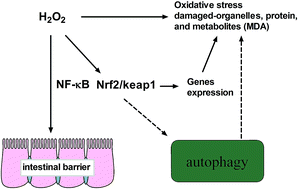
Our experiments proved that administration of H2O2 suppressed the activity of antioxidant enzymes and caused oxidative stress. Meanwhile, the intestinal permeability, morphology, and barrier function also changed after exposure to H2O2 in piglets. All these imbalances might induce autophagy via activation of NF-κB and Nrf2/Keap1 signaling pathways (Fig. 7). Further studies should be conducted to completely determine the mechanism in H2O2 induced autophagy.
image file: c4ra13557a-f7.tif
Fig. 7 H2O2 induced oxidative stress triggers autophagy, which may be associated with NF-κB and Nrf2/Keap1 signaling pathways.

Author contributions

T.L. and Y.Y. conceived and designed the experiments; J.Y., J.D., and Z.C. performed the experiments; J.Y. and W.R. analyzed the data; J.Y. wrote the paper.

Conflict of interest

All authors have no conflict of interest.

Acknowledgements

This study was supported by the National Natural Science Foundation of China (no. 31272463) and Hunan Provincial Natural Science Foundation of China (no. 12JJ2014).

References

  1. J. Yin, W. K. Ren, X. S. Wu, G. Yang, J. Wang, T. J. Li, J. N. Ding, L. C. Cai and D. D. Su, J. Food, Agric. Environ., 2013, 11, 132–139 Search PubMed.
  2. J. Yin, M. M. Wu, H. Xiao, W. K. Ren, J. L. Duan, G. Yang, T. J. Li and Y. L. Yin, J. Anim. Sci., 2014, 92, 612–619 CrossRef CAS PubMed.
  3. A. M. Mohamadin, O. M. Ashour, N. A. El-Sherbeny, A. M. Alahdal, G. M. Morsy and A. B. Abdel-Naim, Clin. Exp. Pharmacol. Physiol., 2009, 36, 367–372 CrossRef CAS PubMed.
  4. E. E. Essick, R. M. Wilson, D. R. Pimentel, M. Shimano, S. Baid, N. Ouchi and F. Sam, PLoS One, 2013, 8, e68697 CAS.
  5. J. Yin, W. Ren, G. Liu, J. Duan, G. Yang, L. Wu, T. Li and Y. Yin, Free Radical Res., 2013, 47, 1027–1035 CrossRef CAS PubMed.
  6. S. Saito and A. Nakashima, Placenta, 2013, 34, S79–S84 CrossRef CAS PubMed.
  7. W. L. Li, S. P. Yu, D. Chen, S. S. Yu, Y. J. Jiang, T. Genetta and L. Wei, Neuroscience, 2013, 244, 16–30 CrossRef CAS PubMed.
  8. A. Kuma, M. Hatano, M. Matsui, A. Yamamoto, H. Nakaya, T. Yoshimori, Y. Ohsumi, T. Tokuhisa and N. Mizushima, Nature, 2004, 432, 1032–1036 CrossRef CAS PubMed.
  9. V. Deretic, M. Delgado, I. Vergne, S. Master, S. De Haro, M. Ponpuak and S. Singh, Curr. Top. Microbiol. Immunol., 2009, 335, 169–188 CAS.
  10. C. Luo, Y. Li, H. Wang, Y. Cui, Z. Feng, H. Li, Y. Li, Y. Wang, K. Wurtz, P. Weber, J. Long and J. Liu, Curr. Cancer Drug Targets, 2013, 13, 625–639 CrossRef CAS.
  11. T. Namikawa, I. Fukudome, H. Kitagawa, T. Okabayashi, M. Kobayashi and K. Hanazaki, Oncology, 2012, 82, 147–152 CrossRef CAS PubMed.
  12. T. Suzuki, Cell. Mol. Life Sci., 2013, 70, 631–659 CrossRef CAS PubMed.
  13. X. Deng, F. Zhang, W. Rui, F. Long, L. Wang, Z. Feng, D. Chen and W. Ding, Toxicol. In Vitro, 2013, 27, 1762–1770 CrossRef CAS PubMed.
  14. K. Sakurai and K. Yamasaki, Jpn. J. Pharmacol., 1994, 64, 229–234 CrossRef CAS.
  15. X. P. Zhou, D. Yuan, M. X. Wang and P. N. He, Am. J. Physiol.: Heart Circ. Physiol., 2013, 304, H82–H93 CrossRef CAS PubMed.
  16. I. Fukudome, M. Kobayashi, K. Dabanaka, H. Maeda, K. Okamoto, T. Okabayashi, R. Baba, N. Kumagai, K. Oba, M. Fujita and K. Hanazaki, Med. Mol. Morphol., 2014, 47, 100–107 CrossRef CAS PubMed.
  17. S. Guo, R. Al-Sadi, H. M. Said and T. Y. Ma, Am. J. Pathol., 2013, 182, 375–387 CrossRef CAS PubMed.
  18. S. Fischer, M. Wiesnet, D. Renz and W. Schaper, Eur. J. Cell Biol., 2005, 84, 687–697 CrossRef CAS PubMed.
  19. M. B. Azad, Y. Chen and S. B. Gibson, Antioxid. Redox Signaling, 2009, 11, 777–790 CrossRef CAS PubMed.
  20. L. Murrow and J. Debnath, Annu. Rev. Pathol.: Mech. Dis., 2013, 8, 105–137 CrossRef CAS PubMed.
  21. R. Kiffin, U. Bandyopadhyay and A. M. Cuervo, Antioxid. Redox Signaling, 2006, 8, 152–162 CrossRef CAS PubMed.
  22. E. E. Essick, R. M. Wilson, D. R. Pimentel, M. Shimano, S. Baid, N. Ouchi and F. Sam, PLoS One, 2013, 8, e68697 CAS.
  23. H. W. Tang, H. M. Liao, W. H. Peng, H. R. Lin, C. H. Chen and G. C. Chen, Dev. Cell, 2013, 27, 489–503 CrossRef CAS PubMed.
  24. H. W. Tang and G. C. Chen, Autophagy, 2011, 7, 778–779 CrossRef.
  25. V. Cianfanelli and F. Cecconi, Cell Cycle, 2012, 11, 436–437 CrossRef CAS PubMed.
  26. M. Zeng, X. Wei, Z. Wu, W. Li, B. Li, Y. Zhen, J. Chen, P. Wang and Y. Fei, Biochem. Biophys. Res. Commun., 2013, 436, 180–185 CrossRef CAS PubMed.
  27. C. C. Su, S. Y. Chien, S. J. Kuo, Y. L. Chen, C. Y. Cheng and D. R. Chen, Mol. Med. Rep., 2012, 5, 1019–1022 CAS.
  28. M. Djavaheri-Mergny, M. Amelotti, J. Mathieu, F. Besancon, C. Bauvy, S. Souquere, G. Pierron and P. Codogno, J. Biol. Chem., 2006, 281, 30373–30382 CrossRef CAS PubMed.
  29. D. Liang, F. Li, Y. Fu, Y. Cao, X. Song, T. Wang, W. Wang, M. Guo, E. Zhou, D. Li, Z. Yang and N. Zhang, Inflammation, 2014, 37, 214–222 CrossRef CAS PubMed.
  30. S. H. Bae, S. H. Sung, S. Y. Oh, J. M. Lim, S. K. Lee, Y. N. Park, H. E. Lee, D. Kang and S. G. Rhee, Cell Metab., 2013, 17, 73–84 CrossRef CAS PubMed.
  31. B. N. Das, Y. W. Kim and Y. S. Keum, Oxid. Med. Cell. Longevity, 2013, 839409 Search PubMed.
  32. L. Zhu, E. C. Barret, Y. Xu, Z. Liu, A. Manoharan and Y. Chen, PLoS One, 2013, 8, e55695 CAS.
  33. W. M. Chen, Z. Sun, X. J. Wang, T. Jiang, Z. P. Huang, D. Y. Fang and D. D. Zhang, Mol. Cell., 2009, 34, 663–673 CrossRef CAS PubMed.
  34. A. Lau, X. J. Wang, F. Zhao, N. F. Villeneuve, T. Wu, T. Jiang, Z. Sun, E. White and D. D. Zhang, Mol. Cell. Biol., 2010, 30, 3275–3285 CrossRef CAS PubMed.
  35. S. Pankiv, T. H. Clausen, T. Lamark, A. Brech, J. A. Bruun, H. Outzen, A. Overvatn, G. Bjorkoy and T. Johansen, J. Biol. Chem., 2007, 282, 24131–24145 CrossRef CAS PubMed.
  36. W. Li, W. Wu, H. Song, F. Wang, H. Li, L. Chen, Y. Lai, J. S. Janicki, K. W. Ward, C. J. Meyer, X. L. Wang, D. Tang and T. Cui, FEBS Lett., 2014, 588, 2115–2124 CrossRef CAS PubMed.
  37. J. Yin, W. Ren, J. Duan, L. Wu, S. Chen, T. Li, Y. Yin and G. Wu, Amino Acids, 2014, 46, 883–892 CrossRef CAS PubMed.

Footnotes

Electronic supplementary information (ESI) available. See DOI: 10.1039/c4ra13557a
These authors contributed equally to this work.

This journal is © The Royal Society of Chemistry 2015