Preparation of layered graphitic carbon nitride/montmorillonite nanohybrids for improving thermal stability of sodium alginate nanocomposites

Lu Liuab, Yongqian Shibc, Bin Yubc, Qilong Tai*bc, Bibo Wangbc, Xiaming Fengbc, Hui Liua, Panyue Wenbc, Bihe Yuanbc and Yuan Hu*bc
aState Key Laboratory of Fire Science, University of Science and Technology of China, 96 Jinzhai Road, Hefei, Anhui 230026, P. R. China. E-mail: yuanhu@ustc.edu.cn; Fax: +86-551-3601664; Tel: +86-551-3601664
bNano Science and Technology Institute, University of Science and Technology of China, 166 Ren'ai Road, Suzhou, Jiangsu 215123, P. R. China
cUSTC-CityU Joint Advanced Research Centre, Suzhou Key Laboratory of Urban Public Safety, Suzhou Institute for Advanced Study, University of Science and Technology of China, 166 Ren'ai Road, Suzhou, Jiangsu 215123, P. R. China. E-mail: qltai@ustc.edu.cn; Fax: +86-512-87181516; Tel: +86-512-87161296

Received 22nd October 2014 , Accepted 7th January 2015

First published on 7th January 2015


Abstract

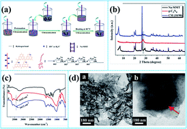
Graphitic carbon nitride (g-C3N4)/montmorillonite (Na-MMT) nanohybrids were successfully fabricated by a facile mixing strategy and then introduced into a sodium alginate host to prepare nanocomposites with different loading levels using a casting technique. The introduction of 4.0 wt% nanohybrids led to the maximal improvement by ca. 149 °C in thermal stability. The improvement of the thermal stability was due to the reason that the mutual intercalation weakened the interlayered interaction of g-C3N4 nanosheets or Na-MMT sheets and enhanced the physical barrier effect that retarded the permeation of heat and the escape of volatile products. This work may provide an approach for exploiting g-C3N4 based materials.


1. Introduction

Graphitic-like carbon nitride (g-C3N4), with a stacked two-dimensional structure, has attracted considerable attention due to its excellent performance.1 The g-C3N4 and its various modifications have been widely studied.2,3 Our group reported that sodium alginate (SA)/g-C3N4 nanocomposites exhibited a remarkable increase of 118.2 °C in the half thermal degradation temperature at 6.0 wt% loadings.4 However, the higher loadings are limited due to presence of severe aggregation. It is necessary to improve the loading levels of nanohybrids in the polymer matrix. Prior reports about graphene based nanohybrids indicated that a synergistic dispersion effect is existent in graphene and its synergists.5,6 Moreover, scarcely any previous work on g-C3N4 based nanohybrids applied to polymer nanocomposites has been reported.

As a typical two-dimensional layered material, montmorillonite (MMT) and its derivatives have been widely utilized to prepare the polymer nanocomposites. Epoxy/OMMT nanocomposites showed an increase of 30 °C in 10 wt% mass loss at 6 phr loading versus pure epoxy matrix.7 More than 100% improvement of tensile strength has been reported for 12 phr addition of Cloisite Na+ in rubber.8 Recently, clay-based nanocomposites have received increasing scientific interest because of its highly desirable qualities including high modulus, significant strength and toughness as well as flame retardancy and low oxygen transmission rate.9 Given the results above, it is expected that combination of g-C3N4 and MMT could contribute to properties improvement of SA nanocomposites. The aim of our present work was to improve the thermal stability of SA nanocomposites.

2. Experimental section

The bulk g-C3N4 was conducted by a procedure similar to that described in prior report.10 Nanohybrids and SA nanocomposites were synthesized by a facile mixing approach. Na-MMT suspension was vigorously stirred for 3 days at room temperature and then mixed with protonated g-C3N4 under ultrasonic agitation. The as-synthesized nanohybrids labeled as CM (x/y) revealing a weight ratio of g-C3N4 to Na-MMT with x/y (x + y = 100), were treated by filtering-washing to get a homogenous distribution. According to the TGA data under air and nitrogen conditions, CM (10/90) shows the best thermal stability and highest char residues among all the nanohybrids (Fig. 1S). The mixture of alginate solution and CM (10/90) suspension was agitated for 30 min under ultrasonication. Each solution was degassed and thrown onto flat dish (10 × 20 cm2) followed by left undisturbed at 40 °C to prepare nanocomposites. These nanocomposites were peeled off from the dish and denoted as SA-0.5, SA-1.0, SA-2.0, SA-4.0 and SA-6.0, corresponding to SA nanocomposites with content of 0.5, 1.0, 2.0, 4.0 and 6.0 wt% nanohybrids, respectively. The schematic representation for the formation of nanocomposites is illustrated in Fig. 1a. For comparison, the identical strategy was used to obtain SA-C-4.0 and SA-M-4.0 corresponding to SA nanocomposites with 4.0 wt% g-C3N4 and Na-MMT, respectively.
image file: c4ra12897d-f1.tif
Fig. 1 Schematic representation for preparation of CM (10/90) and SA nanocomposites (a), XRD patterns (b) and FT-IR spectra (c) of g-C3N4 (up), Na-MMT (middle) and CM (10/90) (down), and TEM images (d) of g-C3N4 (left) and CM (10/90) (right).

X-rays diffraction (XRD) patterns were provided by Japan Rigaku Dmax X-ray diffractometer equipped with graphite monochromatized high-intensity Cu-Kα radiation (λ = 1.54178 Å). Fourier transform infrared (FT-IR) spectroscopy was conducted by Nicolet 6700 FT-IR spectrophotometer with scanning from 4000 to 400 cm−1. Transmission electron microscopy (TEM) was performed by JEOL 2010 with an acceleration voltage of 200 kV. Thermo Gravimetric Analyzer (TGA) curves were provided by Q5000 thermal analyzer (TA Co., USA) heated from 50 to 800 °C at 10 °C min−1 under air with flow rate of 100 ml min−1. The morphology of the fracture surface of nanocomposites was investigated by scanning electron microscopy (SEM) (AMRAY1000B, Beijing R&D Center of the Chinese Academy of Sciences, China).

3. Results and discussion

Structural phase of as-obtained nanohybrids is identified using XRD. As can be seen in Fig. 1b, Na-MMT exhibits a characteristic peak (d001) at ca. 7.07° (2θ). The peak at 2θ = 27.65° is assigned to stacking of the conjugated aromatic system for g-C3N4.11 The in-plane repeating unit with a period of 0.672 nm is visible at 13.16°. In the case of nanohybrids, the d001 is increased from 12.6 to 15.9 Å, indicating the successful intercalation of g-C3N4 nanosheets into interlayers of Na-MMT. Moreover, the peak at 27.65° is invisible, implying increased interlayer spacing of the g-C3N4 nanosheets due to intercalation of Na-MMT sheets.

FT-IR is employed to investigate the microstructure of the nanohybrids, as shown in Fig. 1c. The peaks at 3000–3500 cm−1 are assigned to N–H stretching vibration and hydrogen bonding interactions.12 The bands at 1000–1800 cm−1 are corresponded to the stretching vibration of connected units; the band at ca. 810 cm−1 to vibration of the triazine ring; the peak at 1034 cm−1 to Si–O stretching vibration for Na-MMT. The nanohybrids exhibit similar bands to those of g-C3N4 and Na-MMT. It is noting that the peak at 3450 cm−1 associated with O–H stretching vibration is broadened and shifted to lower wavenumbers, suggesting the existence of interaction between g-C3N4 nanosheets and Na-MMT sheets. These results further confirm the mutual intercalation of the two end members.

The morphology of as-prepared nanohybrids was studied by TEM. Pure g-C3N4 shows a rippled paper-like structure (Fig. 1d). Thin nanosheets on the surface of Na-MMT (marked with red arrow) are observed, indicating the presence of g-C3N4 nanosheets for the nanohybrids.13,14

The XRD traces of the nanocomposites were plotted in Fig. 2a. For virgin SA, the broad peak at 2θ = 13.34° shows a rather amorphous structure.15 In the case of nanocomposites, the peak at 26.63° assigns to Na-MMT strengthens with increasing nanohybrids. The structure of SA is not affected by the nanohybrids, indicating that physical interactions primarily occurred in the interface of the materials.16


image file: c4ra12897d-f2.tif
Fig. 2 XRD patterns (a) and FT-IR spectra (b).

Fig. 2b depicted the FT-IR spectra of the nanocomposites. For untreated SA, the peak at 3440 cm−1 is associated with O–H stretching vibration; the bands at 1611 and 1437 cm−1 to symmetric and asymmetric COO stretching vibrations; and the peak at 1050 cm−1 to the stretching vibration of C–O–C groups.17 In addition, a shoulder peak at 1710 cm−1 is attributed to –COOH group due to the partial hydrolysis of carboxylate salt. These –OH or –COOH groups provide active sites for hydrogen bonding with –NH2, –NH or –OH of CM (10/90). For the nanocomposites, the peak corresponding to –OH groups is shifted to 3420 cm−1, suggesting the interaction of SA chains and CM (10/90) nanosheets through hydrogen bonding.18 However, there is no change at 1710 cm−1 with increase of the nanohybrids. As stated in our previous work, the interaction is very weak. Alternatively, the hydrogen bonding may have induced good interfacial adhesion at the interface SA/CM (10/90), and thus resulted in improving thermal stability of the nanocomposites.

SEM was employed to determine the microstructure of the nanocomposites. The smooth surface is observed for pure SA (Fig. 3a). In the case of the nanocomposites, excellent dispersion is maintained at loadings as high as 2.0 wt% (Fig. 3b–d). However, slight agglomeration occurs at the nanohybrids content exceeding 2.0 wt% (Fig. 3e and f). The SEM results indicate that good distribution is achieved at high loading levels due to the fact that the mutual intercalation effect weakens the interlayered interaction of g-C3N4 nanosheets or Na-MMT sheets. Further investigation of the dispersion of the nanohybrids in SA host was performed by TEM measurement. From Fig. 4, it is evident that the agglomeration is observed from SA-6.0, which is supported by SEM results.


image file: c4ra12897d-f3.tif
Fig. 3 SEM images of SA and its nanocomposites.

image file: c4ra12897d-f4.tif
Fig. 4 TEM ultrathin section image of SA-6.0.

The thermal stability of the nanocomposites was evaluated by TGA, as can be observed in Fig. 5 and Table 1. For neat SA, the degradation terminated at 220 °C is caused by the traces of moisture. T−5 (the temperature at 5 wt% mass loss) is visible at 133.1 °C. Degradation of SA containing hydroxyl, carboxyl groups and polymer chains occurs at 200–580 °C.19 The temperature at half degradation is estimated using T−50. Thermal-oxidized degradation of SA backbone is in the range of 580–700 °C. The nanocomposites are marked by high T−5. All the nanocomposites exhibit significantly increased T−50 rivalled only by pure SA. The thermal stability of SA-4.0 is improved by 149.8 and 31.7 °C, compared with virgin SA and the results of our prior work, respectively.11 However, the thermal stability of the nanocomposites declines upon the content higher than 4.0 wt%. It is likely that the thermal stability is deteriorated by serious aggregation weakening the physical barrier effect that retards the permeation of heat and the escape of volatile products. To confirm the assumption that layered barrier effect contributes to the improvement in the thermal stability of the materials, g-C3N4 along with Na-MMT is incorporated into SA matrix to prepare SA-C-mix-M-4.0 for comparison. It is found that SA-4.0 shows the highest thermal stability with respect to the others, indicating that the mutual intercalation is crucial to the barrier effect.


image file: c4ra12897d-f5.tif
Fig. 5 TGA curves of SA and its nanocomposites.
Table 1 The related TGA data of SA and its nanocomposites under air condition
Sample Air
T−5 (°C) T−10 (°C) T−50 (°C) Residues at 800 °C (wt%)
SA 135.1 213.4 243.8 19.4
SA-0.5 147.2 212.7 337.4 31.4
SA-1.0 147.6 212.9 344.0 31.3
SA-2.0 154.8 215.7 363.4 33.0
SA-4.0 163.7 219.6 393.6 34.7
SA-6.0 156.2 218.1 387.7 31.4
SA-C-4.0 144.4 210.7 329.0 24.7
SA-M-4.0 155.9 210.3 358.9 31.3
SA-C-mix-M-4.0 160.8 210.2 347.4 30.2


4. Conclusions

The CM (10/90) nanohybrids and SA nanocomposites were prepared by the mild mixing strategy and the casting technique, respectively. TGA results indicated that the thermal stability of the nanocomposites was improved significantly, especially, T−50 increased by approximately 149.8 °C. Lower than 4.0 wt%, most of CM (10/90) nanosheets were isolated in SA matrix, resulting in improved thermal stability; higher than 4.0 wt%, the aggregation was present in SA matrices leading to decreased thermal stability due to the weakened interaction between SA chains and CM (10/90) nanosheets. The work may contribute to development of g-C3N4 based hybrids for polymer composites.

Acknowledgements

This work was financially supported by National Natural Science Foundation of China(no. 51403196), National Natural Science Foundation of China (no. 51303167), Natural Science Foundation of Jiangsu Province. (no. BK20130369), China Postdoctoral Science Foundation(no. 2013M531523), National Key Technology R&D Program(no. 2013BAJ01B05).

Notes and references

  1. X. C. Wang, K. Maeda, A. Thomas, K. Takanabe, G. Xin, J. M. Carlsson, K. Domen and M. Antonietti, Nat. Mater., 2009, 8, 76–80 CrossRef CAS PubMed.
  2. Y. Wang, X. C. Wang and M. Antonietti, Angew. Chem., Int. Ed., 2012, 51, 68–89 CrossRef CAS PubMed.
  3. K. Myllymaa, S. Myllymaa, H. Korhonen, M. J. Lammi, H. Saarenpaa, M. Suvanto, T. A. Pakkanen, V. Tiitu and R. Lappalainen, J. Mater. Sci.: Mater. Med., 2009, 20, 2337–2347 CrossRef CAS PubMed.
  4. Y. Q. Shi, S. H. Jiang, K. Q. Zhou, C. L. Bao, B. Yu, X. D. Qian, B. B. Wang, N. N. Hong, P. Y. Wen, Z. Gui, Y. Hu and R. K. K. Yuen, ACS Appl. Mater. Interfaces, 2014, 6, 429–437 CAS.
  5. Y. Y. Liang, Y. G. Li, H. L. Wang, J. G. Zhou, J. Wang, T. Regier and H. J. Dai, Nat. Mater., 2011, 780–786 CrossRef CAS PubMed.
  6. Y. X. Xu, H. Bai, G. W. Lu, C. Li and G. Q. Shi, J. Am. Chem. Soc., 2008, 130, 5856–5857 CrossRef CAS PubMed.
  7. B. C. Guo, D. M. Ha and C. G. Cai, Eur. Polym. J., 2004, 40, 1743–1748 CrossRef CAS PubMed.
  8. N. I. N. Ismail, A. Ansarifar and M. Song, Polym. Eng. Sci., 2013, 53, 2603–2614 CAS.
  9. A. D. Liu and L. A. Berglund, Carbohydr. Polym., 2012, 87, 53–60 CrossRef CAS PubMed.
  10. Y. Q. Shi, S. H. Jiang, K. Q. Zhou, B. B. Wang, B. A. Wang, Z. Gui, Y. Hu and R. K. K. Yuen, RSC Adv., 2014, 4, 2609–2613 RSC.
  11. X. C. Wang, K. Maeda, X. F. Chen, K. Takanabe, K. Domen, Y. D. Hou, X. Z. Fu and M. Antonietti, J. Am. Chem. Soc., 2009, 131, 1680–1684 CrossRef CAS PubMed.
  12. Y. Qiu and L. Gao, Chem. Commun., 2003, 2378–2379 RSC.
  13. S. M. Paek, J. U. Jang, S. J. Hwang and J. H. Choy, J. Phys. Chem., 2006, 67, 1020–1023 CAS.
  14. T. S. Yu, J. P. Lin, J. F. Xu, T. Chen and S. L. Lin, Polymer, 2005, 46, 5695–5697 CrossRef CAS PubMed.
  15. S. G. Kim, G. T. Lim, J. Jegal and K. H. Lee, J. Membr. Sci., 2000, 174, 1–15 CrossRef CAS.
  16. D. L. Han, L. F. Yan, W. F. Chen and W. Li, Carbohydr. Polym., 2011, 83, 653–658 CrossRef CAS PubMed.
  17. C. Sartori, D. S. Finch, B. Ralph and K. Gilding, Polymer, 1997, 38, 43–51 CrossRef CAS.
  18. M. Ionita, M. A. Pandele and H. Iovu, Carbohydr. Polym., 2013, 94, 339–344 CrossRef CAS PubMed.
  19. P. Rani, S. Mishra and G. Sen, Carbohydr. Polym., 2013, 91, 686–692 CrossRef CAS PubMed.

Footnote

Electronic supplementary information (ESI) available. See DOI: 10.1039/c4ra12897d

This journal is © The Royal Society of Chemistry 2015
Click here to see how this site uses Cookies. View our privacy policy here.