Roles of hydrogen bonds and π–π stacking in the optical detection of nitro-explosives with a luminescent metal–organic framework as the sensor

Lei Liuab, Juanyuan Haoa, Yantao Shia, Jieshan Qiua and Ce Hao*a
aState Key Laboratory of Fine Chemicals, Dalian University of Technology, Dalian 116024, China. E-mail: haoce@dlut.edu.cn
bState Key Laboratory of Molecular Reaction Dynamics, Dalian Institute of Chemical Physics, Chinese Academy of Sciences, Dalian 116023, China

Received 21st October 2014 , Accepted 28th November 2014

First published on 28th November 2014


Abstract

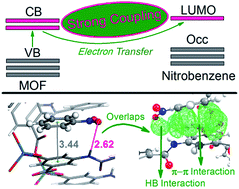
With the aids of density functional theory (DFT) and time-dependent density functional theory (TD-DFT), the explosives-detection mechanism of a typical luminescent metal–organic framework (MOF) sensor has been comprehensively studied by investigating the interactions between the framework and two analytes, namelty, benzene and nitrobenzene. By studying both the periodic crystal models and cluster models we obtained an in-depth understanding of the detecting mechanism from the viewpoint of electronic coupling between the analyte and sensor. Intermolecular electron transfer from the conduction bands of the framework to the LUMO of nitrobenzene is demonstrated to induce the luminescence quenching phenomenon observed in previous experiments. π–π stacking and hydrogen bonding interactions are found to play essential roles in this intermolecular electron transfer process. π–π stacking provides large fragment orbital overlaps between the unoccupied orbitals of the analyte and sensor, which serves as a highly efficient electron transfer bridge. Hydrogen bonds alone cannot provide enough overlaps for electron transfer but are found to reinforce the π–π stacking interactions. The cooperation of the two interactions induces facile intermolecular electron transfer which strongly quenches the luminescence of the MOF sensor. This work sheds light on the analyte–sensor interactions inside the MOF sensors and would provide valuable insights into the design of high efficient explosives-detecting MOF sensors.


Introduction

Due to the high surface area, strong mechanical stability, extraordinary porosity and designable architecture,1–5 metal–organic frameworks (MOFs) have emerged as a new group of promising candidates in molecular storage,6–9 gas separation,10–12 drug delivery,13 and catalysis.14–16 Some MOFs exhibit tunable electrical and optical properties which enable them to be potential candidates such as active materials for hybrid solar cells, quantum dots, and organic semiconducting devices.17–27 Herein, the studies of the optical properties for MOF materials are of special interest. Moreover, the coordination of metal centers with emissive organic-ligands always render MOFs with useful luminescent properties such as high luminescence quantum yield, large stroke shift and guest molecule based emission spectrum changes.28–31 The combination of their unique structural properties with their multiple optical characteristics endorses MOFs to be platforms for sensing applications.32–34 Their pore windows and the chemical environments inside can be delicately tuned by selecting appropriate metal ions and ligands. This will primarily eliminate the relatively bulky interference molecules and pre-concentrate the analytes. Consequently, the guest–host interaction is optimized which influences the luminescence of the framework and achieves specific analyte detection. Based on this, many luminescent MOFs (LMOFs) have been synthesized for the detections of metal cations, anions and small molecules, making MOFs the research frontier in optical sensors.32–34

One of the most significant application amongst is their ability for sensing nitroaromatics,35 which plays a crucial role in anti-terrorism and homeland security. Since the pioneering work of Li,40 dozens of new frameworks have been designed and synthesized for explosives-detection purposes.36–48 Versatile metal centers and organic–ligands make it possible to design numerous LMOFs for explosives-detection purposes, which is an apparent advantage for MOF-based sensors. However, this kind of versatility also makes it hard for one to select the judicious building blocks from the vast collection of candidates to achieve specific analyte detection. Thus, a clear understanding of the detecting mechanism of LMOFs should be essential.

LMOF Zn(L)(HDMA)2(DMF)(H2O)6 (1) (H4L = bis-(3,5-dicarboxy-phenyl)terephthalamide), a typical and newly synthesized LMOF has been reported by Wang and co-workers44 to show good ability in sensing nitroaromatics (Fig. 1, detailed structural information in ref. 44). The strong emission of LMOF 1 is remarkably quenched by nitrobenzene while unaffected by alcohols, ketones, chloroalkanes and even other aromatic compounds. This luminescence quenching phenomenon of LMOFs in the presence of nitroaromatics is very useful in explosives-detection and syntheses of quite a few analogous frameworks have been reported.36–48 Taking the electron-withdrawing nature of nitroaromatics into consideration, this kind of luminescence quenching is generally attributed to the photo-induced electron transfer from the valence bands (VB) to the conduction bands (CB) of the electron-rich MOF then to the LUMO of the electron-deficient nitroaromatics.36–39,49 The positive energy gap between CB and LUMO drives this electron transfer process. This mechanism is quite reasonable but somewhat too brief. As a typical optic sensor, its interaction with the analyte molecule should be the most vital factor that directly affects the intermolecular electron transfer process. Without considerable intermolecular interactions, electron transfer is not likely to occur even in the presence of proper driving forces.36 As is known, electron transfer would be greatly favored via molecular orbital overlap between donor and acceptor.50–52 This kind of overlap is facilitated by intermolecular weak forces which have been studied in the cases of small fluorophores as well as bio-molecules.53–60 In the cases of LMOFs, to our surprise, this issue has been barely studied.


image file: c4ra12835d-f1.tif
Fig. 1 Structural features of LMOF 1. Top view (a); side view (b).

Hydrogen bond and π–π stacking are the most fundamental and widespread weak interactions. Hydrogen bonds play essential roles in the photo-physical and photo-chemical processes which are closely related to the fluorescence emission quenching/enhancing, intersystem crossing and photo induced proton/electron transfer processes from small organic dyes to complicated metal–organic clusters.53–57,61–67 π–π stacking interactions have been demonstrated to facilitate electron transfer in DNAs, metal–organic complexes and so on.58–60 In the cases of LMOFs, their ligands usually contain potential hydrogen bonding sites as well as π–π stacking sites. Nitroaromatics contain nitro groups as hydrogen accepting sites and phenyl rings as π–π stacking sites. When nitroaromatics interact with LMOFs, these two weak forces may coexist. These two interactions will influence the intermolecular electron transfer processes and ultimately coordinate the luminescent properties of LMOFs. However, little is known about the roles of the two weak forces during the luminescent sensing processes of LMOFs. Also, which of them plays a more significant role still remains uncovered.

Based on the above considerations, we perform comprehensive studies on the explosives-detection mechanism of LMOF 1 by using DFT and TD-DFT methods. The interactions between 1 and two analytes from two molecular groups (benzene as hydrocarbon, nitrobenzene as nitroaromatic) are studied. Periodic models are used to study the adsorption of analytes as well as their effects on the electronic structures of LMOF 1. Cluster models are then employed to further look into the electronic couplings between the analytes and sensor.

Methods

For the geometry optimization and single point energy calculation of the periodic structures, all the calculations are performed using the CASTEP68 module implemented in the Material Studio 6.0 program suite. DFT calculations are performed using periodic plane-wave methods with a kinetic energy cutoff of 600 eV. The exchange–correlation functional is treated within the generalized gradient approximation (GGA) parametrized by Perdew, Burke, and Ernzernhof (PBE).69 Ultrasoft pseudo-potentials are applied throughout the computation. A criterion of 5.0 × 10⁻⁷ eV per atom is placed on the self-consistent convergence of the total energy. Atomic positions are considered converged until forces are less than 0.01 eV Å−1. Firstly, the unit cell structure of LMOF 1 is fully optimized with the Brillouin-zone sampling restricted to the Γ point. The calculated lattice parameters are in good agreement with the experimental data44 (Table S1, ESI) which confirms the validity of our computational methods. Then, unit cell of LMOF 1 is doubled in the b direction and optimized to get the supercell structure before simulating the adsorption of different analytes. Two different analytes namely benzene and nitrobenzene are put inside the large pores of LMOF 1. Nitrobenzene is found to have two potential binding sites inside the pores (namely site 1 and site 2) which lead to two different binding patterns after optimization. These supercell structures optimizations are carried out with respect to all atomic positions considering fixed lattice parameters and brillouin-zone sampling are restricted to the Γ point. Analyte–sensor interaction energies are then calculated using the following equations:
 
Einteraction = ELMOF + EanalyteELMOF+analyte (1)

For all the calculations, dispersion corrections are systematically included to energies and geometries (forces) within the DFT-D method of Grimme.70 Local density of states (LDOS) projected on the analytes are then performed based on the optimized structures with the k-point grid of 2 × 2 × 1 (this setting is found to be adequate to give preliminary insights into the electronic coupling between analytes and sensor) using the Monkhorst-Pack scheme. In order to further test the performance of our theoretical methods, GGA/PW91 (ref. 71) functional is also applied in the unit cell optimization with the same settings.

For the clusters calculations, we have first investigated the excitation processes by calculating the low-lying excitation energies for all the clusters using TD-DFT method as implemented in Gaussian 09 software package.72 Molecular orbitals correlated to the excitation processes are then analyzed from the view of fragment orbital interactions. The fragment orbitals involved in the excited processes are thus obtained. Wavefunctions of these fragment orbitals are generated and multiplied with each other using Multiwfn 3.2.1 program73,74 to obtain the intermolecular orbital overlap integral values between analytes and the framework. The overlap value can reflect the electronic coupling between the two orbitals and is used in this contribution to visually and quantitatively measure the intermolecular interactions between MOF and nitrobenzene. All the above calculations are performed at the wb97xd75/genecp level of theory with the self-consistent reaction field (SCRF) method in a polarizable continuum model (PCM) with acetonitrile as solvent.76,77 The Lanl2dz78(Zn)/6-31G**(C, H, O, N) basis set are used throughout. Wb97xd75 functional is a long-range corrected hybrid functional including empirical dispersion which has given very good performances when dealing with weak intermolecular interactions such as hydrogen bonding and π–π stacking interactions.79–81 Thus, we have enough confidence that the level of theory used for the clusters calculations is good enough.

Results and discussions

An overview of the LMOF 1 structure

Having a good understanding of the crystal structure of the MOF sensor, especially the potential binding sites for analytes, will be essential before looking into the detailed sensing mechanism. As shown in Fig. 1, each Zn is coordinated with four oxygen atoms from the linkers. Two kinds of pores are formed with different pore sizes. The small pores are rather jammed which will not provide enough space for the adsorption of analytes. On the contrary, the large pores show enough space for analyte adsorption. As revealed in Fig. 1, the large pores contain imide groups and phenyl rings which provide possible binding sites for analytes. Thus, analyte–sensor interactions inside these pores should be investigated.

Binding models and binding energies of different analytes

Herein, we perform full investigations on the analyte–sensor interactions inside the large pores by using benzene and nitrobenzene as analytes. DFT-D methods are used throughout the optimization of the “analyte-containing” supercell structures. Three binding models are obtained after optimizations, one for the binding of benzene (Ben-MOF) the other two for the bindings of nitrobenzene (NB1-MOF, NB2-MOF). Subsequently, the binding energy of each binding model is obtained (Table 1). As shown in Fig. 2a, the interaction between MOF and benzene is of π–π stacking feature with a centroid distance of 3.38 Å. This may provide chances of intermolecular electron transfer. However, the binding energy is quite small (0.764 eV) due to the lack of intermolecular hydrogen bonds. Also, the electron-rich nature of benzene will prohibit the electron transfer process. Thus, benzene would not quench the luminescence of LMOF 1 which is in accordance with the experimental results.
Table 1 Binding energies for the three binding modelsa
Structure Binding energy (eV)
a Detailed information of the binding energies have been listed in Table S3 (ESI).
Ben-MOF 0.764
NB1-MOF 0.995
NB2-MOF 0.959


image file: c4ra12835d-f2.tif
Fig. 2 Geometries of LMOF 1 optimized by DFT-D calculations for the adsorption of (a) benzene, (b) nitrobenzene on site 1 and (c) nitrobenzene on site 2. Right sides are the corresponding magnifications. Hydrogen bond lengths and centroid distances between aromatic rings are given. All the distances units are in angstrom (Å).

In the case of nitrobenzene, two binding patterns are present inside the large pore. Fig. 2b shows the NB1-MOF binding model (binding site 1). A strong hydrogen bond (1.81 Å) and a moderate hydrogen bond (2.54 Å) form between the analyte and sensor. The total binding energy for this model is 0.995 eV, considerably larger than that of Ben-MOF. The other binding model (NB2-MOF, binding site 2), as revealed in Fig. 2c, shows the co-existence of a moderate hydrogen bond (2.62 Å) and a π–π stacking interaction (centroid distance 3.44 Å). Binding energy for this model is 0.959 eV, which is close to the case of NB1-MOF. The large binding energies of NB1-MOF and NB2-MOF indicate strong analyte–sensor binding strengths. This may increase the degree of electronic coupling between the analyte and sensor which facilitates the electron transfer and induces the experimentally observed luminescence quenching. Thus, the electronic couplings between the analytes and sensor should be investigated.

A primary view of the electronic coupling

A preliminary view of the electronic couplings between the analytes and sensor is indicated by calculating the electronic density of states (DOS) as well as local density of states (LDOS) projected onto the adsorbates. As displayed in Fig. 3b, the adsorption of benzene into the pores of LMOF 1 induces a clear overlap of the main peak in the region −1–0 eV. This indicates that the occupied frontier orbitals of benzene may have strong interactions with the top VB of LMOF 1. However, there are no obvious overlap zones in the regions of CB. This will make the electron transfer from CB to LUMO hard to take place which further confirms that benzene cannot quench the luminescence of LMOF 1.
image file: c4ra12835d-f3.tif
Fig. 3 The total electronic density of states (TDOS) for (a) LMOF 1, (b) Ben-MOF, (c) NB1-MOF and (d) NB2-MOF. The local DOS projected onto the analytes are also plotted (filled areas under DOS curves).

For the cases of nitrobenzene, the two binding models exhibit very similar DOS/LDOS diagrams. As shown in Fig. 3c and d, the adsorptions of nitrobenzene into the two sites all bring about overlaps near the first main peaks in the VB zones. Besides, new peaks appear just around the bottom of the CB of the framework (in region 1.8–2.5 eV) for both cases, indicating the possible existence of strong interactions between the frontier unoccupied orbitals of the analyte (nitrobenzene) and bottom CB of the sensor (LMOF 1). Thus, we can get that, the electronic structure of LMOF 1 is quite sensitive for the presence of nitrobenzene. The strong electronic coupling between nitrobenzene and LMOF 1 increases the chances of electron transfer from CB to LUMO which consequently induces luminescence quenching.

As discussed in the above sections, we have investigated the analyte–sensor interactions from the view of binding models to inherent electronic couplings. The overlap of DOS between the analyte and the framework around the bottom of the CB is found to be a prerequisite for intermolecular electron transfer. Nitrobenzene shows two binding models with LMOF 1. These models have very different intermolecular interactions. NB1-MOF has two hydrogen bonds while NB2-MOF has both hydrogen bonding and π–π stacking interactions. Intermolecular hydrogen bond and π–π stacking seem to facilitate this electronic coupling and play important roles. As hydrogen bond and π–π stacking are intrinsically different in bonding patterns and electronic interactions, the different binding models of nitrobenzene with LMOF 1 may induce very different intermolecular electron transfer features. However, both the analyses of bonding energies and DOS cannot distinguish these two binding models. To get a detailed picture on how these two weak forces function and which of them plays a more significant role during the electron transfer process, we take further steps into the electronic properties of the above mentioned structures. In the sections below, cluster models of these structures have been applied to study these issues.

Descriptions of the cluster models

In the studies of infinite MOFs crystal structures, truncated cluster models from the periodic structures have been successfully applied.82–89 The finite clusters should be delicately selected to best represent their original local chemical environment. In this contribution, four clusters namely CL-LMOF 1, CL-Ben-MOF, CL-NB1-MOF and CL-NB2-MOF are obtained by truncating from the corresponding optimized periodic structures. H atom is used to balance the overall charges such that the truncated clusters remain neutral. Detailed truncating procedures are given in Fig. S1 ESI. Truncated structures are shown in Fig. S2 (ESI).

Analyses of the excitation processes

Excitations of LMOFs from ground states to electronic excited states frequently trigger electron transfer. The presence of analyte molecules would affect these electron transfer processes and induce changes of luminescence. Thus, a good understanding of the excitation process will provide useful insights into the luminescence quenching mechanism of LMOF 1. Herein, TD-DFT calculations are performed to investigate the excitation processes using the cluster models. Based on the fact that the emission spectra are obtained by exciting the samples at the 330 nm absorption peak,44 the electronic transition states around this excitation wavelength are studied. Detailed information on the excitation processes have been given in Table S4 (ESI).

As shown in Fig. 4a, the excitation of LMOF 1 at 330 nm in the experiment corresponds to the S0 → S99 electronic transition (calculated value at 321 nm) of the corresponding cluster model. This transition exhibits ligand to ligand charge transfer (LLCT) feature coupled with intra-ligand charge transfer (ILCT) feature, indicating the excitation of LMOF 1 is linker based. For the case of Ben-MOF, the excitation at 330 nm corresponds to the S0 → S95 (calculated value 325 nm) electronic transition. The molecular orbitals involved in the excitation (HOMO-72, LUMO) show that this process still localizes inside the MOF structure (LLCT, ILCT). This is further confirmed by the molecular orbital composition analyses. As shown in Fig. S3 (ESI), LUMO of the cluster is mainly (99.7%) composed of the LUMO of the framework. Thus, benzene does not participate during the excitation process which is in accordance with the LDOS results (no overlaps are observed between the unoccupied orbitals of benzene and CB of MOF). Also, the LUMO energy of benzene is much higher than that of LMOF 1 owing to its electron-rich nature (Fig. S3, ESI). All these results demonstrate that benzene cannot accept the electrons from LMOF 1 which brings negligible effects on LMOF 1's luminescence.


image file: c4ra12835d-f4.tif
Fig. 4 Molecular orbitals involved in the excitation processes for (a) CL-LMOF 1, (b) CL-Ben-MOF, (c) CL-NB1-MOF and (d) CL-NB2-MOF. Corresponding excitation wavelengths/energies and excitation features are shown in this picture. LLCT represents ligand to ligand charge transfer. ILCT represents intra-ligand charge transfer. “MOF → Analyte” represents electron transfer from MOF to analyte molecule.

The case of nitrobenzene may be quite different as nitrobenzene usually serves as a good electron acceptor. However, as revealed in Fig. 4c, the excitation process of the NB1-MOF cluster features LLCT. Nitrobenzene is not involved in the excitation process with this “pure hydrogen binding model”. For the NB2-MOF cluster (Fig. 4d), the excitation of this “hybrid binding model” exhibits a clear electron transfer process from LMOF 1 to nitrobenzene. As shown in Fig. S4 and S5 (ESI), LUMO energy of nitrobenzene is very close to the unoccupied orbital of LMOF 1 (MOF fragment orbital 278 in both cases) for both models. Nevertheless, the electron transfer processes are essentially different. This disparity may originate from the different nature of hydrogen bond and π–π stacking which leads to different analyte–sensor interactions. To give an intuitive study on the effects of the two intermolecular interactions, the compositions of the molecular orbitals involved in the excitation processes (HOMO-57 and LUMO + 1 for CL-NB1-MOF, HOMO-36 and LUMO + 2 for CL-NB2-MOF) are analyzed. Overlaps between the fragment orbitals are also investigated in the section below. Motivation for the selection of the molecular orbitals are fully addressed below Table S4 (ESI).

Fragment orbital compositions and intermolecular orbital overlaps

Firstly, we have investigated the compositions of the molecular orbitals involved in the excitation processes. The CL-NB2-MOF structure is divided into two fragments namely the MOF fragment and the NB2 (nitrobenzene) fragment. Consequently, we obtain the fragment orbitals interaction diagram (see detailed information in Fig. S4, ESI) between the two fragments. Compositions of the molecular orbitals related to the S0 → S102 electronic transition (orbital number 270, 309) are analyzed. As shown in Fig. 5a, the 309 unoccupied molecular orbital is composed from two fragment orbitals. MOF fragment orbital 278 contributes 2.2% while NB2 fragment orbital 33 contributes as much as 93.7%. This indicates strong intermolecular electron transfer occurs from MOF to nitrobenzene with this “hybrid binding model”. The 278 fragment orbital and the 33 fragment orbital are closely related to the electron transfer process. The corresponding orbitals are also plotted in Fig. 5a. Both fragment orbital 278 and fragment orbital 33 exhibits π* features. These two fragments are close in space with the aromatic rings nearly parallel to each other. This would significantly render fragment orbital overlaps between 278 and 33 which facilitate intermolecular electron transfer. Herein, this overlap integral value is obtained which is as large as 0.0121. Also, the overlapping zones are visually obtained and shown in Fig. 5b. π–π stacking are found to be the main factors which facilities the electron transfer process as the overlapping zones between 278 and 33 mainly locate between the two aromatic rings. Besides, the overlap is also present at the hydrogen bonding zone. This indicates hydrogen bonding interaction also facilities intermolecular electron transfer but is not the major factor. Based on the above analyses, intermolecular electron transfer from CB of MOF to LUMO of nitrobenzene is proved to be a fairly favorable process.
image file: c4ra12835d-f5.tif
Fig. 5 (a) Fragment orbital interaction diagram for CL-NB2-MOF. Intermolecular orbital overlap zone between (b) 278 orbital of fragment MOF and 33 orbital of fragment NB; (c) 239 orbital of fragment MOF and 30 orbital of fragment NB. The green surfaces represent the overlap zones between unoccupied fragment orbitals and the blue surfaces represent the overlap zones between occupied fragment orbitals. Contour value for the orbital overlaps is set to 0.00002.

The occupied molecular orbital 270 is also composed from these two fragments. MOF fragment orbital 239 contributes 60.1% and NB2 fragment orbital 30 contributes 15.0%. This indicates a strong electronic coupling between 239 and 30 which may also induce intermolecular transfer between the two fragments. In this case, electrons may be directly photo-excited from VB of MOF to the LUMO of nitrobenzene. As plotted in Fig. 5a, the 239 fragment orbital has mixed features. The aromatic ring in the middle shows a clear π feature whereas the imide group shows an n feature. Likewise, for fragment orbital 30, the aromatic ring shows a π feature and the nitro group shows an n feature. Thus, we can expect the π feature would induce overlaps between the aromatic rings while the n feature would induce overlaps between nitro and imide groups. Consequently, the overlap zones are plotted in Fig. 5c which is in accordance with the above prediction. However, the overlap integral value is 0.0033, much smaller than that between 278 and 33. Based on this, electrons directly photo-excited from the MOF fragment to LUMO of nitrobenzene is quite unlikely to happen.

Compositions of the molecular orbitals involved in the S0 → S96 excitation process (orbital number 249 and 308) of CL-NB1-MOF are then investigated. In this case, both the 249 orbital and 308 orbital are solely composed from the fragment orbitals of the MOF fragment. As shown in Fig. S5 (ESI), MOF fragment orbital 223 contributes 97.2% for the 249 molecular orbital. MOF fragment orbital 276 contributes 94.5% for the 308 molecular orbital. These indicate that the “pure hydrogen bonding model” is not favorable in transferring electrons. To provide further evidence, the fragment orbital overlap between the MOF fragment orbital 278 and NB1 fragment orbital 33 are obtained and shown in Fig. 6. The overlaps are found to locate at the two hydrogen bonding zones with an integral value of 0.0018. This value is much smaller than the case of the “hybrid binding model”, suggesting hydrogen bond, alone, cannot provide enough overlaps for intermolecular electron transfer.


image file: c4ra12835d-f6.tif
Fig. 6 Fragment orbital overlap between 278 orbital of MOF fragment and 33 orbital of NB1 fragment. Contour value for the orbital overlaps is set to 0.00002.

From the orbital overlap analyses we can conclude that hydrogen bond is less efficient for electron transfer due to the small overlap values. On the contrary, π–π stacking shows higher efficiency for electron transfer. π–π stacking interaction seems to outperform hydrogen bonding interaction in the explosive detecting process of nitrobenzene with this MOF sensor. However, as revealed by the binding energies (Table 1), the “hybrid bonding model” has a binding energy 0.2 eV larger than that of Ben-MOF. This “extra binding strength” is originated from the hydrogen bond, which increases the chances of nitrobenzene binding to the MOF sensor. Thus, hydrogen bond, although less efficient in electron transfer, cooperates with the π–π stacking. The collaboration of the two interactions induces strong luminescence quenching and achieves the detection of explosives.

Conclusions

To sum up, the explosives detecting mechanism for LMOF 1 has been comprehensively studied by analyzing the analyte–sensor interactions. Nitrobenzene-induced luminescence quenching of LMOF 1 is demonstrated to stem from the intermolecular electron transfer from the MOF sensor to the analyte. The electrons transfer from the CB of the MOF to the LUMO of nitrobenzene upon photo-excitation due to the electron withdrawing ability of nitrobenzene. Intermolecular interactions between the framework and nitrobenzene are found to play essential roles in this electron transfer process which are directly responsible for the luminescence quenching of LMOF 1. π–π stacking interaction provides large overlaps between the analyte and sensor which serves as the electron transfer bridge. Hydrogen bond, alone, cannot provide enough overlaps for electron transfer but is found to reinforce the π–π stacking interaction. The cooperation of the two interactions leads to significant luminescence quenching of LMOF 1 in the presence of nitrobenzene. In the design of nitroaromatics-detecting MOF sensors, the analyte–sensor interaction can be optimized by introducing hydrogen binding sites (namely imide group, amino group) as well as π–π stacking sites into the MOF ligands. This provides a piece of useful guidance for the experimentalists.

Conflict of interest

The authors declare no competing financial interest.

Acknowledgements

The authors would like to thank the National Natural Science Foundation of China for the financial support (Grant nos 21137001, 21036006, 21373042).

Notes and references

  1. O. M. Yaghi, M. O'Keeffe, N. W. Ockwig, H. K. Chae, M. Eddaoudi and J. Kim, Nature, 2003, 423, 705–714 CrossRef CAS PubMed.
  2. S. L. James, Chem. Soc. Rev., 2003, 32, 276–288 RSC.
  3. S. Kitagawa, R. Kitaura and S. Noro, Angew. Chem., Int. Ed., 2004, 43, 2334–2375 CrossRef CAS PubMed.
  4. J. R. Long and O. M. Yaghi, Chem. Soc. Rev., 2009, 38, 1213–1214 RSC.
  5. H. C. Zhou, J. R. Long and O. M. Yaghi, Chem. Rev., 2012, 112, 673–674 CrossRef CAS PubMed.
  6. M. Eddaoudi, J. Kim, N. Rosi, D. Vodak, J. Wachter, M. O'Keeffe and O. M. Yaghi, Science, 2002, 295, 469–472 CrossRef CAS PubMed.
  7. J. Rowsell and O. M. Yaghi, Angew. Chem., Int. Ed., 2005, 44, 4670–4679 CrossRef CAS PubMed.
  8. W. Zhou and T. Yildirim, J. Phys. Chem. C, 2008, 112, 8132–8135 CAS.
  9. T. A. Makal, J. R. Li, W. Lu and H. C. Zhou, Chem. Soc. Rev., 2012, 41, 7761–7779 RSC.
  10. A. Demessence and J. R. Long, Chem.–Eur. J., 2010, 16, 5902–5908 CrossRef CAS PubMed.
  11. J. R. Li, J. Sculley and H. C. Zhou, Chem. Rev., 2012, 112, 869–932 CrossRef CAS PubMed.
  12. Q. Yang, D. Liu, C. Zhong and J. R. Li, Chem. Rev., 2013, 113, 8261–8323 CrossRef CAS PubMed.
  13. P. Horcajada, C. Serre, M. Vallet-Regí, M. Sebban, F. Taulelle and G. Férey, Angew. Chem., Int. Ed., 2006, 118, 6120–6124 CrossRef.
  14. J. Lee, O. K. Farha, J. Roberts, K. A. Scheidt, S. T. Nguyen and J. T. Hupp, Chem. Soc. Rev., 2009, 38, 1450–1459 RSC.
  15. D. Dang, P. Wu, C. He, Z. Xie and C. Duan, J. Am. Chem. Soc., 2010, 132, 14321–14323 CrossRef CAS PubMed.
  16. Z. Xie, C. Wang, K. E. DeKrafft and W. Lin, J. Am. Chem. Soc., 2011, 133, 2056–2059 CrossRef CAS PubMed.
  17. L. Yang, P. Vajeeston, P. Ravindran, H. Fjellvåg and M. Tilset, Inorg. Chem., 2010, 49, 10283–10290 CrossRef CAS PubMed.
  18. L. Yang, P. Vajeeston, P. Ravindran, H. Fjellvåg and M. Tilset, Phys. Chem. Chem. Phys., 2011, 13, 10191–10203 RSC.
  19. L. Yang, P. Vajeeston, P. Ravindran and M. Tilset, RSC Adv., 2012, 2, 1618–1631 RSC.
  20. L. Yang, P. Ravindran, P. Vajeeston and M. Tilset, Phys. Chem. Chem. Phys., 2012, 14, 4713–4723 RSC.
  21. L. Yang, P. Ravindran, P. Vajeeston and M. Tilset, J. Mater. Chem., 2012, 22, 16324–16335 RSC.
  22. L. Yang, P. Ravindran and M. Tilset, Inorg. Chem., 2013, 52, 4217–4228 CrossRef CAS PubMed.
  23. L. Yang, P. Ravindran, P. Vajeeston, S. Svelle and M. Tilset, Microporous Mesoporous Mater., 2013, 175, 50–58 CrossRef CAS PubMed.
  24. L. Yang, Microporous Mesoporous Mater., 2014, 183, 218–233 CrossRef CAS PubMed.
  25. L. Yang, G. Fang, J. Ma, E. Ganz and S. S. Han, Cryst. Growth Des., 2014, 14, 2532–2541 CAS.
  26. L. Yang, E. Ganz, S. Svelle and M. Tilset, J. Mater. Chem. C, 2014, 2, 7111–7125 RSC.
  27. J. Gascon, M. D. Hernández-Alonso, A. R. Almeida, G. P. M. van Klink, F. Kapteijn and G. Mul, ChemSusChem, 2008, 1, 981–983 CrossRef CAS PubMed.
  28. M. D. Allendorf, C. A. Bauer, R. K. Bhakta and R. J. T. Houk, Chem. Soc. Rev., 2009, 38, 1330–1352 RSC.
  29. Y. Cui, Y. Yue, G. Qian and B. Chen, Chem. Rev., 2012, 112, 1126–1162 CrossRef CAS PubMed.
  30. Y. Cui, H. Xu, Y. Yue, Z. Guo, J. Yu, Z. Chen, J. Gao, Y. Yang, G. Qian and B. Chen, J. Am. Chem. Soc., 2012, 134, 3979–3982 CrossRef CAS PubMed.
  31. J. C. G. Bunzli and C. Piguet, Chem. Soc. Rev., 2005, 34, 1048–1077 RSC.
  32. B. L. Chen, S. Xiang and G. Qian, Acc. Chem. Res., 2010, 43, 1115–1124 CrossRef CAS PubMed.
  33. L. E. Kreno, K. Leong, O. K. Farha, M. Allendorf, D. R. P. Van and J. T. Hupp, Chem. Rev., 2012, 112, 1105–1125 CrossRef CAS PubMed.
  34. B. Liu, J. Mater. Chem., 2012, 22, 10094–10101 RSC.
  35. Z. C. Hu, B. J. Deibert and J. Li, Chem. Soc. Rev., 2014, 43, 5815–5840 RSC.
  36. S. C. Pramanik, X. Zheng, T. Zhang, J. Emge and J. Li, J. Am. Chem. Soc., 2011, 133, 4153–4155 CrossRef CAS PubMed.
  37. D. Banerjee, Z. Hu, S. Pramanik, X. Zhang, H. Wang and J. Li, CrystEngComm, 2013, 15, 9745–9750 RSC.
  38. B. Gole, A. K. Bar and P. S. Mukherjee, Chem.–Eur. J., 2014, 20, 2276–2291 CrossRef CAS PubMed.
  39. G. L. Liu, Y. J. Qin, J. Li, G. Y. Wei and H. Li, Chem. Commun., 2013, 49, 1699–1701 RSC.
  40. A. Lan, K. Li, H. Wu, D. H. Olson, T. J. Emge, W. Ki, M. Hong and J. Li, Angew. Chem., Int. Ed., 2009, 48, 2334–2338 CrossRef CAS PubMed.
  41. A. Lan, K. Li, H. Wu, L. Kong, N. Nijem, D. H. Olson, T. J. Emge, Y. J. Chabal, D. C. Langreth, M. Hong and J. Li, Inorg. Chem., 2009, 48, 7165–7173 CrossRef CAS PubMed.
  42. G. Y. Wang, L. L. Yang, Y. Li, H. Song, W. J. Ruan, Z. Chang and X. H. Bu, Dalton Trans., 2013, 42, 12865–12868 RSC.
  43. D. X. Ma, B. Y. Li, X. J. Zhou, Q. Zhou, K. Liu, G. Zeng, G. H. Li, Z. Shi and S. H. Feng, Chem. Commun., 2013, 49, 8964–8966 RSC.
  44. G. Y. Wang, C. Song, D. M. Kong, W. J. Ruan, Z. Chang and Y. Li, J. Mater. Chem. A, 2014, 2, 2213–2220 CAS.
  45. Z. Feng, W. B. Tan, M. L. Feng, A. J. Lana and X. Y. Huang, J. Mater. Chem. A, 2014, 2, 6426–6431 Search PubMed.
  46. B. L. Chen, S. Ma, F. Zapata, E. B. Lobkovsky and J. Yang, Inorg. Chem., 2006, 45, 5718–5720 CrossRef CAS PubMed.
  47. T. K. Kim, J. H. Lee, D. Moon and H. R. Moon, Inorg. Chem., 2013, 52, 589–595 CrossRef CAS PubMed.
  48. Z. Hu, S. Pramanik, K. Tan, C. Zheng, W. Liu, X. Zhang, Y. J. Chabal and J. Li, Cryst. Growth Des., 2013, 13, 4204–4207 CAS.
  49. S. Pramanik, Z. Hu, X. Zhang, C. Zheng, S. Kelly and J. Li, Chem.–Eur. J., 2013, 19, 15964–15971 CrossRef CAS PubMed.
  50. L. Lawrence and H. Roald, J. Am. Chem. Soc., 1974, 96, 1370–1383 CrossRef.
  51. L. C. Gerhard, P. Piotr, M. M. Jean and R. F. Graham, J. Am. Chem. Soc., 1988, 110, 2652–2653 CrossRef.
  52. G. Fruzsina, V. Siim, H. Felix, S. Jacob, B. Marian, K. Adam, D. Michel and B. Jochen, J. Chem. Theory Comput., 2014, 10, 4653–4660 CrossRef.
  53. P. J. Rege, S. A. Williams and M. J. Therien, Science, 1995, 269, 1409–1413 CrossRef.
  54. G. J. Zhao, J. Y. Liu, L. C. Zhou and K. L. Han, J. Phys. Chem. B, 2007, 111, 8940–8945 CrossRef CAS PubMed.
  55. A. P. Demchenko, K. C. Tang and P. T. Chou, Chem. Soc. Rev., 2013, 42, 1379–1408 RSC.
  56. D. R. Weinberg, C. J. Gagliardi, J. F. Hull, C. F. Murphy, C. A. Kent, B. C. Westlake, A. Paul, D. H. Ess, D. G. McCafferty and T. J. Meyer, Chem. Rev., 2012, 112, 4016–4093 CrossRef CAS PubMed.
  57. O. S. Wenger, Acc. Chem. Res., 2013, 46, 1517–1526 CrossRef CAS PubMed.
  58. D. B. Hall, R. E. Holmlin and J. K. Barton, Nature, 1996, 382, 731–735 CrossRef CAS PubMed.
  59. S. O. Kelley and J. K. Barton, Chem. Biol., 1998, 5, 413–425 CrossRef CAS.
  60. J. Christoph, J. Chem. Soc., Dalton Trans., 2000, 3885–3896 Search PubMed.
  61. G. J. Zhao and K. L. Han, Acc. Chem. Res., 2012, 45, 404–413 CrossRef CAS PubMed.
  62. K. L. Han and G. J. Zhao, Hydrogen Bonding and Transfer in the Excited State, John Wiley & Sons Ltd., Chichester, U.K., 2011, vol. I & II Search PubMed.
  63. G. J. Zhao and K. L. Han, J. Phys. Chem. A, 2009, 113, 14329–14335 CrossRef CAS PubMed.
  64. Y. H. Liu and S. C. Lan, Commun. Comput. Chem., 2013, 3, 235–243 Search PubMed.
  65. D. P. Yang, Y. G. Yang and Y. F. Liu, Commun. Comput. Chem., 2013, 3, 205–215 Search PubMed.
  66. Y. H. Liu and S. C. Lan, Commun. Comput. Chem., 2013, 1, 1–7 CrossRef.
  67. N. Agmon, J. Phys. Chem. A, 2005, 109, 13–35 CrossRef CAS PubMed.
  68. S. J. Clark, M. D. Segall, C. J. Pickard, P. J. Hasnip, M. J. Probert, K. Refson and M. C. Z. Payne, Z. Kristallogr., 2005, 220, 567–570 CrossRef CAS.
  69. J. Perdew, K. Burke and M. Ernzerhof, Phys. Rev. Lett., 1996, 77, 3865 CrossRef CAS.
  70. S. Grimme, J. Comput. Chem., 2006, 27, 1787–1799 CrossRef CAS PubMed.
  71. J. P. Perdew, J. A. Chevary, S. H. Vosko, K. A. Jackson, M. R. Pederson, D. J. Singh and C. Fiolhais, Phys. Rev. B, 1992, 46, 6671–6687 CrossRef CAS.
  72. M. J. Frisch, G. W. Trucks, H. B. Schlegel, G. E. Scuseria, M. A. Robb, J. R. Cheeseman, G. Scalmani, V. Barone, B. Mennucci, G. A. Petersson, H. Nakatsuji, M. Caricato, X. Li, H. P. Hratchian, A. F. Izmaylov, J. Bloino, G. Zheng, J. L. Sonnenberg, M. Hada, M. Ehara, K. Toyota, R. Fukuda, J. Hasegawa, M. Ishida, T. Nakajima, Y. Honda, O. Kitao, H. Nakai, T. Vreven; J. A. Montgomery Jr, J. E. Peralta, F. Ogliaro, M. Bearpark, J. J. Heyd, E. Brothers, K. N. Kudin, V. N. Staroverov, R. Kobayashi, J. Normand, K. Raghavachari, A. Rendell, J. C. Burant, S. S. Iyengar, J. Tomasi, M. Cossi, N. Rega, N. J. Millam, M. Klene, J. E. Knox, J. B. Cross, V. Bakken, C. Adamo, J. Jaramillo, R. Gomperts, R. E. Stratmann, O. Yazyev, A. J. Austin, R. Cammi, C. Pomelli, J. W. Ochterski, R. L. Martin, K. Morokuma, V. G. Zakrzewski, G. A. Voth, P. Salvador, J. J. Dannenberg, S. Dapprich, A. D. Daniels, Ö. Farkas, J. B. Foresman, J. V. Ortiz, J. Cioslowski and D. J. Fox, Gaussian 09, revision C.01, Gaussian, Inc., Wallingford, CT, 2009 Search PubMed.
  73. T. Lu and F. W. Chen, J. Comput. Chem., 2012, 33, 580–592 CrossRef CAS PubMed.
  74. T. Lu and F. W. Chen, J. Mol. Graphics Modell., 2012, 38, 314–323 CrossRef CAS PubMed.
  75. J. Chai and M. H. Gordon, J. Chem. Phys., 2008, 128, 084106 CrossRef PubMed.
  76. S. Miertuš, E. Scrocco and J. Tomasi, Chem. Phys., 1981, 55, 117–129 CrossRef.
  77. R. Cammi and J. Tomasi, J. Comput. Chem., 1995, 16, 1449–1458 CrossRef CAS.
  78. P. J. Hay and W. R. Wadt, J. Chem. Phys., 1985, 82, 299–310 CrossRef CAS PubMed.
  79. P. Gu, R. Q. Lu, D. Liu, Y. K. Lu and S. T. Wang, Phys. Chem. Chem. Phys., 2014, 16, 10531–10538 RSC.
  80. T. B. Tai and M. T. Nguyen, Chem. Commun., 2013, 49, 913–915 RSC.
  81. S. Panigrahi, A. Bhattacharya, S. Banerjee and D. Bhattacharyya, J. Phys. Chem. C, 2012, 116, 4374–4379 CAS.
  82. L. Grajciar, A. D. Wiersum, P. L. Llewellyn, J. S. Chang and P. Nachtigall, J. Phys. Chem. C, 2011, 115, 17925–17933 CAS.
  83. A. L. Dzubakl, L. C. Lin, J. H. Kim, J. A. Swisher, R. Poloni, S. N. Maximoff, B. Smit and L. Gagliardi, Nat. Chem., 2012, 4, 810–816 CrossRef PubMed.
  84. M. Ji, X. Lan, Z. P. Han, C. Hao and J. S. Qiu, Inorg. Chem., 2012, 51, 12389–12394 CrossRef CAS PubMed.
  85. X. Sui, M. Ji, X. Lan, W. H. Mi, C. Hao and J. S. Qiu, Inorg. Chem., 2013, 52, 5742–5748 CrossRef CAS PubMed.
  86. M. Ji, C. Hao, D. D. Wang, H. Li and J. S. Qiu, Dalton Trans., 2013, 42, 3464–3470 RSC.
  87. M. X. Zhang, W. H. Mi and C. Hao, Commun. Comput. Chem., 2013, 1, 269–281 Search PubMed.
  88. S. H. Kang, Z. Q. Liu, J. J. Tan, C. Hao and J. S. Qiu, Commun. Comput. Chem., 2014, 2, 69–87 Search PubMed.
  89. Y. Yao, X. Song, J. Qiu and C. Hao, J. Phys. Chem. A, 2014, 118, 6191–6196 CrossRef CAS PubMed.

Footnote

Electronic supplementary information (ESI) available: Fig. S1 shows the truncating of the cluster models; Fig. S2 shows the structure of the cluster models. Fig. S3–S5 show the detailed information of the fragment orbital interactions between the framework and analytes. Table S1 shows the calculated crystal lattice parameters as well as the corresponding experimental data. Table S2 tests the selecting of k-points. Table S3 gives the binding energies for the adsorbates in detail. Table S4 reports the calculated excitation energies for the clusters. See DOI: 10.1039/c4ra12835d

This journal is © The Royal Society of Chemistry 2015