DOI:
10.1039/C4RA12042F
(Paper)
RSC Adv., 2015,
5, 5038-5045
In situ formation of gold/silver bi-metal nanodots on silica spheres and evaluation of their microbicidal properties†
Received
9th October 2014
, Accepted 9th December 2014
First published on 9th December 2014
Abstract
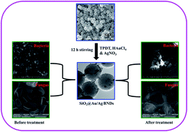
An environmentally benign synthetic method of seedless and one-step growth of 2–4 nm sized gold/silver (Au/Ag) bi-metal nanodots (BNDs) on preformed amine functionalized silica (SiO2) spheres via co-reduction of metal precursors at room temperature and their antimicrobial activity are reported. The resulting BNDs on SiO2 spheres (SiO2@/Au/Ag) exhibited a distinctive surface plasmon resonance (SPR) band between the Ag and Au nanoparticle (NP) SPR absorption regions. The deposition of BNDs on SiO2 spheres was confirmed from the high resolution transmission electron microscopy (HRTEM) and X-ray photoelectron spectroscopy (XPS) analyses. The SiO2@Au/Ag BNDs with different concentrations of Au and Ag (SiO2@Au25/Ag75, SiO2@Au50/Ag50 and SiO2@Au75/Ag25 BNDs) exhibited a broad-spectrum of antimicrobial activities involving significant growth inhibition of Gram negative bacterial strains (Escherichia coli, Pseudomonas aeruginosa), a Gram positive bacterial strain (Bacillus thuringiensis) and a fungal strain (Trichoderma longibrachiyatum MKU-1). The mechanism of interaction between the BNDs and microbial strains was analyzed through scanning electron microscopy (SEM). The results confirmed that the treated microbial cells were damaged and have pits on the cell surface. The results evidently demonstrated the antimicrobial activity of the SiO2@Au/Ag BNDs even at lower concentrations of Ag (SiO2@Au75/Ag25), which may be suitable for the formation of potential microbicidal materials.
Introduction
Metal nanostructured materials with controlled size and shape have received great scientific and technological interest in the fields of catalysis and sensing over the years due to their unique optical, electronic and catalytic properties.1–3 Recently, bi-metal nanoparticles (NPs), in particular gold/silver (Au/Ag) were found to show numerous potential applications due to their distinctive catalytic properties compared to their mono-metal counterparts.4,5 A number of synthetic procedures have been explored for the synthesis of Au/Ag bi-metal NPs by various research groups.6–9 Huang et al.10 reported the preparation of luminescent Au/Ag bi-metal nanodots (BNDs) protected by 11-mercaptoundecanoic acid. Similarly, the syntheses of hybrid bi-metal Au/Pt11 and bi-metallic Au/Ag NPs supported on silica (SiO2)4,12 and their catalytic applications were discussed. The SiO2 supported metal NPs are gaining significant interest due to their stability and enhanced optical and catalytic properties.13,14 However, only limited reports have been published about the synthesis of Au/Ag BNDs and their applications. Generally, seed-mediated growth method is preferred for the synthesis of Au/Ag bi-metallic NPs.15 In this, Au seeds are produced first by reducing the corresponding metal ions with suitable reducing agent like NaBH4 and in the subsequent step AgNO3 is introduced along with suitable surfactants which lead to the formation of Au/Ag nanomaterials via a two-step process. The resulting nanomaterials are surrounded by surfactants that can hinder the direct interaction of analytes especially for biological applications. In addition, the use of reagents like NaBH4 and surfactants are not in accordance with the principles of green chemistry. Hence, in the present work a single step facile route was employed to prepare sub-4 nm sized Au/Ag BNDs on amine functionalized SiO2 spheres (SiO2@Au/Ag BNDs). A modified Stober method16 was employed for the preparation of amine functionalized SiO2 spheres and on this SiO2, Au/Ag BNDs were deposited by in situ method in the presence of amine functionalized silicate sol–gel (SSG) matrix. Herein, N-[3-(trimethoxysilyl) propyl] diethylenetriamine (TPDT) acts as both reducing as well as stabilizing agent for the in situ growth of Au/Ag BNDs.
Interestingly, the bi-metallic core–shell nanostructures and other classes of core–shell NPs have been explored for widespread applications in bio-imaging, drug delivery, cell labelling and catalysis.17 In addition, bi-metallic Au/Ag NPs have been known as potential antimicrobial agents.18 At the same time, the antimicrobial properties of BNDs containing Ag supported on SiO2 need to be explored, because the infection caused by microbes, emergence of new pathogens, increasing number of multiple antibiotic/drug resistant (MDR) strains and the continuing emphasis on health care cost led the scientists to develop new effective antimicrobial agents. Based on these view points, the SiO2@Au/Ag BNDs were prepared and the studies showed excellent antimicrobial effects against Gram negative bacterial strains (Escherichia coli, Pseudomonas aeruginosa), Gram positive bacterial strain (Bacillus thuringiensis) and fungal strain (Trichoderma longibrachiyatum MKU-1). The selected organisms have great ecological importance and the risk associated with them is highly relevant to practical applications.
Materials and methods
Chemicals, microorganisms and cultivation conditions
Gold(III) chloride hydrate (HAuCl4), silver nitrate (AgNO3), tetraethylorthosilicate (TEOS) and TPDT were received from Sigma-Aldrich. Bacterial strains Escherichia coli DH5α, Pseudomonas aeruginosa MTCC no. 7925, Bacillus thuringeinsis MTCC no. 6905 and fungus Trichoderma longibrachiyatum MKU-1 from Department of Genetics, School of Biological Sciences, Madurai Kamaraj University were used as model organisms in antimicrobial studies. Cells were cultivated at 37 °C in 2 L Erlenmeyer flasks containing 100 mL of Lysogeny Broth (LB) (which is composed of 10 g L−1 of peptone and NaCl and 5 g L−1 of yeast extract) supplemented with 2% (w/v) bacto agar as solidifying agent. Fungal organism Trichoderma longibrachiyatum MKU-1 was grown on Potato Dextrose Agar (PDA) at 28 °C for 72 h. The Milli-Q water was used for preparation of solutions (all glassware used for the preparation of SiO2@Au/Ag BNDs was thoroughly cleaned with aqua regia and rinsed extensively with doubly distilled water prior to use).
Synthesis of SiO2@Au/Ag BNDs
The TPDT functionalized SiO2 spheres were prepared by modified Stober method.16 Briefly, ammonium hydroxide (1.7 mL; 25–28%) was added to ethanol (50 mL) along with TEOS (1.5 mL) and water (1 mL). The solution was vigorously stirred and after 3 h an additional 1 mL of TEOS was added. The SiO2 spheres were obtained after 12 h of stirring and then TPDT (0.4 mL) was added to the solution and the stirring was continued for another 6 h. The TPDT functionalized SiO2 particles were purified twice by centrifugation and re-dispersion in water (40 mL). For the preparation of SiO2@Au/Ag BNDs, about 5 mg of preformed amine functionalized SiO2 was added to 5 mM TPDT silane present in 5 mL of water and the solution was sonicated for 5 min. To this solution, HAuCl4 and AgNO3 (each 0.5 mM final concentration for Au50/Ag50 BNDs) were added and the solution was stirred for 12 h. The observed of brownish red color indicated the formation of Au/Ag BNDs on SiO2 spheres (SiO2@Au50/Ag50 BNDs). The same synthetic procedure was followed for the preparation of SiO2@Au25/Ag75 and SiO2@Au75/Ag25 BNDs.
Characterization of the samples
Absorption spectra of the SiO2@Au/Ag BNDs were recorded on an Agilent Technologies 8453 spectrophotometer. The HRTEM analysis was performed on a JEOL JEM 2100 instrument operating at 200 kV. Specimen for HRTEM analysis was prepared by placing a drop of fresh BNDs solution on carbon coated copper grid and then evaporating the solvent under vacuum. The Au and Ag contents present in the Au/Ag BNDs were measured using inductively coupled plasma spectrometry (ICP-MS, PerkinElmer Optima 5300DV). X-ray photoelectron spectroscopy (XPS) analysis was performed using a Kratos AXIS Ultra DLD X-ray photoelectron spectrometer with a monochromatic Al Kα X-ray source (hν = 1486.69 eV) and a hemispherical analyzer. Survey spectrum was obtained with 160 eV pass energy and 0.7 eV step size. High resolution spectra were collected with 40 eV pass energy and 0.05 eV step size. The analysis chamber was maintained at pressure in the 1 × 10⁻⁸ Torr range throughout the data collection. The spectra were collected at a photoelectron take-off angle of 90°. The binding energies were aligned with reference to the C 1s hydrocarbon carbon peak at 285 eV. The SEM analysis of microorganisms treated with the BNDs was performed on Quanta250 FEG and VEGA3 TESCAN instruments. Microorganisms treated with 100 μL (1 mM Au/Ag BNDs) of as prepared BNDs were harvested by centrifugation at 8000 rpm for 10 min at 4 °C after overnight incubation. The pellet was washed twice with 0.5 M NaCl and sterile doubly distilled water. The washed cells were subjected to 30, 50, 70 and 90% absolute ethanol treatment. Finally, the pellet was suspended in 95% absolute ethanol. Then the samples were drop-casted on indium titanium oxide (ITO) coated glass plate and dried at room temperature prior to SEM analysis.
Antimicrobial activity by well diffusion method
Antimicrobial activity of SiO2@Au/Ag BNDs (SiO2@Au25/Ag75, SiO2@Au50/Ag50 and SiO2@Au75/Ag25) was experimentally verified by disc diffusion method. The sterile filter paper discs of 10 mm were dripped with 100 μL (1 mM Au/Ag BNDs) of SiO2@Au/Ag BNDs solution and allowed to dry completely. The pure sub-cultured Gram negative (E.coli, P.aeruginosa), Gram positive (B.thuringeinsis) and fungal strain (T.longibrachiyatum MKU-1) were swabbed uniformly on the individual LB and PDA plates using sterile cotton swab and then the disks with BNDs were placed, opposing the side with BNDs to the microorganism. Sterile water (3 μL) was added above the disk in order to stick them to the agar. Petri plates swabbed with microbes and the filter disk with sterile water alone was used as negative control. The respective bacterial and fungal plates were incubated at 37 °C/28 °C for 24 h after which the different levels of zone of inhibition surrounding the disk were determined. Each experiment was performed at least three times to confirm the reproducibility.
Preservation of microbial cultures
The bacterial cultures to be preserved were streaked on nutrient agar slant and incubated at 37 °C overnight and then refrigerated at 4 °C with routine sub-culturing for every 1 or 2 months for further use. The purity of the strain was ascertained by standard microbiological methods.19 The fungal culture storage was done at −80 °C.
Results and discussion
TEM, absorption spectra and XPS analyses of SiO2@Au/Ag BNDs
The morphology of SiO2@Au/Ag BNDs was analyzed by HRTEM. The HRTEM images recorded for SiO2@Au50/Ag50 BNDs at different magnifications (Fig. 1A–C) revealed that the Au50/Ag50 BNDs are effectively attached and uniformly distributed on the SiO2 spheres. The approximate size of SiO2 spheres was found as 250 nm. The average particle size of Au50/Ag50 BNDs was estimated to be 3.6 nm and they were comfortably accommodated on the TPDT functionalized SiO2 surface due to their interaction with the –NH2 groups present in TPDT. The SiO2 provides stable accommodation sites for the Au50/Ag50 BNDs and prevents the aggregation among them and leads to the formation of uniformly distributed Au50/Ag50 BNDs. Furthermore, the EDX analysis (Fig. 1D) showed the signatures of the elemental Au and Ag. The well identified peaks observed in the EDX spectrum confirm the presence of Au and Ag in Au50/Ag50 BNDs on SiO2. From the ICP-MS analysis, the ratio of Au-to-Ag present in the Au50/Ag50 BNDs was found to be 48.79
:
51.2. The HRTEM images help us to analyse whether the BNDs are in alloy or core–shell form. Usually, TEM images of bimetallic Au/Ag core–shell NPs show a banding in electron density with the dark and bright regions for gold and silver, respectively.20 The HRTEM images of BNDs (Fig. 1C) prepared in this work did not show any banding and showed uniform contrast on BNDs. This feature points out that the scattering was distributed evenly throughout the particle volume, which is only possible if the BNDs are alloy. The HRTEM images for SiO2@Au25/Ag75 and SiO2@Au75/Ag25 BNDs were obtained at different magnifications and are shown in Fig. S1.† From the images, it is observed that the Au25/Ag75 and Au75/Ag25 also formed as BNDs on SiO2 spheres and the average particle sizes were found to be 3.3 and 3.2 nm for Au25/Ag75 and Au75/Ag25 BNDs, respectively.
 |
| Fig. 1 HRTEM images of SiO2@Au50/Ag50 BNDs at different magnifications (A, B and C) and the corresponding EDX spectrum (D). | |
The absorption spectra for mono-metallic SiO2@Au NPs, SiO2@Ag NPs and bi-metallic SiO2@Au/Ag BNDs with different molar ratios of Au and Ag are shown in Fig. 2A. The SPR absorption bands observed at 533, 420 and 475 nm correspond to SiO2@Au NPs, SiO2@Ag NPs and SiO2@Au50/Ag50 BNDs, respectively. The position of the SPR band for SiO2@Au50/Ag50 BNDs was observed in between the SPR band positions of SiO2@Au NPs and SiO2@Ag NPs. The distinctive SPR band and noticeable color change of the solution (inset of Fig. 2) suggest that the Au50/Ag50 BNDs are actually formed and they are not the physical mixtures of the mono-metallic Au and Ag NPs. Furthermore, a single SPR band observed for SiO2@Au50/Ag50 BNDs indicates that the formed Au50/Ag50 BNDs may be in alloy form.21 From the previous reports,22,23 it is well understood that the observation of two peaks in the absorption spectrum is due to the formation of either mixed, single metal or core–shell metal NPs. Moreover, a single absorption peak is the indication for the formation of either single metal NPs or bimetallic alloy NPs. Moreover, the position of the SPR band of SiO2@Au50/Ag50 BNDs (475 nm) demonstrated that the obtained Au50/Ag50 BNDs are in alloy form. In this report, both Au and Ag precursors are co-reduced by the –NH2 groups present in the TPDT and the SSG matrix provides a good stabilization matrix for the BNDs and no other external reducing or stabilizing agents were used for the growth of BNDs. The prepared BNDs were stable for more than a month without any noticeable change in the intensity of SPR band. For the first time, we report a facile and environmentally benign synthetic method to decorate SiO2 with Au/Ag BNDs without adding any external reducing and protecting reagents.
 |
| Fig. 2 Surface plasmon absorption spectra of SiO2@Au NPs, SiO2@Ag NPs and SiO2@Au/Ag BNDs with different% molar compositions of Au and Ag (A) and the plot of SPR band against different SiO2@Au/Ag BNDs (B). | |
The formation mechanism of Au/Ag BNDs on SiO2 via seedless one-step process may be explained as follows: the Au3+ ions are first reduced in the mixture of AuCl4− and Ag+ when compared to Ag+ ions due to the lower reduction potential of AuCl4−/Au (0.99 vs. SCE for AuCl4−/Au, 0.80 vs. SCE for Ag+/Ag). In a mixture, Ag+ also gets reduced by the competitive reduction and the formed Ag NPs may favor the reduction of AuCl4− by the following galvanic replacement reaction.24
3Ag(s) + HAuCl4(aq) → Au(s) + 3AgCl(s) + HCl(aq) |
The formed AgCl may cause the morphological transformation to produce irregular shaped NPs. In other way, the formation of AgCl is inhibited by the formation of ammine chloride complex between AuCl4− and amine groups present in the TPDT SSG matrix.25 By this way, the growth of BNDs is controlled in the present system. The reduction of both Au and Ag ions occurs in a competitive fashion and the formed BNDs may be in alloy form. When Au and Ag ions are reduced simultaneously in the mixture solution, Au/Ag alloy particles formation occurs.26 The alloy nature of the BNDs is further supported by the SPR band shift with respect to the concentrations of Au and Ag.21,26,27 The SPR band of SiO2@Au NPs was blue shifted by the influence of Ag (Fig. 2B). This blue shift confirmed the formation of Au/Ag alloy BNDs on the SiO2 spheres. The schematic representation of Au/Ag alloy BNDs formation on SiO2 spheres is shown in Scheme S1.†
The existence and the chemical states of elements present in the SiO2@Au50/Ag50 BNDs were further analysed by XPS and the results are shown in Fig. 3. The XPS peaks observed at 84.6 and 88.3 eV (Fig. 3A) are assigned to the Au 4f7/2 and Au 4f5/2 peaks, respectively for metallic Au28 and the peaks at 368.7 and 372.3 eV (Fig. 3B) are assigned to the Ag 3d5/2 and Ag 3d3/2 peaks, respectively for metallic Ag.28 These results demonstrated that Au and Ag are present in the form of zero-valent Au and Ag in SiO2@Au50/Ag50 BNDs. The Si 2p spectrum (Fig. 3C) showed two peaks at 99 and 102.5 eV corresponding to Si 2p and particularly, the peak observed at 102.5 eV is due to presence of Si in SiO2.29 The peak observed at 402 eV in N 1s spectrum (Fig. 3D) is assigned to the ‘N’ present in the TPDT SSG matrix. The XPS analysis showed that the signals of Au 4f and Ag 3d peaks are stronger than that of N 1s and Si 2p signals. From this observation, it is understood that the Au50/Ag50 BNDs are present at the top of the SiO2 surface. Since the N 1s spectrum showed a relatively stronger signal than Si 2p spectrum it confirmed the functionalization of TPDT SSG on the SiO2 surface. The –NH2 groups present in the TPDT served as anchoring sites for BNDs and through which the BNDs bind with SiO2 surface. The survey of XPS analysis of SiO2@Au50/Ag50 BNDs is shown in Fig. S2.†
 |
| Fig. 3 XPS analysis of Au 4f (A), Ag 3d (B), Si 2p (C) and N 1s (D) recorded for SiO2@Au50/Ag50 BNDs. | |
Antimicrobial activity of SiO2@Au/Ag BNDs
Antibacterial activity. The antibacterial activity of SiO2@Au/Ag BNDs (SiO2@Au25/Ag75, SiO2@Au50/Ag50 and SiO2@Au75/Ag25) was evaluated against Gram negative (E.coli and P.aeruginosa) and Gram positive (B.thuringiensis) bacterial strains, and their effect on bacterial growth is shown in Fig. 4. Indeed as presented in the Fig. 4, clearing zone around Gram negative bacterial growth was more prominent when compared to Gram positive strain upon treatment with SiO2@Au25/Ag75, SiO2@Au50/Ag50 and SiO2@Au75/Ag25 BNDs. The SiO2@Au25/Ag75 BNDs exhibited maximum antibacterial activity against E.coli, P.aeruginosa and B.thuringiensis with the clearing zone values of 17, 20 and 16 mm, respectively due to the presence of higher concentration of Ag.30,31 The antibacterial activity of SiO2 spheres, SiO2@Ag and SiO2@Au NPs against both Gram negative and Gram positive bacterial strains was also evaluated and are shown in Fig. S3.† As the Ag NPs are known to possess antibacterial activity32,33 the SiO2@Ag NPs showed clearing zones around Gram negative and Gram positive bacterial growth. The SiO2@Au NPs showed a minimum activity against E.coli only. Interestingly, the SiO2@Au/Ag BNDs showed the antibacterial activity against both the bacterial strains even at lower concentration of Ag (SiO2@Au75/Ag25 BNDs). The clearing zone values of SiO2@Au75/Ag25 BNDs against E.coli, P.aeruginosa and B.thuringiensis bacterial strains growth were measured as 10, 13 and 10 mm, respectively.
 |
| Fig. 4 Images of untreated (control) and treated E.coli, P.aeruginosa and B.thuringeinsis with SiO2@Au25/Ag75, SiO2@Au50/Ag50 and SiO2@Au75/Ag25 BNDs (arrows indicate the growth inhibition zone). | |
To further confirm the antibacterial activity and the morphological changes of bacterial cells, the SEM analysis was performed using bacterial strains treated with SiO2@Au25/Ag75, SiO2@Au50/Ag50 and SiO2@Au75/Ag25 BNDs (Fig. 5). The SEM micrographs demonstrated that the control cells (bacterial cells not treated with the BNDs) were rod-shaped and cell surface was intact. On the other hand, the cells treated with SiO2@Au/Ag BNDs exhibited irregular shape (flattened and pits on the cell surface) instead of rod shaped cells. Also, SEM studies demonstrated that the BNDs attached to the bacterial surface which caused membrane damage leading to cell death. Previous reports33,34 reveal the mode of action of Ag NPs on bacterial strains is that the Ag NPs get attached to the sulfur-containing proteins on the cell wall, leading to increased permeability of the membrane, finally causing cell death. Further, the action of BNDs against Gram negative cells was more prominent than Gram positive cells. This can be explained as the Gram negative cell envelope consists of outer membrane, thin peptidoglycan layer and cell membrane whereas Gram positive cell consists of lipoteichoic acid containing thick peptidoglycan layer and cell membrane.35 Therefore, the BNDs can more easily damage Gram negative cells having a few nanometers thick peptidoglycan layer than Gram positive cells having 30 to 100 nm thickness.
 |
| Fig. 5 SEM micrographs of untreated (control) and treated E.coli, P.aeruginosa and B.thuringeinsis with SiO2@Au25/Ag75, SiO2@Au50/Ag50 and SiO2@Au75/Ag25 BNDs (arrows indicate the adherence of BNDs on the cell surface). | |
Antifungal activity. The antifungal activity of SiO2@Au/Ag BNDs (SiO2@Au25/Ag75, SiO2@Au50/Ag50 and SiO2@Au75/Ag25) was also evaluated against T.longibrachiyatum, and their effect on fungal growth is shown in Fig. 6. The highest antifungal activity was observed for SiO2@Au75/Ag25 BNDs with the clearing zone of 10 mm around fungal growth when compared to SiO2@Au25/Ag75 and SiO2@Au50/Ag50 BNDs with clearing zone values of 6 and 7 mm, respectively. The SiO2@Ag NPs (Fig. S4b†) exhibited a minimum antifungal activity while SiO2 spheres (Fig. S4a†) and SiO2@Au NPs (Fig. S4c†) did not show any activity. The observed antifungal activity in SiO2@Au/Ag BNDs can be ascribed to the combination of Au and Ag metals and more specifically to the formation of Au/Ag BNDs. The SEM analysis of fungal strain treated with SiO2@Au/Ag BNDs showed the presence of BNDs on the cells, the cell membrane was well fragmented with many pits and gaps (Fig. 7). All the three different compositions of Au and Ag (SiO2@Au25/Ag75, SiO2@Au50/Ag50 and SiO2@Au75/Ag25) exhibited good antifungal activity. On the other hand, the control cells were smooth and intact and some filaments around the cells were obvious and clear (Fig. 7(control)). The control plates (SiO2@Ag and SiO2@Au NPs) demonstrated the absence of clearing zone because of the strong cell wall composition of fungus (Fig. S4†). The cell wall of the fungus consists of structural components of chitin microfibrils, chitosan in Zygomycota, β-linked glucans and Mannoproteins.36 This strong cell wall provides a very few anchoring sites for NPs thus leading to difficulty in penetration. But in the case of SiO2@Au/Ag BNDs, the sub-4 nm sized Au/Ag BNDs formed on SiO2 might anchor the fungus walls and caused the cell damage. Moreover, due to the presence of SiO2 support the release time of Au/Ag BNDs can be delayed for a long time so that SiO2@Au/Ag BNDs have great effects for antimicrobial activity.37
 |
| Fig. 6 Images of untreated (control) and treated T.longibrachiyatum MKU-1 with SiO2@Au25/Ag75 (a), SiO2@Au50/Ag50 (b) and SiO2@Au75/Ag25 (c) BNDs (arrows indicate the growth inhibition zone). | |
 |
| Fig. 7 SEM micrographs of untreated (control) and treated T.longibrachiyatum with SiO2@Au25/Ag75 (a), SiO2@Au50/Ag50 (b) and SiO2@Au75/Ag25 (c) BNDs. | |
In the present work, an eco-friendly SiO2 sphere was used as support material for the accommodation of Au/Ag BNDs on its surface and also it helps to avoid the aggregation of metal nanoparticles. The amine functionalized TPDT matrix provided anchoring sites for BNDs and the TPDT acted as reducing and stabilizing agent for the formation of BNDs. Interestingly, the Au/Ag bimetal was formed as BNDs on SiO2 sphere in a single step without employing any external reagents or multi-step synthetic protocols. The so formed SiO2@Au/Ag BNDs showed microbicidal property not only against Gram negative and Gram positive bacterial strains but also against fungal strain.
Conclusion
In conclusion, a facile and environmentally benign synthetic method for the formation of 2–4 nm sized Au/Ag BNDs on amine functionalized SiO2 spheres was presented. The amine functionalized silicate served as both reducing and stabilizing agent for the in situ formation of BNDs. The presence of Au/Ag BNDs on the SiO2 spheres was confirmed by the HRTEM images, EDX and XPS analyses. The formed BNDs were confirmed as alloy of Au and Ag by absorption spectral studies. The SiO2@Au/Ag BNDs were tested for the antimicrobial activity against Gram negative, Gram positive bacterial strains and fungal strain. The BNDs showed excellent antimicrobial effect on the growth of bacterial and fungal strains. The SEM micrographs evidently demonstrated the antimicrobial action of the present BNDs. Also, the Au/Ag BNDs supported on SiO2 spheres were able to inhibit the growth of microorganisms even at lower concentration of Ag (Au75/Ag25). Therefore, the newly synthesized SiO2@Au/Ag BNDs of the present study are an excellent biocide to eliminate Gram negative, positive and fungal strains which can endanger human beings at short time.
Acknowledgements
RR acknowledges the financial support from the Science and Engineering Research Board (SERB) (Grant no. SB/S1/IC-03/2013), New Delhi. The authors thank the Co-ordinator UGC-NRCBS, School of Biological Sciences, Madurai Kamaraj University, Madurai for providing infra-structural facility to carry out antimicrobial studies and Dr P. Manisankar, Alagappa University, Karaikudi for SEM analysis.
References
- Y. Xia, Y. Xiong, B. Lim and S. E. Skrabalak, Angew. Chem., Int. Ed., 2009, 48, 60 CrossRef CAS PubMed.
- D. V. Talapin and C. B. Murray, Science, 2005, 310, 86 CrossRef CAS PubMed.
- M. C. Daniel and D. Astruc, Chem. Rev., 2004, 104, 293 CrossRef CAS PubMed.
- X. Y. Liu, A. Q. Wang, X. F. Yang, T. Zhang, C. Y. Mou, D. S. Su and J. Li, Chem. Mater., 2009, 21, 410 CrossRef CAS.
- S. Manivannan and R. Ramaraj, J. Chem. Sci., 2009, 121, 735 CrossRef CAS PubMed.
- C. Gao, Y. Hu, M. Wang, M. Chi and Y. Yin, J. Am. Chem. Soc., 2014, 136, 7474 CrossRef CAS PubMed.
- H. Zhang, M. Haba, M. Okumura, T. Akita, S. Hashimoto and N. Toshima, Langmuir, 2013, 29, 10330 CrossRef CAS PubMed.
- Q. Zhang, J. Lee, J. Yang, C. Boothroyd and J. Zhang, Nanotechnology, 2007, 18, 245605 CrossRef.
- S. Pande, S. K. Ghosh, S. Praharaj, S. Panigrahi, S. Basu, S. Jana, A. Pal, T. Tsukuda and T. Pal, J. Phys. Chem. C, 2007, 111, 10806 CAS.
- C. C. Huang, H. Y. Liao, Y. C. Shiang, Z. H. Lin, Z. Yang and H. T. Chang, J. Mater. Chem., 2009, 19, 755 RSC.
- S. Guo, J. Zhai, Y. Fang, S. Dong and E. Wang, Chem.–Asian J., 2008, 3, 1156 CrossRef CAS PubMed.
- C. W. Yen, M. L. Lin, A. Wang, S. A. Chen, J. M. Chen and C. Y. Mou, J. Phys. Chem. C, 2009, 113, 17831 CAS.
- R. D. Jean, K. C. Chiu, T. H. Chen, C. H. Chen and D. M. Liu, J. Phys. Chem. C, 2010, 114, 15633 CAS.
- D. Gajan, K. Guillois, P. Delichere, J. M. Basset, J. P. Candy, V. Caps, C. Coperet, A. Lesage and L. Emsley, J. Am. Chem. Soc., 2009, 131, 14667 CrossRef CAS PubMed.
- Y. Ma, W. Li, E. C. Cho, Z. Li, T. Yu, J. Zeng, Z. Xie and Y. Xia, ACS Nano, 2010, 4, 6725 CrossRef CAS PubMed.
- P. Rameshkumar, S. Manivannan and R. Ramaraj, J. Nanopart. Res., 2013, 15, 1639 CrossRef.
- N. Sounderya and Y. Zhang, Recent Pat. Biomed. Eng., 2010, 1, 34 CrossRef.
- M. Banerjee, S. Sharma, A. Chattopadhyay and S. S. Ghosh, Nanoscale, 2011, 3, 5120 RSC.
- N. J. Palleroni, Introduction to the family Pseudomonadaceae, 1992, p. 3071 Search PubMed.
- I. Srnova-Sloufova, F. Lednicky, A. Gemperle and J. Gemperlova, Langmuir, 2000, 16, 9928 CrossRef CAS.
- Z. Y. Li, J. P. Wilcoxon, F. Yin, Y. Chen, R. E. Palmer and R. L. Johnston, Faraday Discuss., 2008, 138, 363 RSC.
- T. Yan, X. Zhong, A. E. Rider, Y. Lu, S. A. Furmanb and K. Ostrikov, Chem. Commun., 2014, 50, 3144 RSC.
- M. P. Mallin and C. J. Murphy, Nano Lett., 2002, 2, 1235 CrossRef CAS.
- X. Liu, A. Wang, L. Li, T. Zhang, C. Y. Mou and J. F. Lee, Progress in Natural Science: Materials International, 2013, 23, 317 CrossRef PubMed.
- S. Manivannan and R. Ramaraj, Chem. Eng. J., 2012, 210, 195 CrossRef CAS PubMed.
- S. Link, Z. L. Wang and M. A. El-Sayed, J. Phys. Chem. B, 1999, 103, 3529 CrossRef CAS.
- K. S. Lee and M. A. El-Sayed, J. Phys. Chem. B, 2006, 110, 19220 CrossRef CAS PubMed.
- L. Shang, L. Jin, S. Guo, J. Zhai and S. Dong, Langmuir, 2010, 26, 6713 CrossRef CAS PubMed.
- S. W. Lin and D. H. Chen, Small, 2009, 5, 72 CrossRef CAS PubMed.
- J. R. Morones, J. L. Elechiguerra, A. Camacho, K. Holt, J. B. Kouri, J. T. Ramírez and M. J. Yacaman, Nanotechnology, 2005, 16, 2346 CrossRef CAS PubMed.
- L. Liu, J. Yang, J. Xie, Z. Luo, J. Jiang, Y. Y. Yang and S. Liu, Nanoscale, 2013, 5, 3834 RSC.
- P. Sanpui, A. Murugadoss, P. V. D. Prasad, S. S. Ghosh and A. Chattopadhyay, Int. J. Food Microbiol., 2008, 124, 142 CrossRef CAS PubMed.
- M. Banerjee, S. Mallick, A. Paul, A. Chattopadhyay and S. S. Ghosh, Langmuir, 2010, 26, 5901 CrossRef CAS PubMed.
- P. Gopinath, S. K. Gogoi, A. Chattopadhyay and S. S. Ghosh, Nanotechnology, 2008, 19, 075104 CrossRef CAS PubMed.
- T. J. Silhavy, D. Kahne and S. Walker, Cold Spring Harbor Perspect. Biol., 2010, 2, 000414 CrossRef PubMed.
- J. M. Willey, L. M. Sherwood and C. J. Woolverton, Prescott, Harley and Klein’s Microbiology, McGraw Hill Higher Education, 7th edn, 2008 Search PubMed.
- Y. H. Kim, D. K. Lee, H. G. Cha, C. W. Kim and Y. S. Kang, J. Phys. Chem. C, 2007, 111, 3629 CAS.
Footnote |
† Electronic supplementary information (ESI) available. See DOI: 10.1039/c4ra12042f |
|
This journal is © The Royal Society of Chemistry 2015 |
Click here to see how this site uses Cookies. View our privacy policy here.