DOI:
10.1039/C4RA11124A
(Paper)
RSC Adv., 2015,
5, 2207-2212
pH responding reversible supramolecular self-assembly of water-soluble amino-imidazole-armed perylene diimide dye for biological applications†
Received
24th September 2014
, Accepted 20th November 2014
First published on 20th November 2014
Abstract
It is extremely important to design stimuli-responsive biomimetic supramolecular materials. Such type of materials require molecular monomers with multiple functionalities. Perylene diimide (PDI) has been considered as one of the most versatile building block units for supramolecular architecture. However, most of the reported PDI derivatives work in organic media, whereas their application in aqueous systems is a challenge due to the pronounced hydrophobicity of their perylene backbones. Here, we report a water-soluble amino-imidazole-armed perylene diimide (AIA-PDI) dye that discloses reversible supramolecular structure and fluorescence emission conversion upon external pH-stimulation. Such characteristics offer a gap of PDI derivatives in the fabrication of a pH-responsive biomimetic system. Successful application for glucose detection, as a proof of concept, further demonstrates this PDI derivative's biological suitability in pH-responsive systems.
1. Introduction
Inspired by the stimulation response in biological tissue,1 the design of a class of artificial smart materials that are able to perceive environmental variations is of importance for biomedical nanotechnology.2,3 Supramolecules, among the various smart materials, have been demonstrated as the most potential one because they can be designed at the molecular level depending on the requirements.4 The supramolecule materials consisting of a multi-functional molecule monomer have the potential to modulate their structures and functionalities5,6 upon external environmental changes such as pH,7 temperature8 and mechanical forces.9
Supramolecule formation is commonly favoured by the self-assembly of its monomers based on the weak interactions such as π–π interaction10 and hydrogen-bonding.11 Among various monomers, perylene diimide (PDI) has been considered as one of the most versatile building block units for supramolecular architecture. PDI consists of five-connected benzene rings that are the intrinsic driving force for self-assembly.12 The rich grafting sites on PDI, such as the bay and imide region,13 offer advantages for designing PDI derivatives monomer to enrich supramolecular materials with specific stimulation functionality such as mechanic-chromic14 and thermo-chromic material.15 However, most of the reported PDI derivatives work in organic media, whereas their application in aqueous systems is a challenge due to the pronounced hydrophobicity of their perylene backbones.16 Many efforts have been made to improve the hydrophilicity by grafting hydrophilic groups to expand the application field, e.g., Würthner's group proposed amphiphilic PDI derivatives and used them to form vesicular nanocapsules as loader in the aqueous system.17
Recently, a few water-soluble PDI derivative molecules have been designed.18–22 For example, Müllen's group proposed ionic PDI dyes with high fluorescence quantum yields and significant photostability, and they were used as fluorescent labels in live cell.23 In addition, the water-soluble PDI derivatives with stimuli-responded functionality were also reported.24,25 For example, Malik's group designed a melamine-responded PDI molecule, which can form luminescent gel material in water.26 It is extremely important to design PDI molecules for fabricating pH-responded supramolecular materials because pH is a critical biological stimulus.27 However, there are very few pH-responding PDI molecules that have been synthesized.28
In this work, we design a new type of PDI derivatives, i.e., N,N-bis-(1-aminopropyl-3-propylimidazol)-3,4,9,10-perylene tetracarboxylic acid diimide (Fig. 1A). This molecule features two arm-like ionized amino-imidazole groups that are symmetrically grafted on the imide-positions of the PDI core (this amino-imidazole-armed PDI is denoted as AIA-PDI in the following description). Positively charged N-centres in each arm significantly improve its solubility in water. Moreover, the π-conjugated PDI core reserves the ability of supramolecular self-assembly. Owing to the introduced amino groups, the AIA-PDI molecule discloses reversible conversion in the fluorescence emission and supramolecular structure upon external pH-stimulation.
 |
| Fig. 1 (A) The molecular structure of AIA-PDI; (B) the image of 0.5 mM AIA-PDI water solution; (C) the UV-Vis absorbance spectra of 1 μM AIA-PDI solution at pH 4.0 (a), 5.0 (b), 6.0 (c) and 7.0 (d). | |
2. Experimental
Chemicals
3,4,9,10-Perylenetetracarboxylic dianhydride (PTCDA, 97%) was obtained from Sigma-Aldrich, USA. N-(3-Aminopropyl)-imidazole (98%) and 3-bromopropylamine hydrobromide (98%) were obtained from Alfa Aesar, USA. 3-Morpholinopropanesulfonic acid (MOPS), 99.5%, was purchased from J&K Chemical Ltd. Glucose Oxidase (GOx) was obtained from Fluka. Other reagents were of analytical grade and used as received. All the aqueous solutions were prepared with ultrapure water from a Millipore Milli-Q Plus (>18 MΩ) system.
Instruments
Ultraviolet-Visible (UV-Vis) absorption spectra were recorded on a Hitachi U-3900 spectrophotometer. Fluorescence emission spectra were recorded using a Hitachi F-4600 fluorescence spectrophotometer with an excitation wavelength of 495 nm. Excitation and emission slit widths were both 10 nm. Transmission electron microscopy (TEM) was conducted using a JEOL 2000 transmission electron microscope at an accelerating voltage of 200 kV. The pH measurements were performed using a pH meter.
Synthesis and preparation
AIA-PDI was obtained from 3,4,9,10-perylenetetracarboxylic dianhydride according to our previous work.29 The synthesis procedure and NMR characterizations of AIA-PDI are explained in ESI.† To prepare AIA-PDI solution with different pH, AIA-PDI was first dispersed in 1 mM 3-morpholinopropanesulfonic acid (MOPS) solution (pH 6.0, tuned by NaOH). Then, 0.1 M HCl was added into this solution to tune the pH to 4.0. Finally, 0.1 M NaOH was added drop by drop to get solutions with various pH. The prepared solutions were then analyzed using UV-Vis, fluorescence and TEM experiment. Successively, glucose detection application was used as a proof of concept. To perform the glucose detection experiment, 40 μL GOx (4 mg mL−1) was first added into a 2 mM phosphate buffered (PB) solution (2 mL, pH 7.0) with different concentrations of glucose. Then, the mixture was allowed to stand at 37 °C for 3 hours. Subsequently, the solution was cooled to room temperature. Finally, 20 μL AIA-PDI solutions (100 μM, pH 7.0) were added. Few minutes later, fluorescence measurement was performed.
3. Results and discussion
In contrast to the water-insoluble PDI, AIA-PDI efficiently disperses in water and forms a clear and stable pink solution (Fig. 1B). This solution can be stable for more than half a year without any sediment being observed. UV-Vis absorption spectroscopy shows three absorption bands, i.e., A0–0 (541 nm), A0–1 (500 nm) and A0–2 (470 nm), which are consistent with the typical absorption characteristics of PDI derivatives. The intensity ratio between A0→0 and A0→1 is 0.5 smaller than 1.6, indicating a dominated aggregation-state of AIA-PDI molecules in this pH solution (pH 4.0).30–32 Upon increasing the pH from 4.0 to 7.0, the absorption bands of A0–0 and A0–1 were readily shifted to 506 nm and 560 nm, respectively. Moreover, the ratio between A0–0 and A0–1 were decreased from 0.5 to 0.45. These variations indicate an increased aggregation-state of AIA-PDI molecules with increasing pH.
Fluorescence measurements were further used to examine the abovementioned pH-dependent behaviour. In the case of the solution of pH 4.0, the fluorescence emission spectrum showed peaks at 549 and 590 nm, which represents typical fluorescence characteristics of AIA-PDI monomers in a solution (Fig. 2Aa). The fluorescence intensity decreased with increasing pH (4.0–7.0). Finally, it disappeared at pH 7.0, indicative of a completely aggregated state (Fig. 2A). PDI derivatives often disclose fluorescence ON in the monomer-state, while OFF in the aggregation-state owing to the intramolecular fluorescence energy transfer.33 The abovementioned pH-dependent fluorescence results thus imply a pH-sensitive molecular structure conversion between monomer and aggregation. Through further quantitative analysis, a good linear relationship between the natural logarithm of the fluorescence intensity (Ln (FL)) and pH (Fig. 2B) was observed, which indicates a tunable molecular state of AIA-PDI. In addition, we examined its fluorescent reversibility upon pH stimulation. It was found that AIA-PDI reversibly switched on at pH 4.0 and off at pH 7.0 (Fig. 2C), in which the fluorescence intensity was maintained to about 80% after 10 cycles. Overall, this AIA-PDI molecule shows reversibly pH responded fluorescent conversion and can give sensitive pH information, which is suitable for continuous pH monitoring as fluorescent probes.
 |
| Fig. 2 (A) The fluorescence emission spectra of 1 μM AIA-PDI solution obtained at different pH from 4.0 to 7.0: 4.05 (a), 4.95 (b), 5.46 (c), 5.68 (d), 5.89 (e), 6.14 (f), 7.05 (g). (B) The corresponding linear relationship between Ln (FL) and pH. (C) The fluorescence intensity changes at pH 4.0 and 7.0 with the number of cycles. | |
To better understand the mechanism behind the abovementioned fluorescence response under pH-stimulation, the corresponding transmission electron microscope (TEM) images at different pH were recorded. In the case of pH 4.0, AIA-PDI molecules self-assembled in the form of short nanobelts with the length of 50 nm (Fig. 3A). With the increase of pH, the nanobelts become long and hybranched structure (Fig. 3B–D). Such structural variation is consistent with the change in the fluorescence intensity. Between pH 4.0 and 5.0 (as well as pH 6.0 and 7.0), the structural change is small; thus, the change in the fluorescence intensity is small. On the contrary, the changes in the structure and fluorescence intensity are large between pH 5.0 and 6.0 (Fig. 2A). Moreover, such pH-dependent structural and fluorescent variation can be explained by intermolecular electrostatic and dispersion interactions.34 According to quantum chemical calculations, the electrostatic and dispersion interactions can largely affect the strong π–π interaction among the AIA-PDI.35,36 The strong intramolecular repulsive interaction arising from the protonation of amine groups at pH 4.0 effectively cuts down the π–π attractive interaction and results in short nanobelts. With the increase of pH, the decrease of intramolecular repulsive interaction, because of the deprotonation, leads to long hybranched nanobelts.37
 |
| Fig. 3 The TEM images of AIA-PDI solution at pH 4.0 (A), 5.0 (B), 6.0 (C), 7.0 (D). | |
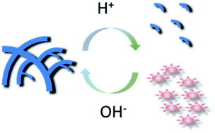
In addition, in accordance with the fluorescence reversibility, the following optical images and TEM images powerfully demonstrate the reversible supramolecular structure conversion upon pH stimuli. In pH modulation cycles, the AIA-PDI solution was always clear at low pH (4.0) and aggregated at high pH (8.0), as shown in Fig. 4A. Moreover, the short nanobelts were observed at pH 4.0 and long hybranched nanobelts were observed at pH 8.0 (Fig. 4B left and right). Therefore, the short nanobelts can self-assemble to form long hybranched nanobelts by modulating pH to basic pH; conversely, the long hybranched nanobelts can disassemble to short nanobelts. The optical images and TEM images clearly disclosed the reversible molecular self-assembly depending upon the pH of the solution (Fig. 4C).
 |
| Fig. 4 (A) Images of 0.5 mM AIA-PDI water solution at low pH (4.0, left) and high pH (8.0, right); (B) TEM images of AIA-PDI at pH 4.0 and pH 7.0; (C) the schematic presentation of AIA-PDI molecules that reversibly self-assemble depending upon pH. | |
According to the above discussion, we have demonstrated that AIA-PDI molecule showed reversible pH-responding fluorescence emission and self-assembly in an aqueous system. Based on the ultrasensitive pH response, we provide a proof of concept using a glucose detection application. In this experiment, AIA-PDI molecule acted as a fluorescence probe. Gluconic acid, one of the compounds formed in the glucose and glucose oxide (GOx) reaction mixture, could cause the pH changes in situ,38,39 and thus induce fluorescence change in AIA-PDI. The detection mechanism is shown in Fig. 5A. The pH values of glucose with different concentrations and GOx reacted mixture are shown in Table S1,† which display that the pH value decrease with the increase of the glucose concentration. The fluorescence emission spectra of AIA-PDI in these solutions were also conducted in Fig. 5B. As expected, with the increase of glucose concentration, the fluorescence intensity increased (Fig. 5B and C) indicating that some aggregated AIA-PDI molecules disassembled to monomers, as shown in Fig. 5A. A good linear relationship between Ln (FL) and glucose concentration was also observed (Fig. 5C, inset). Despite that, we also performed a control experiment using interference agents such as fructose, saccharose, as well as dopamine (DA), ascorbic acid (AA) and uric acid (UA). The results show that there is little fluorescent response to these interference agents (Fig. S1†). The stability of the AIA-PDI molecule is an important parameter for evaluating the glucose biosensor. To investigate the stability of AIA-PDI in glucose detection, the fluorescence stability of AIA-PDI molecule incubated in the mixtures of GOx and glucose for 20 minutes was examined. There is no obvious fluorescence change (Fig. S2†). The reproducibility of glucose detection was examined by three parallel measurements. The small error bars in Fig. 5C indicate a good reproducibility for the glucose sensing. Here, 1 mM glucose detection was taken as an example to calculate the relative standard error. The fluorescence spectra of the three parallel experiments showed that the intensity values are 2.77, 2.57 and 2.66, respectively. A relative standard deviation (RSD) is 3.75%. Such small value demonstrates a high reproducibility for the glucose detection. The glucose detection experiment further demonstrates the PDI derivative's biological suitability in a pH-responsive system.
 |
| Fig. 5 (A) The scheme of glucose detection using AIA-PDI molecules as fluorescence probes. (B) The corresponding fluorescence spectra of AIA-PDI incubated in the reaction mixture of GOx and glucose with different concentration. 0.8 mM (black), 1.0 mM (orange), 1.2 mM (blue), 1.5 mM (dark cyan), 1.8 mM (magenta) glucose and (inset) 0 mM (black), 0.5 mM (red) glucose. (C) Fluorescence intensity plotted as a function of glucose concentration and the linear region from 0.5 to 1.5 mM glucose (inset). | |
4. Conclusions
In this work, we designed a new type of PDI derivatives, which featured two arm-like ionized amino-imidazole groups that significantly improve its solubility in water. Moreover, the π-conjugated PDI core reserves the ability of supramolecular self-assembly. Owing to the introduced amino groups, this PDI derivative exhibits reversible conversions in the fluorescence emission and supramolecular structures upon external pH-stimulation. Based on the ultrasensitive pH response, we provide a proof of concept using a glucose detection application, which further demonstrates this PDI derivative's biological suitability in a pH-responsive system. We believe this PDI derivative has a great potential for application in pH region of biological interest such as imaging pH gradients within live tumour models and for probing intracellular microenvironments.40
Acknowledgements
The authors are most grateful to the NSFC (no. 21205112, 21105096, 21175130, 21225524 and 21375124), Ministry of Science and Technology of China (2012YQ170003) and Department of Science and Technology of Jilin Province (no. 201215091 and 20120308).
References
- Y. Qiao, W. Zhang, P. Tian, F. Meng, H. Zhu, X. Jiang, X. Liu and P. K. Chu, Biomaterials, 2014, 35, 6882–6897 CrossRef CAS PubMed.
- T. Berbasova, M. Nosrati, C. Vasileiou, W. Wang, K. S. S. Lee, I. Yapici, J. H. Geiger and B. Borhan, J. Am. Chem. Soc., 2013, 135, 16111–16119 CrossRef CAS PubMed.
- S. F. Buchsbaum, G. Nguyen, S. Howorka and Z. S. Siwy, J. Am. Chem. Soc., 2014, 136, 9902–9905 CrossRef CAS PubMed.
- X. Ji, Y. Yao, J. Li, X. Yan and F. Huang, J. Am. Chem. Soc., 2012, 135, 74–77 CrossRef PubMed.
- Y. Sagara, T. Komatsu, T. Terai, T. Ueno, K. Hanaoka, T. Kato and T. Nagano, Chem.–Eur. J., 2014, 20, 10397–10403 CrossRef CAS PubMed.
- G.-P. Yong, B. Zhang, Y.-M. Zhang and G.-S. Li, J. Mater. Chem., 2012, 22, 13481–13483 RSC.
- H. Frisch, J. P. Unsleber, D. Lüdeker, M. Peterlechner, G. Brunklaus, M. Waller and P. Besenius, Angew. Chem., Int. Ed., 2013, 52, 10097–10101 CrossRef CAS PubMed.
- R. J. Williams, A. M. Smith, R. Collins, N. Hodson, A. K. Das and R. V. Ulijn, Nat. Nanotechnol., 2009, 4, 19–24 CrossRef CAS PubMed.
- J. M. A. Carnall, C. A. Waudby, A. M. Belenguer, M. C. A. Stuart, J. J.-P. Peyralans and S. Otto, Science, 2010, 327, 1502–1506 CrossRef CAS PubMed.
- E.-M. Schön, E. Marqués-López, R. P. Herrera, C. Alemán and D. D. Díaz, Chem.–Eur. J., 2014, 20, 10720–10731 CrossRef PubMed.
- M. C. Young, L. R. Holloway, A. M. Johnson and R. J. Hooley, Angew. Chem., Int. Ed., 2014, 53, 9832–9836 CrossRef CAS PubMed.
- H. J. Karmel, J. J. Garramone, J. D. Emery, S. Kewalramani, M. J. Bedzyk and M. C. Hersam, Chem. Commun., 2014, 50, 8852–8855 RSC.
- W. Jiang, L. Ye, X. Li, C. Xiao, F. Tan, W. Zhao, J. Hou and Z. Wang, Chem. Commun., 2014, 50, 1024–1026 RSC.
- Y. Sagara, T. Komatsu, T. Ueno, K. Hanaoka, T. Kato and T. Nagano, Adv. Funct. Mater., 2013, 23, 5277–5284 CrossRef CAS.
- N. Mizoshita, T. Tani and S. Inagaki, Adv. Mater., 2012, 24, 3350–3355 CrossRef CAS PubMed.
- L. M. Daffy, A. P. de Silva, H. Q. N. Gunaratne, C. Huber, P. L. M. Lynch, T. Werner and O. S. Wolfbeis, Chem.–Eur. J., 1998, 4, 1810–1815 CrossRef CAS.
- X. Zhang, S. Rehm, M. M. Safont-Sempere and F. Würthner, Nat. Chem., 2009, 1, 623–629 CrossRef CAS PubMed.
- Z. Xu, B. He, J. Shen, W. Yang and M. Yin, Chem. Commun., 2013, 49, 3646–3648 RSC.
- T. Heek, C. Fasting, C. Rest, X. Zhang, F. Wurthner and R. Haag, Chem. Commun., 2010, 46, 1884–1886 RSC.
- B. Gao, H. Li, H. Liu, L. Zhang, Q. Bai and X. Ba, Chem. Commun., 2011, 47, 3894–3896 RSC.
- C. Backes, U. Mundloch, C. D. Schmidt, J. N. Coleman, W. Wohlleben, F. Hauke and A. Hirsch, Chem.–Eur. J., 2010, 16, 13185–13192 CrossRef CAS PubMed.
- J. Schönamsgruber, L. Zeininger and A. Hirsch, Chem.–Eur. J., 2014, 20, 2529–2536 CrossRef PubMed.
- J. Qu, C. Kohl, M. Pottek and K. Müllen, Angew. Chem., Int. Ed., 2004, 43, 1528–1531 CrossRef CAS PubMed.
- F. Biedermann, E. Elmalem, I. Ghosh, W. M. Nau and O. A. Scherman, Angew. Chem., Int. Ed., 2012, 51, 7739–7743 CrossRef CAS PubMed.
- Y. Sun, Z. Li and Z. Wang, J. Mater. Chem., 2012, 22, 4312–4318 RSC.
- P. K. Sukul, D. Asthana, P. Mukhopadhyay, D. Summa, L. Muccioli, C. Zannoni, D. Beljonne, A. E. Rowan and S. Malik, Chem. Commun., 2011, 47, 11858–11860 RSC.
- Y. Bae, S. Fukushima, A. Harada and K. Kataoka, Angew. Chem., Int. Ed., 2003, 42, 4640–4643 CrossRef CAS PubMed.
- A. Datar, K. Balakrishnan and L. Zang, Chem. Commun., 2013, 49, 6894–6896 RSC.
- Y. Hu, K. Wang, Q. Zhang, F. Li, T. Wu and L. Niu, Biomaterials, 2012, 33, 1097–1106 CrossRef CAS PubMed.
- W. Wang, J. J. Han, L.-Q. Wang, L.-S. Li, W. J. Shaw and A. D. Q. Li, Nano Lett., 2003, 3, 455–458 CrossRef CAS.
- W. Wang, L. Wang, B. J. Palmer, G. J. Exarhos and A. D. Q. Li, J. Am. Chem. Soc., 2006, 128, 11150–11159 CrossRef CAS PubMed.
- A. D. Q. Li, W. Wang and L.-Q. Wang, Chem.–Eur. J., 2003, 9, 4594–4601 CrossRef CAS PubMed.
- W. E. Ford, J. Photochem., 1987, 37, 189–204 CrossRef CAS.
- D. Görl, X. Zhang and F. Würthner, Angew. Chem., Int. Ed., 2012, 51, 6328–6348 CrossRef PubMed.
- H.-M. Zhao, J. Pfister, V. Settels, M. Renz, M. Kaupp, V. C. Dehm, F. Würthner, R. F. Fink and B. Engels, J. Am. Chem. Soc., 2009, 131, 15660–15668 CrossRef CAS PubMed.
- R. F. Fink, J. Seibt, V. Engel, M. Renz, M. Kaupp, S. Lochbrunner, H.-M. Zhao, J. Pfister, F. Würthner and B. Engels, J. Am. Chem. Soc., 2008, 130, 12858–12859 CrossRef CAS PubMed.
- S. Rehm, V. Stepanenko, X. Zhang, T. H. Rehm and F. Würthner, Chem.–Eur. J., 2010, 16, 3372–3382 CrossRef CAS PubMed.
- W. Li, L. Feng, J. Ren, L. Wu and X. Qu, Chem.–Eur. J., 2012, 18, 12637–12642 CrossRef CAS PubMed.
- W. Zhao, H. Zhang, Q. He, Y. Li, J. Gu, L. Li, H. Li and J. Shi, Chem. Commun., 2011, 47, 9459–9461 RSC.
- J. Madsen, I. Canton, N. J. Warren, E. Themistou, A. Blanazs, B. Ustbas, X. Tian, R. Pearson, G. Battaglia, A. L. Lewis and S. P. Armes, J. Am. Chem. Soc., 2013, 135, 14863–14870 CrossRef CAS PubMed.
Footnote |
† Electronic supplementary information (ESI) available: The synthesis procedure and NMR characterizations of AIA-PDI; the pH values of reaction mixture of GOx and different concentrations of glucose, the fluorescence response of AIA-PDI to interfering agents. See DOI: 10.1039/c4ra11124a |
|
This journal is © The Royal Society of Chemistry 2015 |
Click here to see how this site uses Cookies. View our privacy policy here.