
Branched ZnO nanotrees on flexible fiber-paper substrates for self-powered energy-harvesting systems†
Y. Qiuac,
D. C. Yangb,
B. Yinac,
J. X. Leiac,
H. Q. Zhangac,
Z. Zhanga,
H. Chend,
Y. P. Lid,
J. M. Biana,
Y. H. Liua,
Y. Zhaoa and
L. Z. Hu*ac
aSchool of Physics and Optoelectronic Technology, Dalian University of Technology, Dalian 116024, People's Republic of China. E-mail: lizhongh@dlut.edu.cn
bDepartment of Electronic Engineering, Dalian Neusoft University of Information, Dalian, 116024, People's Republic of China
cThe Key Laboratory for Micro/Nano Technology and System of Liaoning Province, Dalian University of Technology, Dalian 116024, People's Republic of China
dFaculty of Vehicle Engineering and Mechanics, Dalian University of Technology, Dalian 116024, People's Republic of China
First published on 5th December 2014
Abstract
In this paper, branched ZnO nanotrees (NTs) have been synthesized on flexible fiber-paper substrates by introducing a multistep hydrothermal approach for realizing high-performance piezoelectric nanogenerators. With this method, a significant enhancement in output voltage of the NGs ranging from 14 mV to 0.1 V was achieved, with a nearly 20 times enhanced power density compared to the vertically grown ZnO NWs. This is the first demonstration of fabricating branched ZnO NTs-coated fiber paper for energy harvesting devices, which may provide guidelines for designing high-performance piezoelectric energy harvesting.
Introduction
Energy harvesting technologies may meet critical demand in areas where alternatives to fossil fuels are required for environmental protection as well as future sustainable development. To alleviate this trend, harvesting energy from our living environment, such as solar, thermal, and mechanical energy,1–6 has attracted intensive interest in the past years to meet these needs. Among these, mechanical energy is one of the most abundant and reliable energy sources in our daily life, which is accompanying us anywhere at any time regardless of the weather. Recently, a great of attention has been paid to fabricating piezoelectric energy harvesting devices, namely piezoelectric nanogenerators (NGs), via using various piezoelectric materials, such as ZnO, GaN, PTZ, and BaTiO3 (BTO).4,7–11In recent years, the application of ZnO as piezoelectric materials in energy-harvesting devices has been explored extensively, primarily because of their coupled piezoelectric and semiconducting properties, easy availability, nontoxic, and abundant in nature. Besides, ZnO also can be tailored to various nanostructures,12–17 which might provide a promising means for optimizing the performance of the nanogenerators. As the nanogenerator evolves more aggressively, structural analysis becomes increasingly important. Recently, ZnO piezoelectric nanogenerators based on various 1D and 2D ZnO nanostructures have been demonstrated.13,18 For example, in 2009, Xi et al.19 first reported a piezoelectric NG based on ZnO nanotube arrays by using a low-temperature solution chemical method. This NG gave an output voltage up to 35 mV. Gupta et al.20 also demonstrated the synthesis of 2D vanadium-doped ZnO nanosheets and their application for high-performance flexible direct current power piezoelectric NGs. Recently, Saravanakumar et al.21 also reported the growth of 2D ZnO nanowall on both sides of the flexible substrate through hydrothermal method and their application toward energy harvesting devices. The fabricated nanowall NG produced the maximum output voltage and current of 2.5 V and 80 nA, respectively. To the best of our knowledge, there is no report on the growth of 3D branched ZnO nanotree (ZnO NT) structures on flexible fiber paper substrate as well as the fabrication of the NG using this kind of structure. In this report, we have developed multi-step hydrothermal process to grow ZnO NTs on the common fiber-paper substrates. The branches directly attached to the main ZnO nanowire (NW) backbones could highly improve piezoelectric power generation. The formation mechanism of the ZnO NTs and the role of the branched ZnO nanostructures in the energy-harvesting devices have been investigated.
Experimental section
Synthesis of ZnO NWs
The paper substrates utilized in this work were cut from a large piece of common packing paper with high flexibility. After being cleaned ultrasonically in acetone, ethanol, and then deionized water in order, each piece of paper substrate (with the desired size of 1 × 0.6 cm2) was first deposited with a thin ZnO seed layer by room temperature radio frequency (RF) magnetron sputtering. The background pressure of the vacuum chamber was 1 × 10⁻⁴ Pa and argon was utilized as sputtering gas with a pressure of 3.5 Pa. The 1D ZnO NWs were then grown from the seeds by immersing the seeded substrates in aqueous solutions containing 30 mmol−1 1![[thin space (1/6-em)]](https://www.rsc.org/images/entities/char_2009.gif) :1 ratio of zinc acetate dehydrate (Zn(CH3COO)2·2H2O) and hexamethylenetetramine (HMTA). The temperature was maintained as 95 °C. After 3 h, the fabricated ZnO NWs-coated paper pieces (named as sample A) were taken out of the solution, rinsed with flowing deionized water and then dried.
:1 ratio of zinc acetate dehydrate (Zn(CH3COO)2·2H2O) and hexamethylenetetramine (HMTA). The temperature was maintained as 95 °C. After 3 h, the fabricated ZnO NWs-coated paper pieces (named as sample A) were taken out of the solution, rinsed with flowing deionized water and then dried.
Construction of branched ZnO NTs
For construction of branched ZnO NTs on fiber paper, the obtained sample A was then immersed in the stirred limpid aqueous solution of 0.5 M NaOH and 0.06 M Zn(AC)2 at RT for 5 min, which was to pre-etch the surfaces of the ZnO NWs before formal ZnO deposition. Finally, it was further immersed in similar aqueous solution (as described above) at 80 °C for 60 min. The resulting ZnO NTs-coated paper piece (named as sample B) was rinsed with deionized water and dried in an oven.Fabrication of ZnO nanogenerators device
For fabricating ZnO-paper based nanogenerators, silver (Ag) paste was deposited at both ends of sample A and B to act as the electrodes. The separation between the two electrodes was approximately 0.4 cm, and the effective working area of both devices was ∼0.32 cm2. After dried at 95 °C for 1 h in an oven, two types of flexible paper-based NGs based on ZnO NWs and NTs (defined as ZWNG and ZTNG, respectively) were finally fabricated.Characterization
The surface morphologies of the as-grown ZnO-paper samples (sample A and B) were characterized by scanning electron microscopy (SEM; FEI Nova NanoSEM). X-ray diffraction (XRD) was used to analyze their crystal structures. And a Keithley 4200 semiconductor characterization system was utilized for their output measurements.Results and discussion
For construction of branched ZnO NTs on fiber paper, we adopted a multistep low-temperature hydrothermal approach. And the schematic diagram of the whole growth process is shown in Fig. 1(a), including ZnO seed layer deposition, 1D ZnO NWs growth, branched ZnO NTs growth. Fig. 1(b) and (c) show the low-magnification SEM images of the paper fibers before and after one-step low-temperature hydrothermal process, from which we can see that the initial paper fibers, with average diameters of ∼12 μm, were very smooth. However, they look very rough after being grown with ZnO NWs and the corresponding diameters increase to ∼20 μm. From Fig. 1(d), we can see that the ZnO NWs grow densely on the surfaces of the fibers after one-step hydrothermal process, the average diameters of which are about 100 nm. Using the ZnO NWs-coated fibers as the substrate, the branched nanostructures were further constructed on the ZnO NWs by using another solution of NaOH and Zn(CH3COO)2·2H2O. As shown in Fig. 1(e), the branches are directly synthesized on the surface of the ZnO NWs to form the NTs, with branch length ranging from 50–80 nm. Here, the first 5 min of etching time is vital for formation of the branched ZnO nanostructures because it was difficult to deposit ZnO seed layer directly on the smooth surfaces of the as-grown ZnO NWs by commonly used methods such as spin coating or drop-casting.22,23 Our solution was to pre-etch the surfaces of the ZnO NWs, which was found to be effective. By using a supersaturated solution of Zn(CH3COO)2·2H2O and NaOH, etch pits are initially formed on the surface of ZnO NWs, which could act as sites for further construction of spines.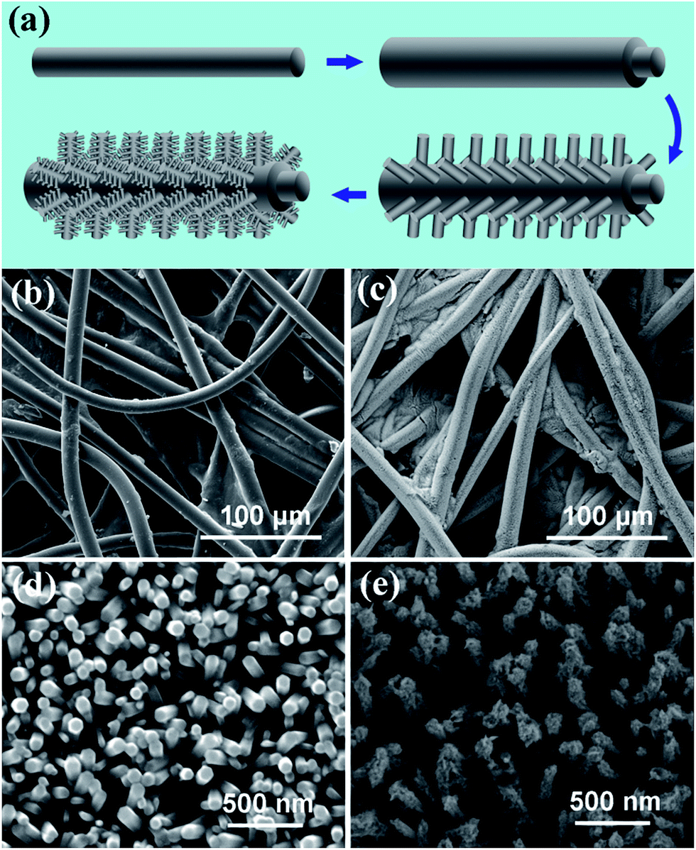 | ||
| Fig. 1 (a) Schematic diagram describing the fabrication process of the ZnO NTs. Low-magnification SEM image of the paper-fibers before (b) and after (c) experiencing one-step hydrothermal approach. High magnification SEM images showing top-view of the ZnO NWs (d) and ZnO NTs (e). | ||
Fig. 2 shows the corresponding XRD patterns of ZnO NWs (sample A) and branched NTs (sample B) synthesized on paper-fiber substrates. All the diffraction peaks can be easily assigned to hexagonal wurtzite phase of ZnO. For sample A, the peak intensity of (002) is much higher than that of NTs, indicating the NW with no branches has more intense orientation on (002) diffraction. For NTs, higher diffraction peaks of (100) and (101) planes are observed, which are associated with the ZnO branches existed on NWs. Besides of this, the mechanical stability of the as-grown samples was also investigated by bending test. And both samples can withstand dozens of bending cycles without any damage (Fig. S1, ESI†), which will benefit the stability of the flexible piezoelectric NGs.
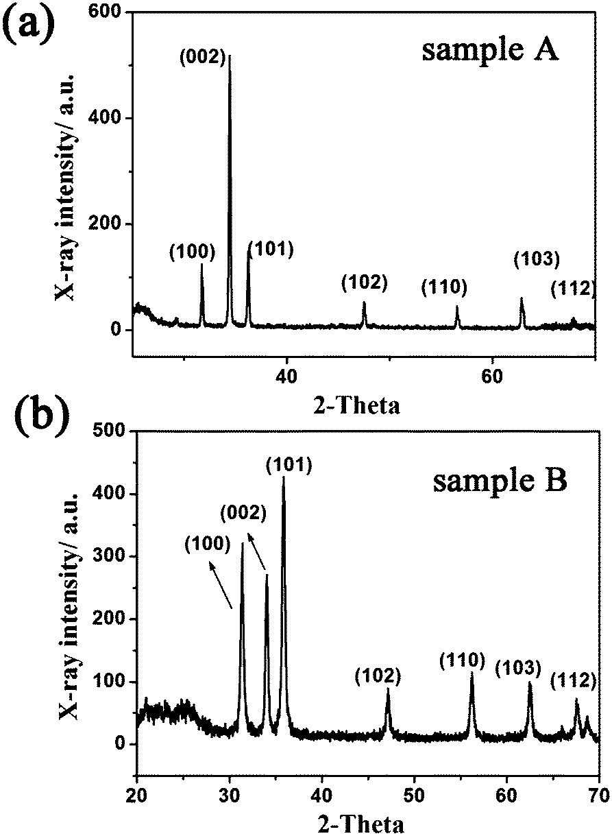 | ||
| Fig. 2 XRD patterns of sample A (a) and sample B (b). | ||
Electrical measurement of output signals generated from both NGs are conducted using specially designed measuring setup, the schematic diagram of which is shown in the inset of Fig. 3. The fabricated NGs were fixed onto the plastic board (PB) with a length of 20 cm, a width of 1.2 cm and a thickness of 0.2 cm. The PB could be easily bent into different curvatures and circular arcs by adjusting the height of the gasket. Before the electromechanical measurements, we first measured the original I–V curves of both ZWNG and ZTNG, as shown in Fig. 3. It was found that both NGs had linear I–V behavior, indicating Ohmic contact formed between ZnO-paper pieces and Ag paste. The resistances of ZWNG and ZTNG were ∼200 and ∼10 MΩ, respectively.
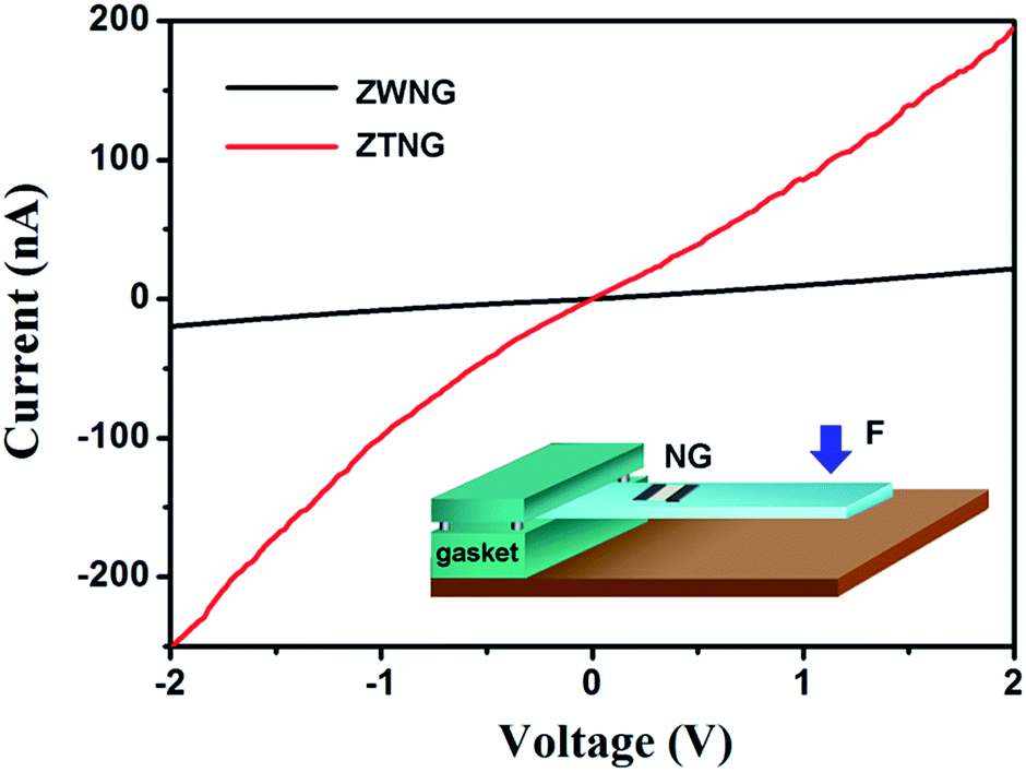 | ||
| Fig. 3 I–V curves of ZWNG and ZTNG; inset is the schematic diagram of the measuring setup during electrical experiment. | ||
To test the electrical output performance of the NGs, a manually applied stress was periodically introduced to deform the PB, so that the NG experienced cycling stretching–releasing deformation process. Fig. 4 shows the output voltage and current of ZWNG and ZTNG being subjected to repeated cycles of fast stretched (FS) and fast released (FR). With the strain of 0.25% (gasket height = 2 cm), the output voltage and current of ZENG were measured to be ∼14 mV and ∼7 nA, respectively (power density: 0.306 nW cm−2), which is similar with the previous reported ZPNG.24 The piezoelectric output of ZTNG was fairly compared under the same experimental and measurement conditions. The measured voltage and current of ZTNG were measured to be ∼100 mV and ∼20 nA, respectively, corresponding to a power density of ∼6.25 nW cm−2, which was nearly 20 times higher than that of the ZWNG. Furthermore, in order to verify that the electric outputs resulted from the piezoelectric property of ZnO-paper rather than for other reasons, we fixed a similar piece of the fiber paper used as the substrate in our study on the PB and deposited the Ag paste electrodes on both ends of the paper to measure its electric output. The results showed that only noise existed in the output voltage and current signals (Fig. S2, ESI†), indicating that the electric outputs of the fabricated NGs were generated from the piezoelectric ZnO NWs on the paper rather than triboelectric effect or measuring system.
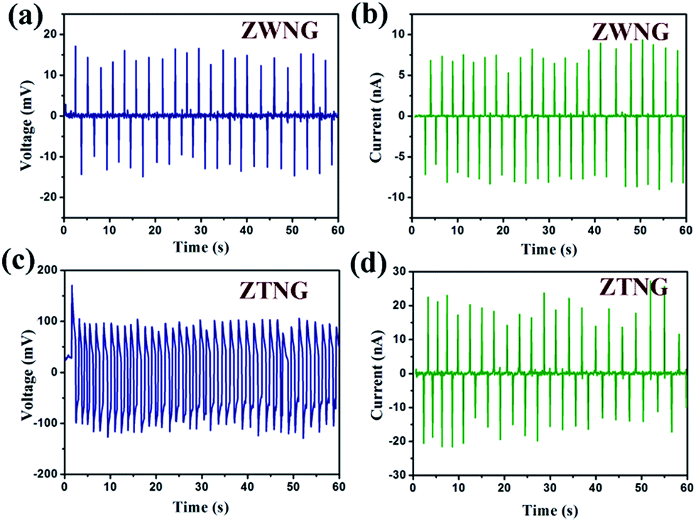 | ||
| Fig. 4 The measured electric outputs of the ZWNG and ZTNG subjected to repeated cycles of FS and FR (gasket height = 2 cm). (a) Voltage output generated from ZWNG. (b) Current output generated from ZWNG. (c) Voltage output generated from ZTNG. (d) Current output generated from ZTNG. | ||
The energy conversion efficiency of the NG can be estimated as the ratio between generated electrical energy and applied mechanical energy. In our study, the output electrical energy generated by stretching and releasing the NG is calculated as:10,24
| We = ∫VIdt |
| Ws = πD2LoEε2/8 |
According to the previous paper,25 when the PB is bent downward, a deformation will take place in the ZnO NWs and cause them to rub against each other. Thus, the piezoelectric bound charges will generate inside the stressed NWs, leading to a piezoelectric potential gradient in the device. And when the PB is released, the generated piezoelectric bound charges will gradually diminish. In this process, the accumulated free charges flow back in the opposite released direction. Therefore, alternating positive and negative electric outputs can be obtained, as shown in Fig. 4. Furthermore, we find that compared to the ZWNG with vertically ZnO NWs, the ZTNG with branched ZnO NTs clearly have better piezoelectric performances. Here, we attribute this phenomenon to the branched structures formed in the secondary grown NWs.
In order to understand the effect of the branched NWs on the piezoelectric potentials, the simulations for both stressed ZnO NWs/NTs were performed by using finite element calculation (COMSOL) as shown in Fig. 5. We can clearly see that the minimum value of calculated piezoelectric potential at the same applied strains, are −10.6 mV for the NG with two NWs and ∼−36.5 mV for the one with two NTs. The obvious piezoelectric potential increase could be found in Fig. 5, indicating that branched NTs provide more effective structure for harvesting externally applied strains. In this case, when the ZTNG was stretched, besides the lateral bending of the backbone ZnO NWs as stated above, the piezoelectric charges would be also generated between the branched structures, which could also contribute to the electric output. What's more, most of these branches were more likely to suffer from vertical compression rather than lateral bending when the deformation was introduced in the branched ZnO NTs, as shown in Fig. 5. It has been confirmed experimentally and theoretically that vertical compression is better than lateral bending under the assumption of negligible free charges,26,27 which may account for the highly enhanced electric outputs of ZTNG. Here, the branches provide not only the possibility of suffering vertical compression but also faster electron transport pathways compared to the smooth ZnO NW. Simulated potential values could not exactly match with the experimental results, because the generated potential is highly influenced by material parameters and applied strain, which are difficult to be exactly defined.
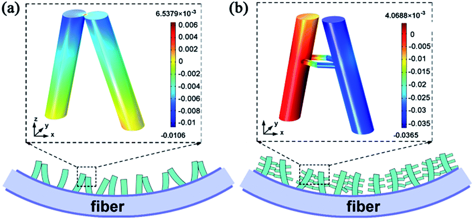 | ||
| Fig. 5 The operation mechanism of the designed ZWNG and ZTNG. The dotted boxes are the corresponding simulations of the piezopotential distribution in the ZnO NWs (a) and in the ZnO NTs (b). | ||
Conclusions
In conclusion, branched ZnO NTs have been synthesized on flexible fiber-paper substrates for realizing high-performance piezoelectric NGs. It was found that the performance of the NTNG with branched NTs was highly enhanced compared with the one with normal NWs. The ZTNG produced the maximum output voltage and current ∼100 mV and ∼20 nA, respectively, corresponding to a power density of ∼6.25 nW cm−2, which was nearly 20 times higher than that of the ZWNG. Experimental and theoretical results showed that the branches of NTs could provide the possibility of suffering vertical compression compared to the smooth ZnO NW, which highly increased the output electric potentials. These results may provide important insight into the facile fabrication method for low-cost and high-performance energy harvesting devices.Acknowledgements
This work was supported by the NSFC (Project no. 60777009); the Natural Science Foundation of Liaoning Province (2013020095); the Fundamental Research Funds for the Central Universities (DUT13LK02, DUT13LAB12); Opening fund of Key laboratory for Micro/Nano Technology and System of Liaoning and System of Liaoning Provence the Fundamental Research Funds for the Central Universities (Project no. DUT11LK46), and Fundamental Research Funds of the Central Universities (DUT14LK35).Notes and references
- B. Tian, X. Zheng, T. J. Kempa, Y. Fang, N. Yu, G. Yu, J. Huang and C. M. Lieber, Nature, 2007, 449, 885 CrossRef CAS PubMed
.
- Z. L. Wang and W. Wu, Angew. Chem., Int. Ed., 2012, 51, 11700 CrossRef CAS PubMed
- Y. Yang, W. Guo, K. C. Pradel, G. Zhu, Y. Zhou, Y. Zhang, Y. Hu, L. Lin and Z. L. Wang, Nano Lett., 2012, 12, 2833 CrossRef CAS PubMed
- Z. L. Wang and J. H. Song, Science, 2006, 312, 242 CrossRef CAS PubMed
- S. Lee, J. I. Hong, C. Xu, M. Lee, D. Kim, L. Lin, W. Hwang and Z. L. Wang, Adv. Mater., 2012, 24, 4398 CrossRef CAS PubMed
- J. H. Lee, K. Y. Lee, M. K. Gupta, T. Y. Kim, D. Y. Lee, J. Oh, C. Ryu, W. J. Yoo, C. Y. Kang and S. J. Yoon, Adv. Mater., 2014, 26, 765 CrossRef CAS PubMed
- C.-T. Huang, J. Song, W.-F. Lee, Y. Ding, Z. Gao, Y. Hao, L.-J. Chen and Z. L. Wang, J. Am. Chem. Soc., 2010, 132, 4766 CrossRef CAS PubMed
- X. Chen, S. Xu, N. Yao and Y. Shi, Nano Lett., 2010, 10, 2133 CrossRef CAS PubMed
- S.-H. Shin, Y.-H. Kim, M. H. Lee, J.-Y. Jung and J. Nah, ACS Nano, 2014, 8, 2766 CrossRef CAS PubMed
- G. Zhu, R. Yang, S. Wang and Z. L. Wang, Nano Lett., 2010, 10, 3151 CrossRef CAS PubMed
- K.-I. Park, S. Xu, Y. Liu, G.-T. Hwang, S.-J. L. Kang, Z. L. Wang and K. J. Lee, Nano Lett., 2010, 10, 4939 CrossRef CAS PubMed
- S. Kar, A. Dev and S. Chaudhuri, J. Phys. Chem. B, 2006, 110, 17848 CrossRef CAS PubMed
- H. Sun, H. Tian, Y. Yang, D. Xie, Y.-C. Zhang, X. Liu, S. Ma, H.-M. Zhao and T.-L. Ren, Nanoscale, 2013, 5, 6117 RSC
- J. Y. Lao, J. G. Wen and Z. F. Ren, Nano Lett., 2002, 2, 1287 CrossRef CAS
- A. Khan, M. Hussain, O. Nur and M. Willander, Chem. Phys. Lett., 2014, 608, 235 CrossRef PubMed
- Z. L. Wang, R. Yang, J. Zhou, Y. Qin, C. Xu, Y. Hu and S. Xu, Mater. Sci. Eng., R, 2010, 70, 320 CrossRef PubMed
- W. L. Hughes and Z. L. Wang, Appl. Phys. Lett., 2005, 86, 043106 CrossRef PubMed
- Y. Yang, H. Tian, H. Sun, R.-J. Xu, Y. Shu and T.-L. Ren, RSC Adv., 2014, 4, 2115 RSC
- Y. Xi, J. Song, S. Xu, R. Yang, Z. Gao, C. Hu and Z. L. Wang, J. Mater. Chem., 2009, 19, 9260 RSC
- M. K. Gupta, J.-H. Lee, K. Y. Lee and S.-W. Kim, ACS Nano, 2013, 7, 8932 CrossRef CAS PubMed
- B. Saravanakumar and S. J. Kim, J. Phys. Chem. C, 2014, 118, 8831 CAS
- Y. Qiu, K. Yan, H. Deng and S. Yang, Nano Lett., 2011, 12, 407 CrossRef PubMed
- H.-M. Cheng, W.-H. Chiu, C.-H. Lee, S.-Y. Tsai and W.-F. Hsieh, J. Phys. Chem. C, 2008, 112, 16359 CAS
- C. Chang, H. Tan, J. Wang, Y. K. Fuh and L. W. Lin, Nano Lett., 2010, 10, 726 CrossRef CAS PubMed
- Y. Qiu, H. Zhang, L. Hu, D. Yang, L. Wang, B. Wang, J. Ji, G. Liu, X. Liu and J. Lin, Nanoscale, 2012, 4, 6568 RSC
- C. Falconi, G. Mantini, A. D'Amico and Z. L. Wang, Sens. Actuators, B, 2009, 139, 511 CrossRef CAS PubMed
- G. Zhu, A. C. Wang, Y. Liu, Y. Zhou and Z. L. Wang, Nano Lett., 2012, 12, 3086 CrossRef CAS PubMed
Footnote |
| † Electronic supplementary information (ESI) available. See DOI: 10.1039/c4ra09163a |
| This journal is © The Royal Society of Chemistry 2015 |