Plasmonic cooperation effect of metal nanomaterials at Au–TiO2–Ag interface to enhance photovoltaic performance for dye-sensitized solar cells

Yang Wanga, Jin Zhai*a and Yanlin Song*ab
aKey Laboratory of Bio-Inspired Smart Interfacial Science and Technology of Ministry of Education, Beijing Key Laboratory of Bio-Inspired Energy Materials and Devices, School of Chemistry and Environment, Beihang University, Beijing 100191, China. E-mail: wangyang@sce.buaa.edu.cn; zhaijin@buaa.edu.cn; ylsong@iccas.ac.cn
bInstitute of Chemistry Chinese Academy of Sciences, 100190, Beijing, P. R. China. E-mail: ylsong@iccas.ac.cn

Received 16th August 2014 , Accepted 14th November 2014

First published on 14th November 2014


Abstract

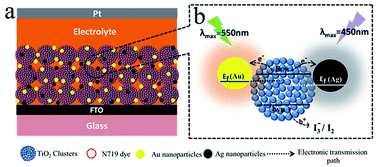
From the plasmonic cooperation effect of metal nanomaterials at a Au–TiO2–Ag interface, Au and Ag used complementary light-harvesting to enhance photovoltaic performance in dye-sensitized solar cells (DSSC). The best efficiency (η) of DSSC reached 7.51%, compared with 6.23% for pure TiO2 electrode. The average energy conversion efficiency and photocurrent density were increased by 20.8% and 29.9% compared with those of pure TiO2 electrodes. Hence, the complementary light-harvesting using different light absorption positions of Au and Ag nanomaterials and plasmonic cooperation effect of Au and Ag together improved the light harvesting, short circuit current density, open circuit voltage and photo-electric conversion efficiency in DSSCs.


Introduction

Dye-sensitized solar cells (DSSCs) are composed of an inorganic semiconducting photo-anode with absorbed dye sensitizers, electrolytes and counter electrode.1,2 These offer an alternative to traditional photovoltaic devices because of their low cost and high efficiency, and there has been considerable development in dye synthesis, preparing novel structures for photo-anodes, counter electrodes and device fabrication technologies.3–6 Currently, for use as an electrode, TiO2 is fabricated as a significant nanomaterial with various structures.4–10 However, a thick TiO2 film works against electrolyte diffusion, resisting smooth flow of electrons and aggravating charge recombination, thus imposing an upper limit on TiO2 film thickness.7 A developing method to solve the problems in DSSCs uses surface plasmon resonance (SPR) and conductivity from metallic nanomaterials introduced into the DSSCs. Single metal nanoparticles (Au or Ag) with surface plasmonic effect to increase light absorption have already been researched, and application of such a surface plasmonic effect has been investigated on DSSCs, with some exciting observations,8–11 including Ag@TiO2,9 SiO2@Ag,10 Au@SiO2,12 and Au@TiO2.13 The nanoparticles have been employed to improve the light adsorption and conversion efficiency of conventional TiO2 photo-anode devices. All of these use single metal nanomaterials to improve the properties in DSSCs. However, Au and Ag nanomaterials, with maximum absorption wavelength at ∼550 nm 14–17 and ∼450 nm,18–20 respectively, have not been considered for their different light absorption positions for complementary light-harvesting and plasmonic cooperation effects to enhance energy conversion efficiency in DSSCs.

Herein, Au and Ag nanomaterials were introduced into photo-anodes for DSSCs. The TiO2 electrodes with micro–nano structure were fabricated by a electro-hydrodynamic (EHD) technique,21,22 and the introduced metal ions (Au and Ag) were reduced by UV light (see Scheme 1a). In this study, the Au–TiO2–Ag interface exhibited complementary light absorption and strong plasmonic cooperation effects (see Scheme 1b). As shown, Au and Ag nanoparticles relatively complemented light absorption because of the different light absorption region between them (λAu = 550 nm and λAg = 450 nm), and greatly improved the light absorption in electrodes. The plasmonic cooperation effect of Au and Ag nanoparticles promoted the electron–hole separation and accelerated the electron transfer. The light to electric energy conversion was obviously improved.


image file: c4ra08753d-s1.tif
Scheme 1 (a) Schematic diagram of DSSCs with Au–TiO2–Ag electrodes; (b) operational principle of the electrodes.

Experimental section

Chemical materials

Silver nitrate and hydrogen tetrachloroaurate(III) hydrate 99.9% were purchased from Sinopharm Chemical Reagent Co., Ltd. Lithium iodide, I2, tert-butylpyridine, 1-propyl-3-methylimidazolium iodide and dye sensitizer cis-bis(isothicyanato) bis(2,2′-bipyridyl-4,4′-discarboxylato)-ruthenium(II)-bis-tetrabutylammonium, (coded as N719) were purchased from Sigma-Aldrich. FTO glass was purchased from Wuhan Georgi & Education Equipment Co., Ltd.

Preparation of anatase titanium dioxide nanoparticles

37 mL of titanium(IV) iso-propoxide (Aldrich) in 1 mL of iso-propanol was dripped slowly into a stirred mixture of 80 mL of glacial acetic acid and 250 mL of deionized water at 0 °C over 30 min. The resulting solution was filtrated and heated to 80 °C for 8 h. After that, it was heated to 230 °C in a Telflon autoclave for 12 h. On removal from the autoclave, the solution was sonicated for 30 min. The TiO2 colloid solution was concentrated to TiO2 0.25 g mL−1.

Preparation of TiO2 films

A dense TiO2 thin layer was deposited on the surface of FTO glass substrate by spin-coating a solution containing 8.5 mL titanium(IV) n-butoxide, 0.9 mL deionized water, 1.6 mL acetylacetone and 60 mL ethanol. The thin layer was kept in air for more than an hour, then used in the following procedure.

TiO2 films were prepared using an electro-spinning technique. The precursor solution was composed of ethanol (5 mL), poly(vinyl alcohol) (PVA, MW = 22[thin space (1/6-em)]000) water solution (1.2 g, 33 wt%), TiO2 colloid solution (2.64 mL, 0.25 g mL−1) and deionized water (3.96 mL). The precursor solution was sprayed onto the conducting FTO glass slides (the electrospinning method of which is described elsewhere21,22). Then the sample was sprayed at an electric field of 30 kV. The distance between the conducting FTO glass slides and the tip of the needle is 13 cm. Finally, the TiO2 films on the conducting FTO glass slides were calcined at 450 °C for 30 min in air.

Preparation of metal Au–TiO2–Ag photo-anodes

A series of electrodes were fabricated using different methods, defined as electrodes 1, 2, 3 and 4. The pure TiO2 film as control group was named electrode 1. Subsequently, the TiO2 film was immersed into a 5 mg mL−1 HAuCl4 or AgNO3 solution. Then, the electrode was washed several times with water to remove excess Au or Ag ions adsorbed on the surface. After reduction under UV light, the doped Au or Ag photo-anodes were immersed into a tetrabutyl titanate solution for an hour and then calcined at 450 °C for 30 min (named electrode 2 and 3) to protect the Au and Ag particles from corrosion in the I3/I solution. Next, electrode 4 was fabricated from electrode 2 immersed in a 5 mg mL−1 AgNO3 solution and reduced under UV light, then treated with tetrabutyl titanate solution and calcined (see Fig. 1 for details). All the prepared electrodes were dried under an N2 stream, then they were irradiated with UV light to convert the Au or Ag ions to metal nanoparticles. In summary, electrode 1: pure TiO2; electrode 2: TiO2–Au; electrode 3: TiO2–Ag; electrode 4: Au–TiO2–Ag. The treated anodes were immersed in a dye N719 (1.68 × 10⁻⁴ M) for 24 h. The integral DSSCs were composed of a sensitized photo-anode, an electrolyte and a platinum counter electrode. The electrolyte was composed of 0.5 M LiI, 0.05 M I2, 0.5 M tertbutylpyridine, and 0.6 M 1-propyl-3-methylimidazolium iodide in 3-methoxypropionitrile.
image file: c4ra08753d-f1.tif
Fig. 1 Schematic design for fabricating Au–TiO2–Ag electrode.

Characterization

The electrodes were characterized using an X-ray diffractometer (XRD) using Cu Ka radiation with scanning speed of 2° min−1. SEM images were obtained using a JEOL JSM-6700F scanning electron microscope at 3.0 kV. UV-vis spectra were recorded on a Hitachi Model U-4100 spectrophotometer. IV characteristics of the cell were measured by an electrochemical analyzer (CHI630A, Chenhua Instruments Co., Shanghai) under solar simulator illumination (CMH-250, Aodite Photoelectronic Technology Ltd, Beijing) at room temperature. The IPCE was measured by illumination with monochromatic light.

Results and discussion

As shown in Fig. 2, the micro–nano structures of TiO2 electrodes using the EHD technique were characterized by scanning electron microscopy (SEM). TiO2 clusters and lots of micro–nano pores existed in the film, while nanoporous morphology and interconnectivity among the TiO2 nanoparticles, with lots of unequal pores distributed in the surface, can be observed. From Fig. 2b (the magnified image), the film was composed of micro and sub-micro-scale near spherical clusters containing TiO2 nanoparticles. This kind of TiO2 electrode with micro–nano structure might be useful for achievement of various photo-electric properties.21
image file: c4ra08753d-f2.tif
Fig. 2 (a) SEM image of TiO2 composite porous films prepared from electro-spinning technique. (b) The magnified image.

TiO2–(Au, Ag or Au and Ag) films were characterized by XRD analyses to determine the presence of metal nanoparticles. Because of the low amount of Au or Ag anchored on the TiO2 films, the diffraction of Au and Ag species could not be detected (see ESI Fig. S1). Therefore, to investigate Au and Ag nanoparticles anchored on TiO2 films, electrode 4 was measured by XPS analysis to judge the presence of Au and Ag nanoparticles and gauge the valence state of those nanoparticles (as shown in Fig. 3). It was found that the differences between the 4f7/2 and 4f5/2 peaks for gold (∼3.7 eV) and between the 3d5/2 and 3d3/2 peaks for silver (∼6.0 eV) were the same as the reported23–25 values of zero valent gold and silver. This observation suggested that the majority of the gold and silver atoms existed in the zero valent state for as-prepared electrodes. The existence of an Au–TiO2–Ag interface was also proved indirectly by SEM, as shown in Fig. S2.


image file: c4ra08753d-f3.tif
Fig. 3 X-Ray photoelectron spectroscopy (XPS) spectrum of the Au and Ag–TiO2 electrode: (a) Au 4f and (b) Ag 3d.

To investigate the properties of photovoltaic devices, the fabricated electrodes were measured with different methods. The thickness of all of the electrodes was the same (∼11 μm). The related parameters are summarized in Table 1, and IV curves are shown in Fig. 4.

Table 1 Parameters for DSSCs based on the different electrodesa
Electrodes Jsc(mA cm−2) Voc (V) FF (%) The averages of values: η (%) The best of values: η (%)
a The active area of the photo electrodes for DSSC is kept at 0.15 cm2. The values shown are from cells, based on averages of five samples.
1 11.7 0.715 70.6 6.01 ± 0.22 6.23
2 14.2 0.672 65.4 6.41 ± 0.29 6.70
3 13.7 0.724 68.3 6.87 ± 0.28 7.15
4 15.2 0.742 63.4 7.26 ± 0.25 7.51


image file: c4ra08753d-f4.tif
Fig. 4 (a) UV-vis absorption of different electrodes. (b) Typical photocurrent–voltage characteristics of different electrodes (the sensitizer is N719. The cell active area is 0.15 cm2, and the light intensity is 100 mW cm−2).

Fig. 4a depicts the absorption spectra for Au and Ag anchored to the micro–nano structure of TiO2 electrodes (electrode 1: pure TiO2; electrode 2: TiO2–Au; electrode 3: TiO2–Ag; electrode 4: Au–TiO2–Ag). Fig. 4a shows no obvious absorption in visible light region for electrode 1. At λ = 550 nm (for electrode 2), the Au–TiO2 electrode showed stronger absorption than that of electrode 1. TiO2 film with anchored Ag (electrode 3) also presented stronger absorption centered in the visible region (λ = 450 nm) than that of electrode 1. However, both Au and Ag adsorbed on TiO2 film (electrode 4) showed the exciting result of a higher value of light absorption in the whole light region, and an improved utilization ratio of light. This indicated that the plasmonic cooperation effect of the Au–TiO2–Ag interface caused a strong complement advantage in light absorption when Au and Ag nanoparticles anchored together on the TiO2 film. Similar results, enhancement of light absorption by localized surface plasmon resonance (LSPR), have been reported previously.26–28 UV-vis absorption spectra can be used to prove the plasmonic cooperation effect of the pure Au, Ag, and mixture (Au and Ag) nanoparticles solutions (see Fig. S3x2020). The positions of Au and Ag nanoparticles' absorption peaks were 540 nm and 420 nm, respectively. When Au and Ag nanoparticles were mixed, there was a broadening phenomenon between 420 nm and 540 nm, which was attributed to a plasmonic cooperation effect of Au and Ag.

The photo-voltage and photocurrent of DSSCs based on these electrodes were characterized under simulated AM 1.5 illumination (100 mW cm−2). From Fig. 4b, a control photo-anode (electrode 1, pure TiO2) was fabricated using only TiO2, and the short-circuit current density (Jsc), open circuit voltage (Voc), energy conversion coefficient (η), and fill factor (FF) were 11.7 mA cm−2, 0.715 V, 6.01% and 70.6%, whereas electrode 2 with Au had higher short circuit current density (14.2 mA cm−2) and lower open circuit voltage (0.672 V). The TiO2 film with anchored Ag (electrode 3) showed lower short circuit current density (13.7 mA cm−2) and higher open circuit voltage (0.724 V) than electrode 2. Electrode 4, TiO2 film with anchored Au and Ag, obtained complementary advantages with improvements in the properties of short circuit current density and open circuit voltage compared with electrodes 2 and 3. For electrode 4 (Au–TiO2–Ag), the cell showed that Jsc, Voc, η and FF were 15.2 mA cm−2, 0.742 V, 7.26% and 63.4%. For comparison, the photo-electric conversion efficiency of electrodes 2 and 3 increased just 6.66% and 14.3% from electrode 1, respectively, whereas the photo-electric conversion efficiency of electrode 4 was increased by 20.8% compared with the value for electrode 1.

This increased efficiency was mainly attributed to two factors: (1) the TiO2 film with anchored Au and Ag improved the visible light absorption, which was consistent with the increased absorption intensity in the UV-vis absorption spectra (Fig. 4a). (2) The LSPR of Au and Ag enhanced light scattering in the visible light range, which would increase the optical path and improve the ability of electron–hole separation. In addition, it was found that the incremental photo-electric conversion efficiency of electrode 4 was approximately the sum of the values for electrodes 2 and 3. This implied that the enhancement of efficiency was triggered by significant cooperation of the surface plasmon resonance effect of the Au–TiO2–Ag interface. The above results were further verified by IPCE and EIS measurements as described in the following sections.

To investigate the origin of the Jsc increase, the incident photon-to-electron conversion efficiency (IPCE) spectrum was measured. The IPCE measurement determined further the enhancement in light harvesting efficiency for DSSCs.

Fig. 5 shows the IPCE spectra of the prepared electrodes for DSSCs. It was found that the IPCE value increased in the order of the pure TiO2 (electrode 1), TiO2–Au (electrode 2), TiO2–Ag (electrode 3) and Au–TiO2–Ag (electrode 4). The Au–TiO2–Ag (electrode 4) showed enhancement in the whole visible region, which was consistent with the strong UV-vis absorption spectrum. This result indicated that integrating Ag and Au for use as a light enhancer could promote light absorption and strengthen this phenomenon via an Au–TiO2–Ag interface plasmonic cooperation effect. In addition, electrode 4 with Au and Ag was better than those using single metals at increasing the photocurrent density.


image file: c4ra08753d-f5.tif
Fig. 5 IPCE spectra of electrodes 1, 2, 3 and 4.

To corroborate the contribution of the plasmonic cooperation effect of Au and Ag in charge separation and the electronic transfer process, the fabricated photo-anodes were investigated by electrochemical impedance spectra (EIS) under illumination of one sun (AM 1.5 G, 100 mW cm−2) at an open circuit bias (Voc, value listed in Table 1). In the Nyquist plots (Fig. 6a), the biggest semicircle located at medium frequencies, which related to the charge transfer resistance (Rw), was attributed to back reaction from the injected electrons to the electrolyte. The other high-frequency region corresponded to the charge transfer resistance (Rct) at the electrolyte/counter electrode interface. The related parameters are summarized in Table 2. The corresponding equivalent circuit of the photo-anodes is inserted in Fig. 6a. Comparing with the charge transfer resistance (Rw) of all electrodes, the semicircle size in the middle frequency for the Au–TiO2–Ag photo-anode (electrode 4) obviously decreased and obtained the smallest charge transfer resistance (24.87 Ω). It was explained that the plasmonic cooperation effect of Au and Ag reduced the recombination rate because of better electron–hole separation.


image file: c4ra08753d-f6.tif
Fig. 6 EIS of the devices under illumination conditions: (a) Nyquist plots and (b) Bode plots. The spectra were measured under the illumination of one sun at open circuit potential.
Table 2 Parameters determined by EISa
Electrodes Rw (Ω) Rk/Rw keff (s−1) τ (ms) Deff (cm2 s−1) Ln (μm)
a EIS was measured with 11 μm thick photo-anode films under 100 mW cm−2.
1 40.48 2.44 34.18 29.26 10.1 × 10⁻⁵ 17.17
2 28.64 2.50 27.71 36.09 8.39 × 10⁻⁵ 17.40
3 28.44 2.55 19.85 50.37 6.12 × 10⁻⁵ 17.56
4 24.87 2.58 19.29 51.84 6.02 × 10⁻⁵ 17.67


This result was also supported by the corresponding Bode plots (Fig. 6b). For comparison, the characteristic frequency peak position of electrodes (1, 2, 3 and 4) was shifted to a lower frequency (from 5.44 Hz to 3.07 Hz). This showed that the shift of peak from high frequency to low frequency revealed a longer electron transport process because of a longer electron lifetime (τ). The middle-frequency (fmid) was related to the inverse of τ as follows: τ = 1/(2πfmid). Using this equation, the longest electron lifetime (51.84 ms) was obtained for the Au–TiO2–Ag photo-anode (electrode 4), compared with 29.26 ms for the DSSC with control TiO2 photo-anode (electrode 1). Therefore, the Nyquist plots and Bode phase plots showed that the TiO2 photo-anode with Au and Ag greatly improved the electrochemical properties through prolonged τ. In addition, according to a previous report, the longer electron lifetime improved the effective carrier diffusion length (Ln), which reflected the competition between charge transport and recombination in DSSCs.29 The value of Ln can be obtained from eqn (1):

 
L2n = Deff × τ (1)
where Deff is the effective electron diffusion coefficient and τ is the electron lifetime. Based on the Bisquert model,30–32 Deff is described by eqn (2),
 
Deff = (Rw/Rk)L2keff (2)
where Rw is the resistance of electron transport in the photo-anode, Rk is the resistance of charge transfer related to recombination, L is the thickness of the fabricated photo-anodes and keff is the constant of effective rate for recombination. The values of parameters were estimated from the central arc of Nyquist and Bode plots. Finally, the expected Rw/Rk, keff, Deff obtained and the resulting Ln are summarized in Table 2. The Au–TiO2–Ag photo-anode had the longest Ln (17.67 μm), showing that Au and Ag using the cooperation of surface plasmon resonance effect not only reduced electron recombination, but also improved the effective carrier diffusion length.

Conclusion

In summary, we successfully introduced Au and Ag nanomaterials into TiO2 electrodes by a photo reduction process. The properties of these electrodes were systematically investigated by optical and electrochemical measurements. The best efficiency for DSSC reached 7.51%, compared with 6.23% for pure TiO2 electrode. The energy conversion efficiency and photocurrent density for Au and Ag–TiO2 photo-anodes were enhanced by about 20.8% and 29.9% compared with those of pure TiO2 cells. Utilizing the plasmonic cooperation effect of the Au–TiO2–Ag interface enhanced complementary light-harvesting, and provides an alternative approach for improving performance of DSSCs.

Acknowledgements

This work was supported by the National Natural Science Foundation of China (91333120), Ph.D. Programs Foundation of Ministry of Education of China (30400002011127001) and National Basic Research Program (2011CB935704, 2012CB720904).

Notes and references

  1. B. Oregan and M. Grätzel, Nature, 1991, 353, 737 CrossRef CAS.
  2. A. Yella, H. W. Lee, H. N. Tsao, C. Yi, A. K. Chandiran, M. K. Nazeeruddin, E. W. G. Diau, C. Y. Yeh, S. M. Zakeeruddin and M. Grätzel, Science, 2011, 334, 629 CrossRef CAS PubMed.
  3. J. C. You, W. J. Sheng, K. K. Huang, C. M. Hou, H. J. Yue, B. Hu, M. Wang, D. L. Wei, Q. W. Li, L. P. Zhao, W. Y. Dong, Z. G. Zhao and Y. J. Li, ACS Appl. Mater. Interfaces, 2013, 5, 2278 CAS.
  4. H. Li, Q. Zhao, W. Wang, H. Dong, D. S. Xu, G. J. Zou, H. L. Duan and D. P. Yu, Nano Lett., 2013, 13, 1271 CrossRef CAS PubMed.
  5. S. W. Lee, K. S. Ahn, K. Zhu, N. R. Neale and A. J. Frank, J. Phys. Chem. C, 2012, 116, 21285 CAS.
  6. Y. C. Park, Y. J. Chang, B. G. Kum, E. H. Kong, J. Y. Son, Y. S. Kwon, T. Park and H. M. Jang, J. Mater. Chem., 2011, 21, 9582 RSC.
  7. S. Ito, S. M. Zakeeruddin, R. Humphry-Baker, P. Liska, R. Charvet, P. Comte, M. K. Nazeeruddin, P. P'echy, M. Takata, H. Miura, S. Uchida and M. Grätzel, Adv. Mater., 2006, 18, 1202 CrossRef CAS.
  8. A. Baba, K. Wakatsuki, K. Shinbo, K. Kato and F. Kaneko, J. Mater. Chem., 2011, 21, 16436 RSC.
  9. J. Qi, X. Dang, P. T. Hammond and A. M. Belcher, ACS Nano, 2011, 5, 7108 CrossRef CAS PubMed.
  10. B. Ding, B. J. Lee, M. Yang, H. S. Jung and J.-K. Lee, Adv. Energy Mater., 2011, 1, 415 CrossRef CAS.
  11. X. Zhou, G. Liu, J. Yu and W. Fan, J. Mater. Chem., 2012, 22, 21337 RSC.
  12. M. D. Brown, T. Suteewong, R. S. S. Kumar, V. D'Innocenzo, A. Petrozza, M. M. Lee, U. Wiesner and H. J. Snaith, Nano Lett., 2011, 11, 438 CrossRef CAS PubMed.
  13. T. Kawawaki, Y. Takahashi and T. Tatsuma, Nanoscale, 2011, 3, 2865 RSC.
  14. J. Du, J. Qi, D. Wang and Z. Tang, Energy Environ. Sci., 2012, 5(5), 6914 CAS.
  15. H. Li, W. Hong, F. Cai, Q. Tang, Y. Yan, X. Hu, B. Zhao, D. Zhang and Z. Xu, J. Mater. Chem., 2012, 22(47), 24734 RSC.
  16. H. Li, K. Yuan, Y. Zhang and J. Wang, ACS Appl. Mater. Interfaces, 2013, 5(12), 5601 CAS.
  17. Y. Li, H. Wang, Q. Feng, G. Zhou and Z.-S. Wang, Energy Environ. Sci., 2013, 6(7), 2156 CAS.
  18. O. Akhavan, J. Colloid Interface Sci., 2009, 336(1), 117 CrossRef CAS PubMed.
  19. M. Lisunova, M. Mahmoud, N. Holland, Z. A. Combs, M. A. El-Sayed and V. V. Tsukruk, J. Mater. Chem., 2012, 22(33), 16745 RSC.
  20. X. Zhang, J. Liu, S. Li, X. Tan, M. Yu and J. Du, RSC Adv., 2013, 3(40), 18587 RSC.
  21. Y. Zhao, J. Zhai, S. X. Tan, L. F. Wang, L. Jiang and D. B. Zhu, Nanotechnology, 2006, 17, 2090 CrossRef CAS.
  22. Y. Zhao, X. L. Sheng, J. Zhai, L. Jiang, C. H. Yang, Z. W. Sun, Y. F. Li and D. B. Zhu, ChemPhysChem, 2007, 8, 856 CrossRef CAS PubMed.
  23. S. W. Han, Y. Kim and K. Kim, J. Colloid Interface Sci., 1998, 208, 272 CrossRef CAS PubMed.
  24. W. Jiang, H. Z. Liu, L. Yin and Y. C. Ding, J. Mater. Chem. A, 2013, 1, 6433 CAS.
  25. H. Zhang, G. Wang, D. Chen, X. J. Lv and J. H. Li, Chem. Mater., 2008, 20, 6543 CrossRef CAS.
  26. T. Bai, J. Sun, R. Che, L. Xu, C. Yin, Z. Guo and N. Gu, ACS Appl. Mater. Interfaces, 2014, 6(5), 3331 CAS.
  27. Y. Ma, W. Li, E. C. Cho, Z. Li, T. Yu, J. Zeng, Z. Xie and Y. Xia, ACS Nano, 2010, 4(11), 6725 CrossRef CAS PubMed.
  28. A. K. Samal, L. Polavarapu, S. Rodal-Cedeira, L. M. Liz-Marzan, J. Perez-Juste and I. Pastoriza-Santos, Langmuir, 2013, 29(48), 15076 CrossRef CAS PubMed.
  29. I. K. Ding, J. Zhu, W. S. Cai, S. J. Moon, N. Cai, P. Wang, S. M. Zakeeruddin, M. Grätzel, M. L. Brongersma, Y. Cui and M. D. McGehee, Adv. Energy Mater., 2011, 1, 52 CrossRef CAS.
  30. M. Adachi, M. Sakamoto, J. Jiu, Y. Ogata and S. Isoda, J. Phys. Chem. B, 2006, 110(28), 13872 CrossRef CAS PubMed.
  31. J. Song, Z. Yin, Z. Yang, P. Amaladass, S. Wu, J. Ye, Y. Zhao, W. Q. Deng, H. Zhang and X. W. Liu, Chem.–Eur. J., 2011, 17(39), 10832 CrossRef CAS PubMed.
  32. F. Xu, J. Chen, X. Wu, Y. Zhang, Y. Wang, J. Sun, H. Bi, W. Lei, Y. Ni and L. Sun, J. Phys. Chem. C, 2013, 117(17), 8619 CAS.

Footnote

Electronic supplementary information (ESI) available. See DOI: 10.1039/c4ra08753d

This journal is © The Royal Society of Chemistry 2015
Click here to see how this site uses Cookies. View our privacy policy here.