
 Open Access Article
 This Open Access Article is licensed under a
 Creative Commons Attribution 3.0 Unported Licence
McCLEC, a robust and stable enzymatic based microreactor platform†
Mayte
Conejero-Muriel
a,
Isaac
Rodríguez-Ruiz
b,
Sergio
Martínez-Rodríguez
c,
Andreu
Llobera
b and
José A.
Gavira
 *a
*a
aLaboratorio de Estudios Cristalográficos, Laboratorio de Estudios Cristalográficos, IACT (CSIC-UGR), Avda de las Palmeras, 4, 18100 Armilla, Granada, Spain. E-mail: jgavira@iact.ugr-csic.es
bInstitut de Microelectronica de Barcelona (IMB-CNM, CSIC), Campus UAB, 08193 Bellaterra, Spain
cDepartamento de Quimica-Fisica, Universidad de Granada Facultad de Ciencias, 18071 Granada, Spain
First published on 20th August 2015
Abstract
A microfluidic chip for cross-linked enzyme crystals (McCLEC) is presented and demonstrated to be a stable, reusable and robust biocatalyst-based device with very promising biotechnological applications. The cost-effective microfluidic platform allows in situ crystallization, cross-linking and enzymatic reaction assays on a single device. A large number of enzymatic reuses of the McCLEC platform were achieved and a comparative analysis is shown illustrating the efficiency of the process and its storage stability for more than one year.
Introduction
The use and development of enzymes as robust biocatalysts is one of the main challenges in biotechnology.1 Enzymes are a specific kind of proteins involved in the catalysis of essential biochemical reactions in life processes. Biocatalysis allows the mild and selective formation of products using isolated enzymes. Compared to chemical methods, biocatalysis shows excellent chemo-, regio- and stereoselectivity while retaining a small environmental footprint. Enzymes are widely applied in many different industries such as food processing (for humans and animals),2 pharmaceuticals,3,4 materials processing (textiles, paper, detergents, etc.), waste treatment and also biofuel production.5 A similar level of development can be found in the application of enzymes in biosensor technologies.6–8 However, there are also drawbacks in enzyme-catalyzed reactions. They can exhibit a slow reaction rate, be unstable in practical operational conditions (such as high temperatures or the use of organic solvents), their storage stability (or shelf life) can be poor and the downstream processing can be complex. At the industrial level these drawbacks are typically overcome by the production of immobilized enzymes to enhance stability, robustness and solubility.9Immobilization is aiming to be a very powerful tool to improve the characteristic properties of enzymes (stability, activity, specificity and selectivity), allowing continuous operation, control of the enzymatic reaction and enzyme recovery and reuse.10,11 Among all the immobilization techniques, the cross-linking of enzymes in their crystalline state, cross-linked enzyme crystals (CLECs), or as aggregates, cross-linked enzyme aggregates (CLEAs), are being explored. Although CLEAs are the easier and economically beneficial way to produce these highly active materials compared to CLECs, their particle size and distribution are difficult to control and they are less robust. CLECs, described for the first time in 1964 by Quiocho and Richards,12 are prepared by the crystallization of the enzyme followed by cross-linking of the micro-crystals produced by using bifunctional reagents.13 In contrast to enzymes immobilized by classical techniques (bounded to a support or encapsulated), carrier-free immobilized enzymes in the form of CLECs present numerous advantages versus carrier-bound or free enzyme: i) volumetric activities 10 to 1000 (U g−1) times higher, as CLECs are basically pure protein, with a high concentration of enzyme per volume unit (any carrier inevitably suffers from dilution of volumetric and specific activities as carriers account for 90% to >99% of the mass or volume of the catalyst;14 ii) higher stability against unnatural conditions (high temperature, organic solvents, etc.), as the enzyme crystal formation and the additional cross-linking process prevent denaturation, unfolding or degradation of the enzyme by proteases;14 and iii) in particular cases even higher selectivity. Furthermore, CLECs can be easily lyophilized and indefinitely stored at room temperature and their insoluble nature facilitates the isolation, recycling and reuse of the enzyme.13 Therefore, the use of CLECs for biosensing applications has a ground-breaking and yet unexplored potential when considering their actual storage limitations (biosensors cannot be stored for long periods due to quick denaturation of the enzymes) and shelf life, which usually lasts just a few days or weeks in the best situation.
In spite of all the advantages mentioned above, crystallization is hitherto identified as the limiting step for the production of CLECs. However, taking advantage of microfluidics, it is possible to provide a unique environment for the production and immobilization of enzyme crystals. Microfluidics offers clear advantages over more conventional systems, such as a dramatic reduction of reagent consumption, shorter analysis time, separation efficiency and high resolution and sensitivity in detection.15–19 Microfluidics has also been applied in protein crystallization20,21 and enzymatic assays, providing sample and reagent volumes smaller than those used in robotic systems,22–24 and exploiting the short diffusion lengths inside the devices to have faster reaction velocities.25–27 Additionally, this science field offers the attractive possibility of combination of chemical processes and biological assays in the same device.22,28 In this way, and due to the reduction of volume reagents (i.e. cost-effective), in terms of costs, microfluidics, or lab-on-a-chip technology,29,30 is offered as a powerful and high-throughput approach in a vast number of applications such as molecular biology, protein analysis, immunoassays, diagnosis and drug development.31–40 Indeed, the use of enzymatic catalytic reactions under microfluidic flow conditions reveals a promising technology with a series of strategic advantages, such as dramatic enhancement of surface/volume ratios and spatial/temporal reaction control, continuous processing at smaller scales, improved energy efficiency and mass transport in multiphase reactions or faster process development, which are putting it into the spotlight of vast and broad applications, speeding up the discovery and development of new biotechnological applications.41
In this work we propose the use of cross-linked enzyme crystals (CLECs) for enzymatic catalytic reactions as a stable, reusable and robust catalyst with very promising applications in the synthesis of high-value products as well as in biosensing. For this purpose, and taking advantage of a common technology widely used in biosensing applications, a microfluidics platform, McCLEC (microfluidic chip for cross-linked enzyme crystals), has been conceived for performing in situ and spatially controlled enzyme crystallization from solution and to cross-link the crystals that can repetitively be used or fed with the appropriated substrate. As a proof of concept, the crystallization process, subsequent cross-linking and in situ enzymatic reaction of two proteins is presented here: lysozyme as a model protein and a formamidase as a protein of interest for the pharmaceutical industry for either the production of high-value products or its use in sensing technologies.
Experimental
Materials and reagents
PDMS Sylgard 184 elastomer kit was supplied by Dow Corning (Midland, MI, USA). SU-8 negative tone photoresist and propylene glycol methyl ether acetate developer (PGMEA) were supplied by MicroChem Corp. (Newton, MA, USA).Chicken egg-white lysozyme (lysozyme) was purchased as a lyophilized powder from Sigma (L6876). Bacillus cereus formamidase (FASE) was produced in a two-step purification in the lab. Glutaraldehyde (G7651) and formamide (F4761) were purchased as solutions from Sigma and 4-nitrophenyl-β-D-N,N′,N′′,-triacetylchitotriose from Santa Cruz Biotechnology (sc-220973A). Ammonia kit was purchased from R-Biopharm (11112732035).
McCLECs design, concept and fabrication
McCLECs were fabricated in one single step by casting of PDMS in a 2-level SU-8 master42 (see the ESI† for details and Fig. S1). Their global design is shown in Fig. 1 and it is described as follows: two different inlet ports (number 1 in Fig. 1a) permit the mixing of up to 2 different solutions if required, which merge downstream in a single channel configured as a passive zigzag mixer (number 3 in Fig. 1a) and which is implemented in the design for providing fast homogenization of the mixtures.43,44 Subsequently, a serpentine channel with a section of 250 μm × 500 μm (number 4 in Fig. 1a) allows the storage of thousands of nanolitre droplets in each microdevice by means of a solution trapping system. A detail of the system, consisting of an array of microwells with a volume of 2 nL each (100 μm × 100 μm section, 200 μm high) can be seen in Fig. 1b. The array is constructed over a main serpentine channel but at a different height (inset in Fig. 1a). The serpentine channel containing these microwell structures provides a total reaction volume of ≈17 μL, with the total McCLEC volume being ≈20 μL. The operation of the trapping system will be described later in the text. Additionally, a filter of PDMS was fabricated in the same single cast-molding step, which defines the microfluidic structure and wells (number 5 in Fig. 1a and b). The filter is located before and after the serpentine channel to prevent any possible non-fixed crystal or crystal aggregate larger than 30 μm to be dragged by the injected solutions, either during the cross-linking of the crystals or during the subsequent enzymatic catalytic reaction. A detail of the PDMS filter is presented in Fig. 1c. Finally the serpentine channel ends up in an outlet port (number 2 in Fig. 1a), which is positioned at the same plane of the inputs to facilitate microscope access to the reactor region.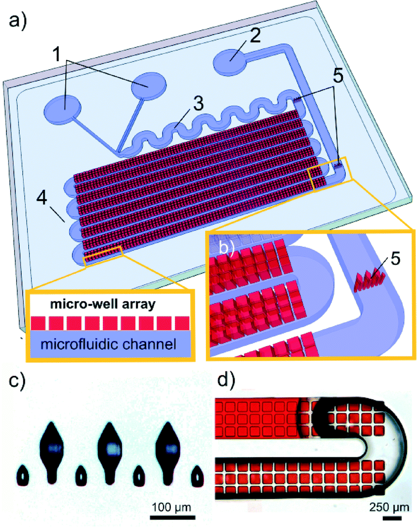 | ||
| Fig. 1 Images and schematics of McCLECs. a) General view: (1) inlet ports; (2) outlet port; (3) passive zigzag mixer; (4) serpentine channel for droplet storage; (5) PDMS filters. Inset on the left: cross section depicting the layout of the solution trapping system (microwell array) over the microfluidic channel. b) Detail of the microwell array and the PDMS filter. c) Image of the PDMS filter. d) Operation of the solution trapping system during the injection of a red dye solution. The meniscus of the flowing solution is observed when emptying the microfluidic device, while the solution trapping system retains nanolitre-sized droplets in the microwell array. | ||
Protein preparation and production
Lysozyme was dissolved and dialyzed in 50 mM sodium acetate, pH 4.5. B. cereus formamidase (FASE) was expressed in E. coli BL21 (DE3) as a fusion protein with a C-terminal hexahistidine tag and purified from bacterial lysates to homogeneity in two steps: i) affinity chromatography (15 ml bed volume, GE Healthcare) in 50 mM Na2HPO4 and 300 mM NaCl buffer and ii) size-exclusion chromatography (Superdex 200 column; GE Healthcare) in 20 mM Tris-HCl, pH 8.0. The protein was then concentrated using a Centricon centrifugation system with a 10 kDa molecular mass cut-off membrane. Based on SDS-PAGE experiments, the purity of the recombinant protein was estimated to be greater than 95%.Crystallization experiments
McCLECs were tested for crystallization with lysozyme as a model protein and FASE as an enzyme with potential biotechnological applications.An initial grid screening for lysozyme crystallization was performed by varying the concentration of the protein from 10 to 50 mg ml−1 and that of precipitant NaCl from 1% to 10% in hanging drop experiments at 293 K. Although all the conditions of the grid produced crystals, the most suitable crystals in terms of size and number were obtained at a protein concentration of 20 mg ml−1 and 5% NaCl as precipitant. Thus, this crystallization condition was adapted for lysozyme crystallization in McCLECs by batch method, obtaining tetragonal lysozyme crystals at a supersaturation (C/CEquilibrium) of 4.2.
Initial crystallization conditions for FASE were obtained by screening in capillaries of 0.3 mm inner diameter using the counterdiffusion technique in pre-filled GCBs.45 In this case, the precipitant was a mixture of PEGs (20% PEG 400, 15% PEG 4000, 10% PEG 8000) in the pH range of 4.0 to 9.0 (KIT PEG448-49, Triana S&T) and the protein was used at a concentration of 28 mg ml−1. Conditions were then adapted to the batch method in two steps (Table S1 and Fig. S2†). Firstly, the microbatch under oil method was used to reduce the protein and precipitant concentration to a desirable nucleation density and crystal size values by varying the protein concentration between 15 to 3.75 mg ml−1 and diluting the precipitant cocktail from 50% to 12.5% with 100 mM sodium acetate, pH 4.0 (Table S1†). Secondly, similar conditions were used in Eppendorf-PCR tubes to produce FASE crystals that could be further cross-linked and recovered (Fig. S2†). The experiments were set in volumes ranging from 10 to 500 μL to study the influence on the final nucleation density and crystal size.
Crystallization experiments in McCLECs were performed by batch technique in different steps. Initially, the McCLEC devices were incubated with a solution containing 1% Triton X-100 surfactant (108643 Merck Millipore) in order to modify the highly hydrophobic PDMS surface, allowing the correct filling of the microwell array. The crystallizing batch solution (enzyme and precipitant) was injected to fill each McCLEC and subsequently a soft vacuum was applied through the outlet port, emptying the main microfluidic channel. The difference in surface energy of both the modified PDMS walls and the injected solutions makes it possible to extract the excess solution while keeping the microwell array filled (Fig. 2, step 1). McCLECs were stored in Petri dishes with water reservoirs to avoid evaporation from the microwell array.
 | ||
| Fig. 2 Schematic representation of the three major steps carried out to prepare CLECs in McCLECs. Step 1 shows the crystallization process inside the device and examples of lysozyme crystals are depicted. Step 2 displays the cross-linking process showing the cross-linked crystals. Finally, steps 3 and 4 respectively show the operation of the chip as an enzymatic reactor and the final spectrophotometric detection for the product of the reaction. | ||
A total of 15–30 μL of crystallizing solution was set up for each protein; 20 mg ml−1 protein and 5% NaCl as precipitant were used in the case of HEWL, whereas 5 mg ml−1 protein and a polyethylene glycol mixture (10% PEG 400, 7.5% PEG 4000, 5% PEG 8000) in 100 mM sodium acetate (pH 4.0) was used to trigger FASE crystallization. All the experiments were performed at 293 K and observed with an optical microscope. Pictures were acquired with a ProgRes® CapturePro 2.8 detector (JENOPTIK optical systems, GmbH).
Cross-linked enzyme crystal production
Crystals of both enzymes were cross-linked by incubating McCLECs for 1 hour at room temperature (RT) with the corresponding crystallizing solution (see above) supplemented with 1% (v/v) glutaraldehyde. Incubation time and cross-linker concentration were both previously optimized using SDS-PAGE (see ESI† for details and Fig. S3). An example of cross-linked lysozyme crystals is shown in Fig. 2, step 2. Subsequently, cross-linked crystals were carefully washed by injecting at least three volumes of water into the McCLECs. Finally the McCLECs containing cross-linked lysozyme and FASE crystals (McCLLCs and McCLFCs, respectively) were dried and stored at RT for several months.Enzyme assays
Spectrophotometric determinations were carried out in a Cary 300 UV-VIS spectrophotometer (Agilent Technologies, Santa Clara, CA) with blank corrections for all measurements. The enzymatic assay for lysozyme was adapted from Osawa T experiments46,47 to be assayed colorimetrically by using nitrophenyl-β-D-N,N′,N′′-triacetylchitotriose as substrate and the p-nitrophenol liberated as a product of the reaction was determined at a wavelength of 400 nm. Enzymatic production of ammonia by FASE was spectrophotometrically determined using a commercially available ammonia determination kit following manufacturer instructions (ammonia kit, R-Biopharm, 11112732035).CLEC activities grown inside McCLECs were determined by reaction with 4-nitrophenyl-β-D-N,N′,N′′-triacetylchitotriose (1 mg ml−1) dissolved in 1 mM sodium phosphate, pH 9.0 (McCLLCs), and 6 mM formamide in 100 mM citrate/citric acid, pH 6.0 (McCLFCs) (Fig. 2, step 3). Substrate-loaded McCLECs were incubated for 2 hours at 323 K in Petri dishes containing a water reservoir to avoid solution evaporation from the devices. The reaction solutions were then recovered from the devices using a micropipette, and product concentrations were determined spectrophotometrically as described above (Fig. 2, step 4).
In order to compare the activity of CLECs produced both inside and outside McCLECs, crystals for the two proteins were produced by the batch methods, cross-linked by adding 1% glutaraldehyde directly into their mother liquor, washed, lyophilized, weighed on a precision balance and added to the corresponding reaction solutions (see the ESI† for details and Fig. S4). For the reuse evaluation experiments, CLECs were extensively washed with water after each run, dried and stored at RT till the next use.
Results and discussion
The McCLECs presented here were specifically designed to allow preferential crystallization into a microwell array while allowing the mixture and flow of a solution along a microfluidic channel that connects the array. This makes possible the subsequent enzymatic reaction and the recovery of the product if required. Although the experiments presented here were carried out by batch method, a continuous solution injection would transform McCLEC batch reactors into continuous flow reactors for the production of a desired product. Additionally, it is worth mentioning that coupling an appropriate detection system to the McCLECs would lead to the creation of a biosensor with the possibility of operating in continuous mode. Although related optofluidic enzymatic biosensors have been previously reported,24 the combination of McCLECs with the multiple path photonic lab on a chip concept48 for the creation of a continuous photonic sensing platform is yet to be reported and represents a substantial advance, since it provides unmatchable advantages in terms of robustness, sensitivity and stability. The validation and proof of feasibility for these biotechnological reactor/sensor approaches are presented below.McCLEC validation
In order to prove the McCLEC concept, lysozyme was selected since it is a highly characterized enzyme from both the crystallization and enzymatic points of view.49–52 Tetragonal lysozyme crystals were grown in McCLEC microwells using the batch method. Crystals appeared within 8–12 h at 293 K (Fig. 3A), and after equilibration (2–4 days) they were cross-linked (Fig. 3B), washed and dried. The devices were stored at RT for more than one year without noticeable loss of activity (see below). | ||
| Fig. 3 Lysozyme crystallization, cross-linking and CLLC reutilization in microfluidic chips. A) Lysozyme crystals grown in 8–12 h at 293 K inside McCLEC microwells. Crystallization experiments were set up by batch method with a supersaturated protein solution at 20 mg ml−1 and 5% NaCl as precipitant. B) CLLCs produced by incubating for 2 hours with 1% glutaraldehyde at RT. D) Results of enzymatic assays with 9 different McCLLCs. Each coloured bar represents the average of the total number of reuses of independent McCLLCs with its standard deviation. Chips 1–6 had been stored for 8 months and dried at room temperature until use for enzymatic assays, and devices 7–9 one year after the initial proofs. | ||
Since the microchip was not conceived to be able to measure the total amount of protein crystallized, several McCLLCs (cross-linked lysozyme crystals in McCLEC) were prepared (chips 1 to 3) and employed to study the three main variables used to carry out the enzymatic reaction, i.e. substrate concentration, temperature and reaction time. Two substrate concentrations (0.5 mg ml−1 and 1 mg ml−1), three temperatures (293 K, 315 K and 325 K) and three reaction times (2 h, 3.5 h and 12 h) were tested and the extension of the reaction followed by the production of p-nitrophenol. Optimal operational conditions (>95% substrate conversion within 2 h) were achieved at 325 K using 1 mg ml−1 substrate.
The robustness of the McCLLCs was evaluated by reutilization of different chips (once a day) by means of two approaches: i) after shelf storage at RT for 1 year (chips 1–3) and ii) with freshly prepared devices (chips 4–9) (Fig. 3C). Both shelf-stored and freshly prepared CLLC devices were fully operative with no detectable loss of activity after 20 reuses (see also Fig. S5A of the ESI†). Observed deviations may be attributed to either the preparation of the substrate or the recovery and determination of the product concentration. Both experiments validate the McCLECs as a cost-effective preparation of a novel type of auto-supported enzymatic system.
McCLECs application
To address the applicability of the microfluidic devices in a potential biotechnological application, formamidase (FASE) was chosen for setting up McCLFCs (cross-linked FASE crystals in McCLEC). FASE is a member of the nitrilase superfamily, a group of enzymes that play a key role in chemical and pharmaceutical engineering and bioremediation.53 Despite the natural function of FASE in the hydrolysis of formamide, this enzyme also shows acyl-transferase activity, allowing the production of acetohydroxamic acid (Lithostat®),54 a potent inhibitor of urease, indicated in patients with chronic urea-splitting urinary infection.FASE crystallization conditions used in McCLFCs were determined from batch experiments at 293 K (5 mg ml−1 protein, 10% PEG 400, 7.5% PEG 4000, 5% PEG 8000, and 100 mM sodium acetate, pH 4.0, as precipitant). Under these conditions, FASE crystals were obtained in 24 h. In this case, the number and size of FASE crystals were more disperse than in the case of tetragonal lysozyme (Fig. 4A–D).
 | ||
| Fig. 4 FASE crystallization, cross-linking and enzymatic assays in microfluidic devices. A) FASE crystals grown in 24 h at 293 K by batch method in McCLECs with a supersaturated protein solution of 5 mg ml−1 and a polyethylene glycol mixture (20% PEG 400, 15% PEG 4000, 10% PEG 8000) in 100 mM sodium acetate (pH 4.0) as crystallizing agent. B) McCLFCs produced by incubation of the crystals for 1 h at room temperature with 1% glutaraldehyde. C) and D) Photographs of CLFCs after 20 enzymatic reuses. E) Results of enzymatic assays with 6 different McCLFCs. Each coloured bar represents an enzymatic reuse of the McCLFCs in each device. Devices 1–3 had been stored for 8 months and dried at room temperature until use for enzymatic assays and devices, 4 and 5–7, 3 months after the initial proofs. | ||
Three different sets of McCLFCs (devices 1–3, 4 and 5–7) were independently prepared to study their reuse capability. Considering an effective trapped solution volume of 4 μL and a protein concentration of 5 mg ml−1, the maximum amount of protein that could be precipitated in each McCLFC was ~20 μg. Devices 1–3 (first set) were initially used to optimize the operational conditions while keeping the substrate concentration constant at 6 mM formamide. The best reproducible results were obtained with 2 h of substrate incubation at 313 K. Following a protocol similar to that with CLLC, devices 1–3 were thoroughly washed and stored at RT for 8 months before the reuse experiments while the other two sets were freshly prepared. From the three devices used to prepare the second set only in one of the devices did the obtained crystals remain stable after the cross-linking. Therefore a third set of McCLFC was prepared from a new purification preparation. The reuse activity of the three sets was determined during 17 cycles (1–3), 23 cycles (4) and 21 cycles (5–7). No difference in the morphology of the crystals was observed after the reuse (Fig. 4C and D). As can be observed in Fig. 4E, device 4 showed a lower activity level than the other two sets. These differences, which should not be surprising when working with different protein preparations, arise as the main cause of the inter-reproducibility of the measured activity (see also Fig. S5B of the ESI†). Further visual inspection of the McCLFCs indicates, as expected, that the number and shape of FASE crystals in each set were different and therefore could be directly related to the catalyst effective concentration translated as a different level of activity. On the other hand, their stability after several reaction cycles and storage does not seem to be affected.
Conclusions
In conclusion, a microfluidic system (McCLEC) is proposed as a simple and unique platform for in situ protein crystallization, cross-linked enzyme crystal production and enzymatic catalytic bioreaction performance. The McCLEC concept has been validated with two different proteins: a commercial protein, lysozyme, which is easy to crystallize and well characterized, and a protein of pharmaceutical interest, a formamidase, produced and purified for this study. CLECs of both proteins were obtained inside McCLECs and their application as a biocatalyst was studied. Reuse of CLECs has been shown possible, thus decreasing the economic costs of the enzymatic reaction, presenting McCLECs as a robust and cost-effective technology. Furthermore, the McCLECs have shown excellent long-term shelf life and storage capabilities at room temperature (up to one year), not only freshly stored but also stored after several reaction cycles, without a decrease in the enzymatic activity. Therefore, McCLECs offer a stable, robust and durable platform for enzymatic catalysis, which can be used not only for enzyme characterization and high-value product synthesis but also as a sensing platform when coupled to a detection system.Acknowledgements
This work has been partly funded by the MICINN (Spain) projects BIO2010-16800 (JAG) and BIO2012-34937 (SMR), the European Commission (Contract No. 317916) under the LiPhos project (AL and IRR), “Factoría Española de Cristalización” Consolider-Ingenio 2010 (JAG and MCM) and EDRF Funds (JAG and AL). MCM thanks the Consejo Superior de Investigaciones Científicas (CSIC, Spain) for a JAE predoc fellowship.References
- J. L. Adrio and A. L. Demain, Biomolecules, 2014, 4, 117–139 CrossRef PubMed.
- Z. S. Olempska-Beer, R. I. Merker, M. D. Ditto and M. J. DiNovi, Regul. Toxicol. Pharmacol., 2006, 45, 144–158 CrossRef CAS PubMed.
- M. Vellard, Curr. Opin. Biotechnol., 2003, 14, 444–450 CrossRef CAS.
- A. Zaks and D. R. Dodds, Drug Discovery Today, 1997, 2, 513–531 CrossRef CAS.
- O. Kirk, T. V. Borchert and C. C. Fuglsang, Curr. Opin. Biotechnol., 2002, 13, 345–351 CrossRef CAS.
- M. Gerard, A. Chaubey and B. D. Malhotra, Biosens. Bioelectron., 2002, 17, 345–359 CrossRef CAS.
- T. Vo-Dinh and B. Cullum, Fresenius' J. Anal. Chem., 2000, 366, 540–551 CrossRef CAS.
- A. Huber, S. Demartis and D. Neri, J. Mol. Recognit., 1999, 12, 198–216 CrossRef CAS.
- S. Datta, L. R. Christena and Y. R. S. Rajaram, 3 Biotech., 2013, 3, 1–9 CrossRef.
- J. Jegan Roy and T. Emilia Abraham, Chem. Rev., 2004, 104, 3705–3722 CrossRef CAS PubMed.
- C. Mateo, J. M. Palomo, G. Fernandez-Lorente, J. M. Guisan and R. Fernandez-Lafuente, Enzyme Microb. Technol., 2007, 40, 1451–1463 CrossRef CAS PubMed.
- F. A. Quiocho and F. M. Richards, Proc. Natl. Acad. Sci. U. S. A., 1964, 52, 833–839 CrossRef CAS.
- T. E. Abraham, J. R. Joseph, L. B. V. Bindhu and K. K. Jayakumar, Carbohydr. Res., 2004, 339, 1099–1104 CrossRef CAS PubMed.
- N. L. St. Clair and M. A. Navia, J. Am. Chem. Soc., 1992, 114, 7314–7316 CrossRef CAS.
- A. Manz, D. J. Harrison, E. M. J. Verpoorte, J. C. Fettinger, A. Paulus, H. Lüdi and H. M. Widmer, J. Chromatogr. A, 1992, 593, 253–258 CrossRef CAS.
- W. Laiwattanapaisal, J. Yakovleva, M. Bengtsson, T. Laurell, S. Wiyakrutta, V. Meevootisom, O. Chailapakul and J. Emneus, Biomicrofluidics, 2009, 3, 14104 CrossRef CAS PubMed.
- J. M. K. Ng, I. Gitlin, A. D. Stroock and G. M. Whitesides, Electrophoresis, 2002, 23, 3461–3473 CrossRef CAS.
- H. Becker and L. E. Locascio, Talanta, 2002, 56, 267–287 CrossRef CAS.
- H. N. Joensson and H. Andersson Svahn, Angew. Chem., Int. Ed., 2012, 51, 12176–12192 CrossRef CAS PubMed.
- C. L. Hansen, E. Skordalakes, J. M. Berger and S. R. Quake, Proc. Natl. Acad. Sci. U. S. A., 2002, 99, 16531–16536 CrossRef CAS PubMed.
- F. Pinker, M. Brun, P. Morin, A.-L. Deman, J.-F. Chateaux, V. Oliéric, C. Stirnimann, B. Lorber, N. Terrier, R. Ferrigno and C. Sauter, Cryst. Growth Des., 2013, 13, 3333–3340 CAS.
- C. Hansen and S. R. Quake, Curr. Opin. Struct. Biol., 2003, 13, 538–544 CrossRef CAS PubMed.
- M. van der Woerd, D. Ferree and M. Pusey, J. Struct. Biol., 2003, 142, 180–187 CrossRef CAS.
- I. Rodríguez-Ruiz, A. Llobera, J. Vila-Planas, D. W. Johnson, J. Gómez-Morales and J. M. García-Ruiz, Anal. Chem., 2013, 85, 9678–9685 CrossRef PubMed.
- A. C. Ng, U. Uddayasankar and A. Wheeler, Anal. Bioanal. Chem., 2010, 397, 991–1007 CrossRef CAS PubMed.
- T. G. Henares, F. Mizutani and H. Hisamoto, Anal. Chim. Acta, 2008, 611, 17–30 CrossRef CAS PubMed.
- J. Křenková and F. Foret, Electrophoresis, 2004, 25, 3550–3563 CrossRef PubMed.
- C. Hoera, S. Ohla, Z. Shu, E. Beckert, S. Nagl and D. Belder, Anal. Bioanal. Chem., 2015, 407, 387–396 CrossRef CAS PubMed.
- C. D. Chin, V. Linder and S. K. Sia, Lab Chip, 2007, 7, 41–57 RSC.
- D. Figeys and D. Pinto, Anal. Chem., 2000, 72, 330 A–335 A CrossRef CAS.
- G. M. Whitesides, Nature, 2006, 442, 368–373 CrossRef CAS PubMed.
- A. Tripathi, J. Riddell and N. Chronis, Sens. Actuators, B, 2013, 186, 244–251 CrossRef CAS PubMed.
- J. Wang, Talanta, 2002, 56, 223–231 CrossRef CAS.
- R. Ehrnstrom, Lab Chip, 2002, 2, 26N–30N RSC.
- I. Inoue, Y. Wakamoto, H. Moriguchi, K. Okano and K. Yasuda, Lab Chip, 2001, 1, 50–55 RSC.
- D. Figeys and D. Pinto, Electrophoresis, 2001, 22, 208–216 CrossRef CAS.
- L. Bousse, S. Mouradian, A. Minalla, H. Yee, K. Williams and R. Dubrow, Anal. Chem., 2001, 73, 1207–1212 CrossRef CAS.
- N. Christodoulides, M. Tran, P. N. Floriano, M. Rodriguez, A. Goodey, M. Ali, D. Neikirk and J. T. McDevitt, Anal. Chem., 2002, 74, 3030–3036 CrossRef CAS.
- A. Bernard, B. Michel and E. Delamarche, Anal. Chem., 2001, 73, 8–12 CrossRef CAS.
- P. Arenkov, A. Kukhtin, A. Gemmell, S. Voloshchuk, V. Chupeeva and A. Mirzabekov, Anal. Biochem., 2000, 278, 123–131 CrossRef CAS PubMed.
- R. Wohlgemuth, I. Plazl, P. Žnidaršič-Plazl, K. V. Gernaey and J. M. Woodley, Trends Biotechnol., 2015, 33, 302–314 CrossRef CAS PubMed.
- J. Vila-Planas, E. Fernandez-Rosas, B. Ibarlucea, S. Demming, C. Nogues, J. A. Plaza, C. Dominguez, S. Buttgenbach and A. Llobera, Nat. Protoc., 2011, 6, 1642–1655 CrossRef CAS PubMed.
- I. Rodríguez-Ruiz, E. Masvidal-Codina, T. Ackermann and A. Llobera, Microfluid. Nanofluid., 2014, 1–10, DOI:10.1007/s10404-014-1526-4.
- H. Song, J. D. Tice and R. F. Ismagilov, Angew. Chem., Int. Ed., 2003, 42, 768–772 CrossRef CAS PubMed.
- F. Otalora, J. Antonio Gavira, J. D. Ng and J. Manuel Garcia-Ruiz, Prog. Biophys. Mol. Biol., 2009, 101, 26–37 CrossRef CAS PubMed.
- T. Osawa and Y. Nakazawa, Biochim. Biophys. Acta, Gen. Subj., 1966, 130, 56–63 CrossRef CAS.
- T. Osawa, Carbohydr. Res., 1968, 7, 217–220 CrossRef CAS.
- I. Rodriguez-Ruiz, M. Conejero-Muriel, T. N. Ackermann, J. A. Gavira and A. Llobera, Lab Chip, 2015, 15, 1133–1139 RSC.
- J. V. Holtje, Experientia, Suppl., 1996, 75, 105–110 CAS.
- P. Jollès, Angew. Chem., Int. Ed. Engl., 1969, 8, 227–239 CrossRef PubMed.
- J. A. Rupley, L. Butler, M. Gerring, F. J. Hartdegen and R. Pecoraro, Proc. Natl. Acad. Sci. U. S. A., 1967, 57, 1088–1095 CrossRef CAS.
- M. D. Scanlon, E. Jennings and D. W. M. Arrigan, Phys. Chem. Chem. Phys., 2009, 11, 2272–2280 RSC.
- A. Banerjee, R. Sharma and U. Banerjee, Appl. Microbiol. Biotechnol., 2002, 60, 33–44 CrossRef CAS PubMed.
- P. Soriano-Maldonado, A. I. Martinez-Gomez, M. Andujar-Sanchez, J. L. Neira, J. M. Clemente-Jimenez, F. J. Las Heras-Vazquez, F. Rodriguez-Vico and S. Martinez-Rodriguez, Appl. Environ. Microbiol., 2011, 77, 5761–5769 CrossRef CAS PubMed.
Footnote |
| † Electronic supplementary information (ESI) available. See DOI: 10.1039/c5lc00776c |
| This journal is © The Royal Society of Chemistry 2015 |