
Neutron computed laminography on ancient metal artefacts
Filomena
Salvemini†
*a,
Francesco
Grazzi
a,
Nikolay
Kardjilov
b,
Ingo
Manke
b,
Francesco
Civita
c and
Marco
Zoppi
a
aConsiglio Nazionale delle Ricerche, Istituto dei Sistemi Complessi, Sesto Fiorentino (FI), Italy. E-mail: filomena.salvemini@ansto.gov.au
bHelmholtz Zentrum Berlin, Hahn-Meitner Pl. 1, 14109 Berlin, Germany
cMuseo Stibbert, Firenze (FI), Italy
First published on 5th November 2014
Abstract
Computed laminography (CL) has been developed as a method to complement computed tomography for three-dimensional imaging of laterally extended specimens. Originally applied for medical purposes, more recently CL has been used as a non-destructive method, not only in the materials science research, but also with rising interest in artwork investigations. Here, we report the implementation of computed laminography with polychromatic and monochromatic neutron radiation carried out on five historical and archaeological metal artefacts pertaining to different periods and technological contexts. The measurements were performed at the CONRAD II neutron imaging facility at the BERII research reactor of Helmholtz-Zentrum Berlin (HZB).
Introduction
Neutron computed tomography (CT) has been demonstrated to be a powerful method for non-destructive investigation of metal artefacts.1–7 However, laterally extended specimens (e.g. plate-like objects) are much less suitable for CT. The transmitted radiation, collected from angular projections in which the plate is oriented parallel, or close, to the incoming beam, can be almost completely attenuated since the beam intercepts a thick material layer with a significant cross-section. In this case, the CT reconstruction can produce serious streaking artefacts, affecting data interpretation.8In order to overcome this drawback, Computed Laminography (CL) was developed as a method to complement computed tomography for three-dimensional imaging of laterally extended specimens. In the typical laminography set-up, the sample rotation axis is tilted with respect to the standard tomographic rotation axis that is oriented perpendicular to the beam. The extended sample plane is then arranged approximately perpendicular to the rotation axis as shown in Fig. 1. The advantage of this scanning scheme is that the integral transmission stays almost constant for the full set of angular projections. As a consequence, the integrity of laterally extended samples can be preserved.9,10
 | ||
| Fig. 1 Scheme of the experimental set-up used for neutron laminography. | ||
Early use of laminography with X-rays was in the field of medical applications.11 Recently neutron computed laminography has been applied also to materials science research12 and the interest in the application of the technique to art or archaeological objects rose since such samples require investigation through non-invasive methods.13,14
However, this technique can be further improved, to achieve material discrimination, by a proper selection of the neutron energy. In fact, monochromatic neutron beams give the possibility of modifying the image contrast for different phases taking advantage of the abrupt change of the attenuation coefficients in the proximity of the so-called Bragg edges.15–17
Considering the potential of laminography for implementing the status of knowledge about ancient technology, the purpose of this study was to examine expanded applications of these non-invasive methods. These studies include the use of different neutron laminographic methods which would allow for reconstruction and imaging of ancient metal artefacts. Neutron white beam laminography (WBL), and for the first time, energy-selective laminography (ESL) were performed on the CONRAD II beam line at HZB.
Experimental set-up
The neutron imaging study was performed using a CONRAD II instrument at the Helmholtz-Zentrum Berlin (HZB). CONRAD II is a multifunctional facility for radiography and tomography with cold neutrons. It is located at the end of a curved neutron guide, which faces the cold-neutron source of the BER-II research reactor.18 The facility provides a cold-neutron beam with wavelengths ranging from 2 Å to 12 Å. The available flux at the sample position is approximately 2 × 10⁷ n cm−2 s−1 at an L/D of 300.19 The cold neutron beam of the instrument allows for enhancing the contrast between different phases of the material and increases the thickness sensitivity, thanks to the increased cross-sections for some elements in comparison to thermal neutrons.Samples and experimental conditions
For the present study a set of ancient artefacts, suitable for the designated aims, were collected as test samples and provided by private collectors. They were two hand-guards of Japanese swords (tsuba), two hilt collars of Japanese sword fittings (fuchi), and an archaeological bronze Celtic spiral (Fig. 2). | ||
| Fig. 2 (a) The iron tsuba, (b) the copper tsuba, (c) the Celtic spiral, (d) the fuchi 01 and (e) the fuchi 02 are reported. | ||
The first tsuba (Fig. 2a) is a slightly elliptical iron one with a rounded rim. The tsuba is carved with a tendril design (sukashi technique), including facing dragons, a jewel, and stylized flowers. The rim is rounded and gilded and there are traces of gilding in other areas. It is not signed but can be dated to the first half of the XVIIIth century, probably from the Nagasaki (Hizen) area, in namban style. The Japanese word namban, translated as “southern barbarian”, was used by Japanese not only to indicate any foreigner who entered their country, but also to define a large number of artworks in which the decorations reflected a foreign influence in their appearance. The diameter of 8 cm leads us to presume that this hand-guard was created for a long sword (katana). It was equipped with a nakago hitsu, the central hole for fitting the tang of the blade, and a kozuka hitsu, the left side hole for accommodating the handle of a small knife called kozuka.
The second Japanese hand-guard was made of copper and copper alloys (Fig. 2b) and is of sanmai type: this is composed of three layers with the central one made of a different alloy with respect to the others. The upper and lower layers are decorated through hammering. Decoration was typically made by acting on the back of the layer and then fastening it to the central layer with adhesive or lacquer. A fukurin, i.e. soft metal, type rim was hammered around to hold the three layers together. The rim profile is rounded and not decorated. The decoration theme is made of waves and imaginary animals (dragons). The piece can be dated either to the end of the Momoyama period or to the beginning of the Edo period. It was probably made in the Kyoto area around the beginning of the XVIIth century. A relief decoration is visible on the surface even though it is in poor conditions of conservation. Detachments are recognizable at the edge and on the decorated surface. As in the previous sample, there is a nakago hitsu and a kozuka hitsu.
The other samples are a bronze Celtic spiral, showing the typical alteration products of copper on its whole surface (Fig. 2c), together with two fuchi (Fig. 2d and e) made of copper and copper alloys.
Bronze was extensively employed by the Celts to make a wide variety of objects and this metal alloy was especially common for jewellery. Brooches, buckles, belt ends, dress pins and rings could be obtained by simply cold-working the metal blank or applying complex inlay techniques.20 In the case of our sample, the spiral, one of the most used patterns by Celts, was probably made out of a bronze wire that was bent to form a spiral into a looped circular shape. The sample was found in England and dated to the 1st millennium BC.
Regarding the fuchi, it has the mechanical function to strengthen the hilt of the blade at the conjunction with the tsuba. The ordinary fuchi consists of a flat band of metal up to half an inch wide that encircles the hilt immediately below the guard. As any component in a Japanese sword mount, it is easily removable. Its base is filled with an oval plaque, usually copper, pierced with a hole to insert the tang. The two fuchi analysed in this work are both manufactured during the Edo period.
In order to place the samples in the beam, an aluminium threaded rod was gently passed through the main hole (nakago hitsu) of the two tsuba and fastened with an iron bolt. Small Al plates were placed between the sample surface and the bolt in order to avoid direct contact that could affect the data processing and damage the objects. Concerning the Celtic spiral, due to its bad conservation conditions and to the shape, this sample was wrapped in aluminium foil and fixed on an Al plate mounted on the top of the rod (Fig. 3a). The very low macroscopic cross-section of aluminium makes this metal almost transparent to neutrons and avoids interference during the measurements. The rod, with the samples fixed along it, was connected to the rotating table and placed on a goniometer. The rotation axis of the samples was tilted 20 degrees towards the detector as shown in Fig. 3b and angular projections were measured at stepwise rotation over 360 degrees. The measured projections were reconstructed by the laminographic method using the software package Octopus21 and visualized using VGStudio Max.22
 | ||
| Fig. 3 The instrumental set-up adopted for performing neutron laminography. (a) The sample fixation and (b) the experimental arrangement (the two tusba and the Celtic spiral prepared for analysis are shown as reference). A tilt angle of 20 degrees toward the detector was used. | ||
The experimental conditions for the performed different experiments are presented in Table 1. White beam laminography was performed only on the two tsuba and the Celtic spiral, instead, the two fuchi were chosen for the energy-selective laminography on the base of following considerations. As evidenced by previous neutron diffraction analysis, these samples have a composite copper and copper-alloy structure. Moreover, a non-homogeneous distribution of the neutron wavelengths inside the acquisition window could be experienced when the neutron beam is monochromatized. This implies a gradient of wavelength which distribution and extension depend on the type and arrangement of the selector device (details will be discussed elsewhere). Considering the high wavelength resolution required to detect the different metal alloys of interest (they have a similar composition and a very close phase lattice parameter), only the two fuchi, whose size is below the range of the wavelength shift in the acquisition field, were analyzed.
| W-B laminography | Radiography E scan | E-S laminography | |
|---|---|---|---|
| Wavelength | Polychromatic | From 2.0 Å to 4.5 Å with a step of 0.02 Å | 4.18 Å/4.21 Å/4.30 Å |
| Camera | Andor DW436N-BV CCD3325 2048 × 2048 16 bit | ||
| Scintillator | LiF 50 μm | LiF 400 μm | LiF 400 μm |
| L/D | 500 | 333 | 333 |
| FOV | 110 × 110 mm | 200 × 200 mm | 111.5 × 111.5 mm |
| Sample–detector distance | 90 mm max. | 20 mm | 90 mm max. |
| Pixel size | 53.8 μm | 106 μm | 54.45 μm |
| Exposure time | 60 s | 150 s | 130 s |
| Projection # | 600 | — | 300 |
| Tomo angle | 0–360° | — | 0–360° |
| Tilt angle | 20° | — | 20° |
| Flat field # | 20 | 1 × wavelength | 10 × wavelength |
| Dark field # | 20 | 10 | 10 |
Results and discussion
White beam laminography
The two tsuba and the Celtic spiral were initially characterized using white beam laminography whose instrumental set-up is reported in the first column of Table 1. The white beam imaging was performed with a polychromatic spectrum of cold neutrons. In this case, the interpretation of the image contrast is obtained by integrating the attenuation coefficients over the whole energy range. As a consequence, the energy-dependent features are lost in the averaging process.The white beam laminography revealed a homogeneous body for the iron tsuba, probably filed starting from a single piece of iron (Fig. 4). As a matter of fact, starting from the Muromachi period, the sukashi technique evolved over time. The iron plate prepared as the ground metal was very uniformly forged and relatively soft in order to cut out the fine designs successfully but hard enough to avoid breakage during use. Any void or significant irregularity in the iron body would have made the shaping of continuous decorative lines impossible. Extra effort in the preparation of the plate would have been necessary, as observed in the inner volume of the samples which was free from any cracks, pores and inclusions.23 Evidence of forge-welding was not identified in the design motifs involving plant and animal figures, apart from the gilded ring outlining the profile of the kozuka hitsu. While the inner brighter areas are reconstruction artefacts, the ones detected on the surface were probably due to the application of a patina or to the early formation of products of alteration.24
 | ||
| Fig. 4 On the bottom left side of the figure, a virtual cross-section of the iron tsuba is reported in false colours (brighter areas related to higher attenuation cross-sections). The red dotted lines define the position of the orthogonal views shown alongside (a–d). The red squares map the area of interest inside the views and their respective enlargements. In the cross-section (a), the evidence of soldering is visible in red-orange tone at the edge of the ring. While images (c and d) show the absence of such soldering and the brighter spots can be related to the patination or alteration of the surface. The view (d) gives an idea of the complicate arabesque-structure made by piercing a single metal disc. The top right image refers to a 3D rendering of the sample. | ||
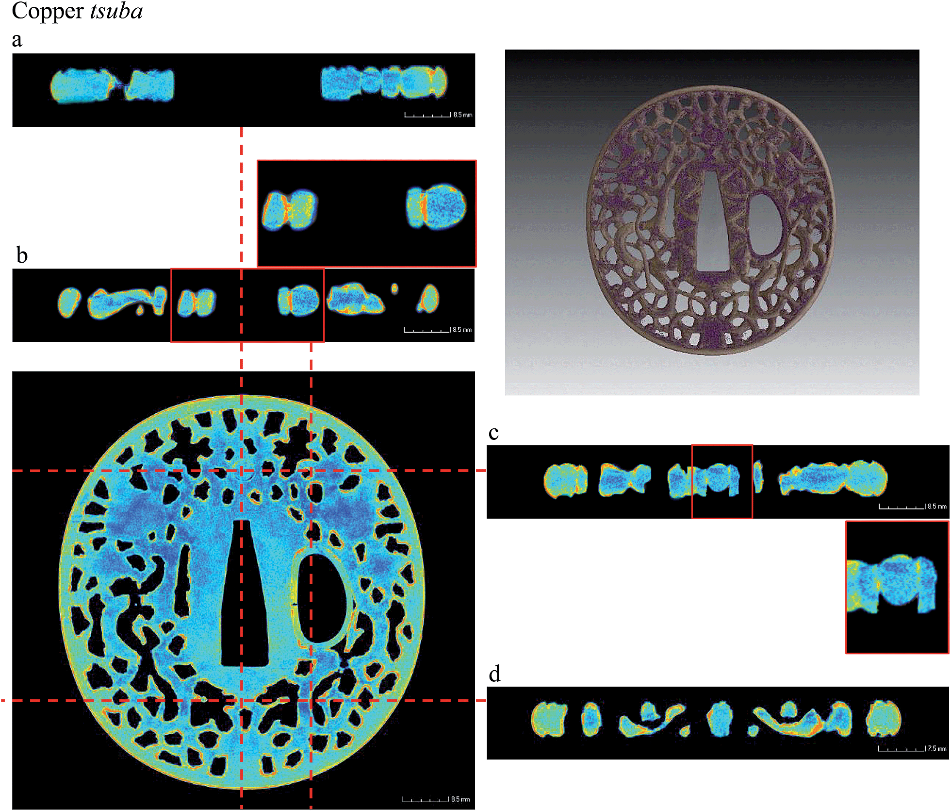
Virtual cross-sections taken at different heights of the copper tsuba revealed three layers of copper and copper alloys (Fig. 5). According to the ancient metal working technique described in the literature,25 the external sheets of metal were probably applied on the two sides of the inner core by heating and slightly hammering, while their rather soft surface was worked with a punch to give texture. All around the rim of the tsuba and the pierced hole, the composite structure was finally tightened by applying a thin ring appearing detached and lost in some areas.
 | ||
| Fig. 5 The three bottom images refer to cross-sections taken at different depths of the copper tsuba, starting from one side of the metal disk and progressively emerging on the other one. The layers closer to the surface (the first and the third radial cross-sections) feature the decorative pattern and the lacking area (frontal cross-sections), enhanced by the false colour visualization. Moreover, the remaining of a ring around the border of the tsuba is visible (red arrow, second image) The red dotted lines map the position for the orthogonal views, reported above, where the decorated layers joined to the bulk sheet and the enclosing ring can be observed. | ||
The laminography allowed reconstruction of the whole inner volume of the Celtic copper spiral, entirely covered by alteration products (Fig. 6). A qualitative elemental characterization of the sample has been performed. Taking into account the neutron wavelength distribution of the probing beam and the image slope value, the total neutron macroscopic cross-sections (absorption and scattering) of Cu and H have been estimated. On the basis of the calculated threshold value between copper and hydrogen, the segmentation of the image histogram allowed us to distinguish the still intact metal, inside the sample, and the alteration, covering the surface and diffusing into the metal. The intact metal is visible inside as well as the dark areas that, according to shape and extension, are probably related to the presence of porosity. The detected structure of the artefact is simple and was obtained by wrapping a wire of copper around itself.
 | ||
| Fig. 6 Here the three orthogonal views acquired by cutting the Celtic spiral along the main directions are shown. The red dotted lines indicate the virtual cutting position. In the false colour visualization, the red-orange color (high attenuation coefficient) refers to the altered area rich in hydroxide-base mineralization, following the profile of the object. In contrast, the blue-green color stands for the still remaining metal inside the volume of the sample. The bottom-right image visualizes in 3D the products of alteration. | ||
Radiographic energy scan
For polycrystalline materials, the neutron attenuation coefficient decreases suddenly for well-defined neutron wavelengths where the conditions for Bragg scattering are no more fulfilled – the so-called Bragg edges. Therefore, the position of the Bragg edges is related to the corresponding dhkl spacing of the analysed phase. Thus, the conventional method can be greatly improved, to achieve phase discrimination in heterogeneous materials, by a proper selection of the neutron energy interval.26,27 In order to study Bragg edges, a polychromatic neutron beam must be monochromatized. A monochromatic neutron beam gives the possibility of modifying the image contrast, for different phases, taking advantage of the abrupt change of the attenuation coefficients at the Bragg edges specific for each phase. This has been accomplished at the CONRAD beam line using a double crystal monochromator device (DCM).28Preliminarily to the ESL, radiographic energy scan was carried out aiming to discriminate the presence of copper and copper alloy materials by exploiting the contrast enhancement obtained using different neutron wavelengths (Table 1, central column). In particular, to fulfil this task, we used neutron wavelengths at values immediately above and below the 111 Bragg-edge for Cu and Cu-alloys,29 according to the lattice parameter defined by previous neutron diffraction analysis (Table 2). We recall that the copper alloys maintain the same lattice structure of pure copper (FCC) while the lattice parameter changes according to the amount and type of solute mixed in it.30
| (a) | |
|---|---|
| hkl | 2d = λ (Å) |
| 111 | 4.18 |
| 200 | 3.62 |
| 220 | 2.56 |
| 311 | 2.18 |
| 222 | 2.09 |
| 400 | 1.81 |
| (b) | ||||
|---|---|---|---|---|
| Cu | Cu-alloy | |||
| a o | Δao | a o | Δao | |
| Tsuba | 3.6158 | 0.0004 | 3.693 | 0.001 |
| Fuchi 01 | 3.61829 | 0.00002 | 3.6279 | 0.0004 |
| Fuchi 02 | 3.61857 | 0.00002 | 3.6251 | 0.0006 |
| (c) | |||
|---|---|---|---|
| a o | d (111) | Bragg-edge (Å) | |
| Cu | 3.617 | 2.088 | 4.176 |
| Cu-alloy | 3.649 | 2.107 | 4.214 |
Energy scans have been performed from 2.0 Å to 4.5 Å with steps of 0.02 Å, crossing the 111 FCC Bragg edge for copper and for copper alloys. Fig. 7 reports the optimum neutron wavelength values above and below the Bragg edges at which the highest intensity variation have been found (i.e. 4.18 Å and 4.22 Å).
 | ||
| Fig. 7 On the left, radiographies of the fuchi taken at different neutron wavelengths (F01 side-oriented and F02 in front-oriented toward the detector). On the right, the graph shows the variation in transmission across the 111 (FFC) Cu Bragg edge. | ||
Energy-selective laminography
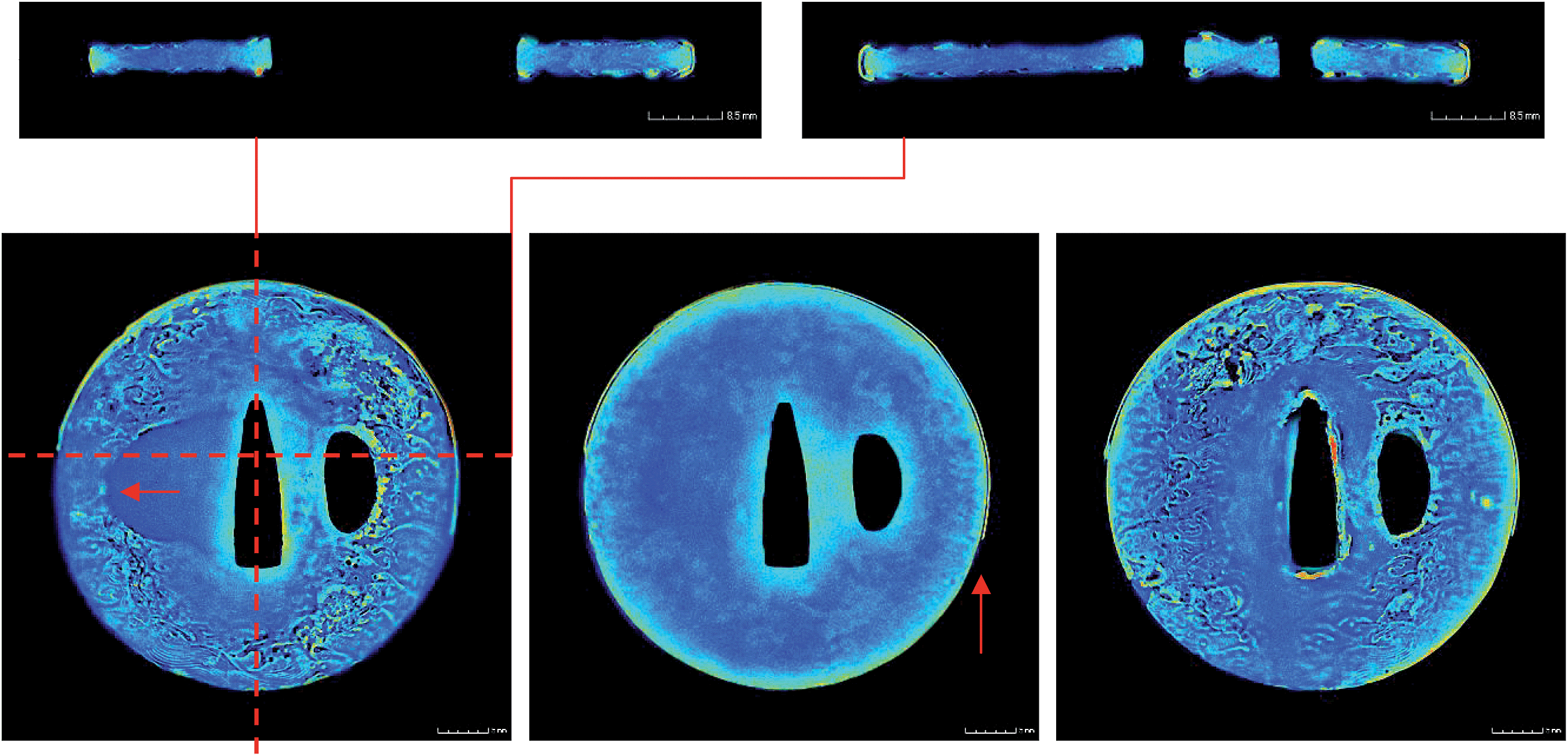
In the part of the experiment dedicated to the energy-selective laminography technique (the instrumental set-up for this configuration is reported in Table 1, third column), the measurement was carried out using three different neutron wavelengths. Thanks to the energy scan and according to the evaluations based on neutron diffraction data, 4.15 Å, 4.21 Å and 4.30 Å were defined as the optimal values for enhancing the image contrast for material discrimination. Similar to the WBL experiment, also in this case, all the samples were fixed to an aluminium threaded rod (Fig. 3b) and laminography measurements were performed by tilting the rotation axis by 20 degrees towards the detector and collecting 300 angular projections over 360 degrees. The reconstructed image stacks by using Octopus software have been further processed by progressively subtracting the latter laminography from the former. In this way, in the resulting image the difference of the attenuation coefficients at the two wavelengths will be presented. The images acquired at 4.15 Å were above the 111 Cu Bragg-edge, while the ones at 4.21 Å and 4.30 Å were below the edge for Cu and Cu-alloy respectively.In order to increase the statistics, each laminographic stack was processed by applying a median filter over 50 slices along the z axis.
According to the neutron diffraction results reported in Table 2c, and the energy scan shown in Fig. 7, the Bragg edges for copper and copper-alloy are approximately located at 4.18 Å and 4.22 Å. Applying the method described in ref. 5, which is omitted from here for the sake of conciseness, the change in the transmission spectrum at the Bragg-edge correspondence has been exploited to recognize the distribution of copper and copper-alloy. The presence of Cu has been evidenced by subtracting the 4.15 (above the Cu 110 Bragg edge) from the 4.21 Å image (below the Cu 110 Bragg edge). Similarly, Cu-alloy-based elements were mapped by subtracting the 4.21 Å (above the Cu-alloy 110 Bragg edge) from the 4.30 Å (below the Cu-alloy 110 Bragg edge) image.
The same procedure was followed for both fuchi. In the case of sample F01, the image resulting from subtracting the stack above and below the copper Bragg edge (Fig. 8, F01, first sequence – image 4.15–4.21 Å) evidenced, in the brighter grey tone, the presence of a flat pale made of Cu. A collar surrounded by decorative elements of a Cu-alloy-based material can be pointed out in the brighter area of the image 4.21–4.30 Å (Fig. 8, F01, second sequence).
 | ||
| Fig. 8 On the left of each sequence, slices taken at different heights of the fuchi F01 and F02 are shown. On the right, we can see the results of the subtraction process at wavelength values above and below the Cu and Cu-alloy Bragg edges, respectively. | ||
Similar results were evidenced for the sample F02, featuring a copper collar outlined by a Cu-alloy material (Fig. 8, F02).
Conclusions
In the present work, we have studied five different historical metal artefacts, chosen as test samples, pertaining to different periods and forging traditions.Neutron white beam and energy-selective laminographies were used for the non-invasive investigation of laterally extended specimens and for showing the validity of the proposed laminographic methods. Moreover, the contrast enhancement induced by the selection of an appropriate neutron wavelength was exploited to increase the image contrast for the different material containing the analyzed specimens.
White beam laminography allowed identification of peculiar characteristics related to the manufacturing methods of these flat objects:
■ the inner structure, resulting from the particular manufacturing process, was analysed all over the samples;
■ the presence of possible forge-welding and soldering was mapped;
■ patination of the surface was established;
■ alterations, originating from the surface and extending to the body of the sample, have been identified for assessing the conditions of conservation.
Concerning the energy-selective laminography, the present measurements demonstrate that the proposed method can allow characterization of the inner structure and composition of metal artefacts, non-destructively, in combination with other neutron inspection techniques.
However, further work is needed to improve the methods. In particular, higher homogeneous distribution in the whole neutron field of view is needed to characterize quantitatively the phase distribution in the case of flat samples made of Cu and Cu alloys.
References
- B. Schillinger, E. Lehmann and P. Vontobel, 3D neutron computed tomography: requirements and applications, Physica B, 2000, 276–278, 59–62 CrossRef CAS.
- B. Schillinger, W. Blümlhuber, A. Fent and M. Wegner, 3D neutron tomography, Nucl. Instrum. Methods Phys. Res., Sect. A, 1999, 424(1), 58–65 CrossRef CAS.
- E. Lehmann, E. Deschler-Erb and A. Ford, Neutron Tomography as a valuable tool for the non-destructive analysis of historical bronze sculptures, Archaeometry, 2010, 52, 272 CrossRef PubMed.
- N. Kardjilov, I. Manke, A. Hilger, M. Strobl and J. Banhart, Neutron imaging in materials science, Mater. Today, 2011, 14(6), 248–2565 CrossRef CAS; F. Salvemini, F. Grazzi, S. Peetermans, F. Civita, R. Franci, S. Hartmann, E. Lehmann and M. Zoppi, Quantitative characterization of Japanese ancient swords through Energy-Resolved Neutron Imaging, J. Anal. At. Spectrom., 2012, 27, 1494 RSC.
- F. Salvemini, F. Grazzi, S. Peetermans, F. Civita, R. Franci, S. Hartmann, E. Lehmann and M. Zoppi, Quantitative characterization of Japanese ancient swords through energy-resolved neutron imaging, J. Anal. At. Spectrom., 2012, 27, 1494 RSC.
- F. Salvemini, F. Grazzi, A. Agostino, R. Iannaccone, F. Civita, S. Hartmann, E. Lehmann and M. Zoppi, Non-invasive characterization through X-ray fluorescence and neutron radiography of an ancient Japanese lacquer, Archaeological and Anthropological Sciences, 2013, DOI:10.1007/s12520-013-0127-6.
- F. Salvemini, F. Grazzi, A. Fedrigo, A. Williams, F. Civita, A. Scherillo, P. Vontobel, S. Hartmann, E. Lehmann and M. Zoppi, Revealing secrets of composite helmets of ancient Japanese tradition, Eur. Phys. J. Plus, 2013, 128, 87, DOI:10.1140/epjp/i2013-13087-y.
- L. Helfen, T. Baumbach, P. Mikulík, D. Kiel, P. Pernot, P. Cloetens and J. Baruchel, Appl. Phys. Lett., 2005, 86, 071915 CrossRef PubMed.
- L. Helfen, T. F. Morgeneyer, F. Xu, M. N. Mavrogordato, I. Sinclair, B. Schillinger and T. Baumbach, Neutron laminography—a novel approach to three-dimensional imaging of flat objects with neutrons, Nucl. Instrum. Methods Phys. Res., Sect. A, 2011, 651(1), 135–139 CrossRef CAS PubMed.
- L. Helfen, T. F. Morgeneyer, F. Xu, M. N. Mavrogordato, I. Sinclair, B. Schillinger and T. Baumbach, Synchrotron and neutron laminography for three-dimensional imaging of devices and flat material specimens, Int. J. Mater. Res., 2012, 170–173 CrossRef CAS.
- D. G. Grant, Tomosynthesis: a three-dimensional radiographic imaging technique, IEEE Trans. Biomed. Eng., 1972, 19, 20 CrossRef CAS.
- L. Helfen, A. Myagotin, A. Rack, P. Pernot, P. Mikulík, M. Di Michiel and T. Baumbach, Synchrotron-radiation computed laminography for high-resolution three-dimensional imaging of flat devices, Phys. Status Solidi A, 2007, 204, 2760 CrossRef CAS.
- K. Krug, L. Porra, P. Coan, G. Tauber, A. Wallert, J. Dik, A. Coerdt, A. Bravin, M. Elyyan, L. Helfen and T. Baumbach, Relics in medieval altarpieces? Combining X-ray tomographic, laminographic and phase-contrast imaging to visualize thin organic objects in paintings, J. Synchrotron Radiat., 2008, 15, 55 CAS.
- H.-Y. Nguyen, Computed Tomography for the Non-destructive Imaging of Cultural Heritage: X-ray, Gamma and Neutron Sources, Thesis Submitted to the Department of Art in Conformity with the Requirements for the Degree of Master of Art Conservation, Queen's University Kingston, Ontario, Canada, 2011 Search PubMed.
- N. Kardjilov, B. Schillinger and E. Steichele, Energy-selective neutron radiography and tomography at FRM, Appl. Radiat. Isot., 2004, 61(4), 455–460 CrossRef CAS PubMed.
- N. Kardjilov, I. Manke, A. Hilger, S. Williams, M. Strobl, R. Woracek, M. Boin, E. Lehmann, D. Penumadu and J. Banhart, Neutron Bragg-edge mapping of weld seams, Int. J. Mater. Res., 2012, 103, 151 CrossRef CAS.
- L. Josic, A. Steuwer and E. Lehmann, Energy selective neutron radiography in material research, Appl. Phys. A, 2010, 99, 515 CrossRef CAS PubMed.
- N. Kardjilov, A. Hilger, I. Manke, M. Strobl, S. Williams, M. Dawson and J. Banhart, Neutron tomography instrument CONRAD at HZB, Nucl. Instrum. Methods Phys. Res., Sect. A, 2011, 651(1), 47–52 CrossRef CAS PubMed.
- N. Kardjilov, M. Dawson, A. Hilger, I. Manke, M. Strobl, D. Penumadu, F. H. Kim, F. Garcia-Moreno and J. Banhart, A highly adaptive detector system for high resolution neutron imaging, Nucl. Instrum. Methods Phys. Res., Sect. A, 2011, 651(1), 95–99 CrossRef CAS PubMed.
- Lloyd Robert Laing, The Archaeology of Celtic Britain and Ireland: C.AD 400 – 1200, Cambridge University Press Jun 29, 2006.
- M. Dierick, B. Masschaele and L. Van Hoorebeke, Meas. Sci. Technol., 2004, 15(7), 1366–1370 CrossRef CAS.
- http://www.volumegraphics.com/en/products/vgstudio-max.html .
- K. Sato, The Japanese Sword: a Comprehensive Guide, Kodansha, 1983, ISBN 0870115626 Search PubMed.
- E. Sugimori, Japanese Patinas, Brynmorgen Press, 2004, ISBN 1-929565-11-9 Search PubMed.
- B. W. Robinson, The Arts of the Japanese Sword, R. MacLehose and Company, 1970 Search PubMed.
- J. R. Santisteban, L. Edwards, A. Steuwer and P. J. Withers, Time-of-flight neutron transmission diffraction, J. Appl. Crystallogr., 2001, 34, 289–297 CrossRef CAS.
- N. Kardjilov, A. Hilger, I. Manke, M. Strobl, M. Dawson, S. Williams and J. Banhart, Neutron tomography instrument CONRAD at HZB, Nucl. Instrum. Methods Phys. Res., Sect. A, 2011, 651, 47–52 CrossRef CAS PubMed.
- W. Treimer, M. Strobl, N. Kardjilov, A. Hilger and I. Manke, A wavelength tunable device for neutron radiography and tomography, Appl. Phys. Lett., 2006, 89(20), 203504 CrossRef PubMed.
- L. Giller, U. Filges, G. Kühne, M. Wohlmuther and L. Zanini, Validation of Monte-Carlo simulations with measurements at the ICON beam-line at SINQ, Nucl. Instrum. Methods Phys. Res., Sect. A, 2008, 586, 59–63 CrossRef CAS PubMed.
- W. B. Pearson and G. H. Vineyard, Reviewer, A Handbook of Lattice Spacings and Structures of Metals and Alloys, Brookhaven National Laboratory, American Institute of Physics, 1958, ISSN 0031-9228 Search PubMed.
Footnote |
| † Current affiliation: Bragg Institute, Australian Nuclear Science and Technology Organisation (ANSTO), New Illawarra Road, Lucas Heights NSW, Australia. |
| This journal is © The Royal Society of Chemistry 2015 |