Finding a suitable separation condition for TLC/FTIR analysis by using multiple-narrow-band TLC technique

Ye Jiangab, Xiaoyan Kangab, Danqing Gaoab, Anqi Hebc, Ran Guob, Xiaokun Fanbd, Yanjun Zhai*a, Jinming Xiae, Yizhuang Xu*bc, Isao Nodabf and Jinguang Wub
aCollege of Pharmacy, Liaoning University of Traditional Chinese Medicine, Dalian 116600, China. E-mail: lnzyzyj@sohu.com
bCollege of Chemistry and Molecular Engineering, Peking University, Beijing 100871, P. R. China. E-mail: xyz@pku.edu.cn
cNinhai Doubly Advanced Martial Co, Ltd., Ninhai, 315602, China
dCollege of Chemistry and Material Science, Hebei Normal University, Shijiazhuang 050024, P. R. China
eTwo Five Eight Health Technology Co. Ltd., Beijing 100045, P. R. China
fDepartment of Materials Science and Engineering, University of Delaware, Newark, DE 19716, USA

Received 31st October 2014 , Accepted 18th February 2015

First published on 18th February 2015


Abstract

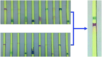
Thin layer chromatography (TLC) coupled with infrared spectroscopic technique has great advantages in the separation and identification of different components in a mixture. However, determination of suitable separation condition is a tedious task. To improve the efficiency of finding suitable separation conditions, we propose a multi-narrow-band TLC approach. In this approach, a TLC plate containing several parallel narrow bands was utilized. In experiment, different pure substance was added on each of the narrow bands and TLC analysis was performed under same condition. Different substance exhibits different migration distance. Thus, it becomes much easier to separate substances with large difference in migration distance. This approach can be used to find multiple mixed sample pairs in a parallel manner. Consequently, a high-throughput method on selection of suitable mobile phase and separation conditions can be established.


1 Introduction

Thin-layer chromatography (TLC) is an efficient, quick and economic approach to separate different components from a given mixture.1–8 Although TLC exhibits many advantages in the separation of mixtures, there are some limitations in the identification of separated components.9 In most traditional TLC experiments, separated components can be identified by color, appropriate UV-Vis absorption or fluorescence signals.10 For those components without color, UV-Vis absorption or fluorescence signals, visualization reagents are often used on a developed TLC plate so that visible spots are formed for their identification. Unfortunately, the utility of visualization reagents is somewhat limited since a visualization reagent may not be suitable for every chemical substance with different structural features.7 Infrared spectra (IR)11,12 may offer a simple and effective approach to identifying separated substances, even if the separated substance lacks color, UV-Vis absorption or fluorescence signals.

Research on TLC/IR appeared in the 1960s. At that time, analysis was carried out via solvent elution transfer approach. This approach uses suitable solvent to dissolve each separated sample out of the TLC plate.13,14 After removing the solvent by vaporization, IR spectrum of the obtained sample is recorded. This approach is tedious and the analysis is under the risk of sample loss and contamination. In 1975, Griffiths et al.15–17 developed in situ TLC/IR technique. This approach is quick, convenient and free of risk of sample loss since IR spectrum is directly obtained from the separated sample spot on a TLC plate. Subsequently, Griffiths et al.,16,17 Zuber et al.,18 Eyring et al.19 and White20 tried to use various spectral techniques to develop in situ TLC/IR techniques. However, the in situ TLC/IR approach suffers from the following problem: stationary phases of traditional TLC plates, such as silica and alumina, exhibit a strong background absorption that can bring about significant interference to sample identification. Danielson et al. proposed to use zirconium oxide that has no mid-IR absorption between 4000 cm−1 and 1000 cm−1 as a new stationary phase for TLC/FTIR analysis.21,22 However, zirconium oxide retains a considerable amount of adsorbed water, and the adsorbed water still brings about strong interference on the resultant FTIR spectra. Consequently, the problem of interference from stationary phase was not adequately addressed.

To break the bottleneck of the TLC/IR method, we propose to use IR-transparent, insoluble inorganic particles as new stationary phases for TLC/IR analysis. In our previous work, we have carried out a series of work to use BaF2, LaF3, AgI and sulfur particles as stationary phases for TLC/IR analysis.23–27 Our preliminary experiments demonstrated that mixed samples can be successfully separated by using the above materials as stationary phase. Moreover, the separated spots can be identified correctly by using FTIR spectroscopic measurement. Our further experiments indicate that BaF2, LaF3 particles tend to retain considerable amount of adsorbed water that is rather difficult to be removed completely.28 However, it is much easier to remove adsorbed water completely for silver iodide particles. Thus, we focus our investigation on TLC/IR analysis by using AgI as stationary phase in the subsequent work. However, silver iodide is rather expensive, and the usage of silver iodide in large amounts will lead to an unacceptable economic cost. To solve this problem, we proposed to use narrow band TLC plate in TLC/IR analysis.29 The results demonstrated that not only the amount of AgI used in the experiment is remarkably reduced, but also the performance of the TLC/IR analysis is significantly improved by using narrow band TLC plate. Furthermore, the development of FTIR spectrometer with a microscope attachment has provided a new chance for the development of TLC/IR technique. We adopted the mapping mode of FTIR microscope in the experiment. As a result, the spectrum of every tiny region on a TLC plate can be recorded automatically. By using this approach, we have used TLC with AgI as the stationary phase to analysis colorless mixtures, such as benzamide/cholesterol mixture and alanine/arginine mixture. Satisfactory results have been obtained.29,30 All these work have demonstrated that TLC/FTIR by using AgI as stationary phase is applicable to real chemical systems.

Silver iodide is still a new stationary phase for TLC/IR analysis. To a large extent, the chromatographic behavior of AgI particles has not been fully exploited. Experimental conditions that have been proved to be successful in separation of a given mixed sample by using silica gel as stationary phase may not be suitable for the same mixture when AgI is utilized as stationary phase. Consequently, a large amount of work has been devoted in the selection of suitable mobile phase and optimization of experimental conditions in the analysis of mixed samples. The entire procedure is time consuming and tedious.

In the present paper, we propose a new approach to improving the efficiency of the selection of mobile phase and optimization of separation condition by using multiple-narrow-bands TLC plate.

In our experiments, different pure chemical substances were loaded on each of the separated narrow band of a TLC plate. Subsequently these substances are observed simultaneously under the same experimental condition. Different substances may exhibit different migration distances. Therefore, suitable separation condition for separating two substances with dramatically different migration distances can be obtained. This approach can be used to find multiple mixed sample pairs in a parallel manner. Consequently, a method with high-throughput selection of suitable mobile phase and separation conditions can be established.

2 Experimental

2.1 Reagents

All reagents are of AR grade. Methanol, ethanol, aqueous ammonia, rhodamine B, silver nitrate, bromophenol blue and potassium iodide were from Beijing Chemical Factory. Bromocresol green and methyl violet were from Beijing Xudong Chemical Plant. Methyl red, malachite green and phenol red are from Sinopharm Chemical Reagent Co. Ltd. Congo red was from Chinese Transport Reagent Factory. Coomassie brilliant blue was from Shanghai Chemical Reagent Company. Methylene blue was from Beijing Chemical Reagent Factory.

2.2 Apparatus

IR spectra were collected on a Thermo-Fisher Nicolet iN10 MX FTIR spectrometer equipped with an IR microscope. All the spectra were recorded under reflection mode at a resolution of 4 cm−1 and 16 scans were co-added. The size of the aperture on the IR microscope is 100–100 μm.

Silver iodide was synthesized by using a reaction between silver nitrate and potassium iodide. The experimental detail of the preparation of silver iodide particles can be found in our previous work.25 The diameters of the silver iodide particles are around 100 nm.

2.3 Procedures

2.3.1 Preparation of mixed sample solutions. Methyl red (0.050 g) was dissolved in 10.00 g ethanol to prepare a 0.5 wt% solution. In a similar way, 0.5 wt% solutions of malachite green, phenol red, Coomassie brilliant blue, methyl violet, rhodamine B, bromocresol green, Congo red, bromophenol blue and malachite green solutions were prepared. Mixed sample solutions of 0.5 wt% rhodamine B and bromocresol green was prepared by dissolving 0.050 g rhodamine B and 0.050 g bromocresol green in 10.00 g ethanol. Then we prepared other mixed sample solutions of bromocresol green/rhodamine B mixture, methyl red/rhodamine mixture and bromophenol blue/rhodamine B mixture by using the similar methods.
2.3.2 Preparation of multiple-narrow-band TLC plate. Multiple-narrow-band TLC plate was prepared according to the following procedure. First, mask a glass plate (100 mm × 100 mm × 1.5 mm) on both sides by using a plastic tape. The plastic tape on the glass plate was burnt by using a computer controlled CO2 laser engraving machine on one side. As a result, multiple narrow rectangle shaped openings (95 mm × 2.0 mm) that are parallel to one another were formed on the glass plate and glass underneath is exposed for further corrosion. The glass plate was soaked in hydrofluoric acid solution (13.3 wt%) under stirring for 40 min. Then the glass plate was taken out from the hydrofluoric acid solution and washed with water. The plastic tape on the glass plate was removed and the plate was washed with distilled water. As a result, glass plate with designed multiple parallel grooves can be obtained.

Silver iodide suspension was prepared by mixing silver iodide and ethanol (the weight ratio between silver iodide and ethanol was 1[thin space (1/6-em)]:[thin space (1/6-em)]5). The suspension was directly dripped into the grooves in the glass plate. After ethanol evaporation, excess silver iodide at the edge of the grooves was removed. Thus, a multiple-narrow-band TLC plate was obtained.

2.3.3 Migration behavior of pure substances on TLC plate developed by different mobile phase. Ten ethanol solutions containing the following pure substances (methyl red, malachite green, phenol red, Coomassie brilliant blue, methyl violet, rhodamine B, bromocresol green, Congo red, bromophenol blue, methylene blue) were manually spotted at one end of multiple-narrow-bands silver iodide TLC plate. In the first experiment, the multiple-narrow-bands TLC plate was developed by using methanol/aqueous ammonia mixture as the mobile phase (the volume ratio of methanol to aqueous ammonia is 15[thin space (1/6-em)]:[thin space (1/6-em)]0.3). The development time was 20 minutes. In the second experiment, the multiple-narrow-bands TLC plate containing the above ten substance was developed by using acetone as the mobile phase, and the development time was 20 minutes.
2.3.4 Separation of a mixture according to the migration behavior of its constituents.
TLC experiment. A mixed solution containing methyl violet and bromocresol green was manually spotted at one end of narrow-band TLC plate and developed for 20 minutes.
FTIR characterization. The developed TLC plate was dried at 100 °C in a vacuum oven to remove residual mobile phase for five hours. Then the TLC plate was examined by using the FTIR microscope.

Reference FTIR spectra of bromocresol green and methyl violet are recorded according to the following procedure: each of the 0.5 wt% ethanol solutions of pure bromocresol green and methyl violet was manually spotted at one end of multiple-narrow-bands TLC plate. Then the TLC plate was developed with the same condition used in bromocresol/methyl violet separation. FTIR spectra on the spot were recorded and used as reference spectra for bromocresol green and methyl violet.

2.3.5 Eliminating the spot tail by a two-step separation process. The mixture was development in methanol/aqueous ammonia mixture (the volume ratio of methanol to aqueous ammonia is 15[thin space (1/6-em)]:[thin space (1/6-em)]0.3) for 20 minutes and spots of bromophenol blue and rhodamine B are completely separated. Then the TLC plate was further developed by using acetone as a mobile phase for 40 minutes.

3 Results and discussion

3.1 Migration behavior of pure substances on TLC plate developed by different mobile phase

To test if the multiple-narrow-bands TLC plate is applicable in practice, ten substances were developed on the multiple-narrow-bands. The experiment procedure in detail is described in Section 2.3.3 of Experimental.

Images of multiple-narrow-bands plate after developed by using methanol/aqueous ammonia mixture as the mobile phase is showed in Fig. 1. Methyl red, phenol red, bromocresol green, Congo red and bromophenol blue exhibited larger migration distances, while malachite green, methyl violet, phenolphthalein and rhodamine B showed lower migration distances. According to the difference in migration speed shown in Fig. 1, we propose that methanol/aqueous ammonia mixture might be used as the mobile phase to separate the following mixtures containing two substances: (methyl red, malachite green), (methyl red, methyl violet), (methyl red, Coomassie brilliant blue), (methyl red, rhodamine B), (phenol red, malachite green), (phenol red, methyl violet), (phenol red, Coomassie brilliant blue), (phenol red, rhodamine B), (bromocresol green, malachite green), (bromocresol green, methyl violet), (bromocresol green, Coomassie brilliant blue), (bromocresol green, rhodamine B), (Congo red, malachite green), (Congo red, methyl violet), (Congo red, Coomassie brilliant blue), (Congo red, rhodamine B), (bromophenol blue, malachite green), (bromophenol blue, methyl violet), (bromophenol blue, Coomassie brilliant blue) and (bromophenol blue, rhodamine B). In Section 3.2 of Result and discussion, we select bromocresol green/methyl violet as an example to test whether methanol/aqueous ammonia mixture is a suitable mobile phase to separate the mixture.


image file: c4ra13537g-f1.tif
Fig. 1 A photograph of a multiple-narrow-band TLC plate after development for 20 minutes by using methanol/ammonium hydroxide mixture (the volume ratio of the two solvents is 15[thin space (1/6-em)]:[thin space (1/6-em)]0.3) as a mobile phase. (1) Methyl red; (2) malachite green; (3) phenol red; (4) Coomassie brilliant blue; (5) methyl violet; (6) rhodamine B; (7) bromocresol green; (8) Congo red; (9) bromophenol blue; (10) methylene blue.

In addition, the above ten substances were also developed by using acetone as the mobile phase and the results are shown in Fig. 2. In this case, methyl red, malachite green, phenol red, rhodamine B, bromocresol green; bromophenol blue exhibited larger migration distances. While other substances show lower migration distance. Another noticeable feature in Fig. 2 is that the spot of rhodamine B, which shows a tail in Fig. 1, become a very tiny spot when acetone is utilized. Based on this feature, a two-step separation process was adopted on the analysis of mixture containing rhodamine B. The large tail on the rhodamine B spot can be successfully eliminated during the analysis of mixtures containing rhodamine B. The detail on this issue can be found in Section 3.3 of Results and discussion.


image file: c4ra13537g-f2.tif
Fig. 2 A photograph of a multiple-narrow-band TLC plate after development by using acetone as a mobile phase. (1) Methyl red; (2) malachite green; (3) phenol red; (4) Coomassie brilliant blue; (5) methyl violet; (6) rhodamine B; (7) bromocresol green; (8) Congo red; (9) bromophenol blue; (10) methylene blue.

3.2 Separation of a mixture according to the migration behavior of its constituents

Based on the dramatically different migration distances shown in Fig. 1, a variety of mixtures might be separated by using methanol/aqueous ammonia mixture as the mobile phase. To test whether above speculate is correct or not, we used methanol/aqueous ammonia mixture (the volume ratio of methanol to aqueous ammonia is 15[thin space (1/6-em)]:[thin space (1/6-em)]0.3) as the mobile phase to separate bromocresol green/methyl violet mixture. The experimental procedure in detail is described in Section 2.3.4 of Experimental. After separation, two isolated spots can be observed (Fig. 3A). To confirm that the above two separated spots are bromocresol green and methyl violet, respectively, FTIR spectroscopic experiments were also conducted. The experimental procedure is described in Section 2.3.4 of Experimental. FTIR spectra of the separated bromocresol green and methyl violet as well as the corresponding reference spectra are shown in Fig. 3B. The spectra of bromocresol green and methyl violet are roughly the same as the corresponding reference spectra. That is to say, the separated spots can be identified by using FTIR spectroscopic method. Therefore, the result of multiple-narrow-band TLC experiment is applicable in the separation of the bromocresol green/methyl violet mixture.
image file: c4ra13537g-f3.tif
Fig. 3 (A) A photograph of a narrow-band TLC plate after separation of bromocresol green/methyl violet mixture. (B) FTIR spectra of separated bromocresol green/methyl violet spots and reference spectra. Trace a: reference FTIR spectrum of bromocresol green; trace b: FTIR spectrum of the separated spot of bromocresol green; trace c: FTIR spectrum of the separated spot of methyl violet; trace d: reference FTIR spectrum of methyl violet.

3.3 Eliminating the spot tail by a two-step separation process

Then we tried to separate bromophenol blue/rhodamine B mixture by using methanol/aqueous ammonia mixture (the volume ratio of methanol to aqueous ammonia is 15[thin space (1/6-em)]:[thin space (1/6-em)]0.3) as the mobile phase. The results of TLC experiment are shown in Fig. 4. Although completely separation on the mixture can be achieved, there remains a problem. A large tail can be observed on the spot of rhodamine B. The same phenomenon can also be found in Fig. 1. This result suggests that the solubility of rhodamine B is not adequate in the methanol/aqueous ammonia mixture. Separated spot with a larger tail means that the separated component has spread in a quite large area. Thus, the content of the separated component in a detection region under an IR microscope (the size of detection region is only 100 μm × 100 μm) will be attenuated accordingly. This tendency may bring about problem in the detection of the separated component in the subsequent FTIR measurement.
image file: c4ra13537g-f4.tif
Fig. 4 A photograph of a narrow-band TLC plate after separation of bromophenol blue/rhodamine B mixture.

Then we study the chromatographic behavior of bromophenol blue and rhodamine B by using acetone as a mobile phase. In this case, the difference in migration distance between bromophenol blue and rhodamine B is much reduced. The results suggest that acetone might not be a suitable mobile phase to separate bromophenol blue/rhodamine B mixture. However, the tail on the spot of rhodamine B cannot be observed any more (Fig. 2).

Based on the different chromatographic behavior by using methanol/aqueous ammonia mixture and acetone as mobile phases, we proposed a separation and elimination of tail strategy. First, bromophenol blue and rhodamine B was firstly developed by using methanol/aqueous ammonia mixture so that a complete separation can be achieved on the bromophenol blue/rhodamine B mixture. Then the TLC plate was further developed by using acetone as mobile phase. Since the rhodamine B spot with a large tail locates at the bottom of the TLC plate, the migration behavior of rhodamine B can be firstly affected by acetone. The large tail is expected to be eliminated. On the other hand, the spot of bromophenol blue locates at the upper part of the TLC plate. Hence, acetone gets contact with the spot of bromophenol blue at later stage. If we control the time of development in acetone, the tail of rhodamine B spot can be eliminated before the spot of rhodamine B catch up with the spot of bromophenol blue. According to this idea, a two-step experiment was conducted. The experimental procedure is described in Section 2.3.5 of Experimental. As shown in Fig. 5A, bromophenol blue/rhodamine B mixture was successfully separated. Moreover, the larger tail on the rhodamine B spot cannot be observed any more. By using the same strategy, we have also separated methyl red/rhodamine B mixture (Fig. 6A).


image file: c4ra13537g-f5.tif
Fig. 5 (A) A photograph of a narrow-band TLC plate after separation of bromophenol blue/rhodamine B mixture by using the two-step separation process. (B) FTIR spectra of separated bromophenol blue/rhodamine B mixture sample spots and reference spectra. Trace a: reference FTIR spectrum of bromophenol blue; trace b: FTIR spectrum of bromophenol blue spot separated from the mixture; trace c: FTIR spectrum of rhodamine B spot separated from the mixture; trace d: reference FTIR spectrum of rhodamine B.

image file: c4ra13537g-f6.tif
Fig. 6 (A) A photograph of a narrow-band TLC plate after separation of methyl/rhodamine B mixture by using the two-step separation process. (B) FTIR spectra of separated methyl/rhodamine B spots and reference spectra. Trace e: reference FTIR spectrum of methyl red; trace f: reference FTIR spectrum of methyl red spot separated from the mixture; trace g: FTIR spectrum of rhodamine B spot separated from the mixture; trace h: reference FTIR spectrum of rhodamine B.

To confirm that the separated spots shown in Fig. 5A and 6A are methyl red, bromophenol blue and rhodamine B, respectively, reference FTIR spectra of the above substances were recorded according to the procedure described in Section 2.3.4 of Experimental. Then FTIR spectra of the separated spots shown in Fig. 5B and 6B were also recorded. FTIR spectra of methyl red, bromophenol blue and rhodamine B are roughly the same as the corresponding reference spectra, respectively. The results confirm that the separated spot can be identified by using FTIR spectroscopic method.

4 Conclusion

In this study, we proposed a new approach to improving the efficiency of the selection of mobile phase and optimization of separation condition by using multiple-narrow-bands TLC plate. Under the guidance of the result of multiple-narrow-band TLC experiment, we have successfully separated the bromocresol green/methyl violet mixture. Furthermore, we adopt two-step separation process by using methanol/aqueous ammonia mixture and acetone as the first and second mobile phase. As a result, bromocresol green/rhodamine B mixture, methyl red/rhodamine mixture and bromophenol blue/rhodamine B mixture can be successfully separated. Moreover, the problem that rhodamine B spot show a large tail has also been addressed. The above results confirm that the use of the multiple-narrow-bands TLC technique proposed in the present work should be helpful in the selection of suitable mobile phase and experimental condition for TLC/FTIR analysis.

Acknowledgements

This work is supported by the National Natural Science Foundation of China (51373003, 30873437) and Ph.D. Programs Foundation of Ministry of Education of China (20112133110001).

References

  1. High-performance thin-layer chromatography (HPTLC), ed. M. Srivastava, Springer, Heidelberg, 2011 Search PubMed.
  2. Method and Application of Planar Chromatography, ed. L.Y. He, Chemical Industry Press, Beijing. 2005 Search PubMed.
  3. J. Sherma, Anal. Chem., 2010, 82, 4895–4910 CrossRef CAS PubMed.
  4. J. Sherma, Anal. Chem., 2008, 80, 4253–4267 CrossRef CAS PubMed.
  5. J. Sherma, Anal. Chem., 2006, 78, 3841–3852 CrossRef CAS PubMed.
  6. S. C. Cheng, M. Z. Huang and J. Shiea, Anal. Chem., 2009, 81, 9274–9281 CrossRef CAS PubMed.
  7. A. Skorupa and A. Gierak, J. Planar Chromatogr.--Mod. TLC, 2011, 24, 274–280 CrossRef CAS.
  8. R. L. White, Anal. Chem., 1985, 57, 1819–1822 CrossRef CAS.
  9. E. Koglin, J. Mol. Struct., 1988, 173, 369–376 CrossRef CAS.
  10. G. Glauninger, K.-A. Kovar and V. Hoffmann, J. Anal. Chem., 1990, 338, 710–716 CAS.
  11. Modern FTIR Spectroscopy Technology and Application, ed. J. G. Wu, Science and Technology Press, Beijing, 1994 Search PubMed.
  12. Fourier Transform Infrared Spectrometer, ed. S. F. Weng, Chemical Industry Press, Beijing, 2nd edn, 2010 Search PubMed.
  13. R. N. McCoy and E. C. Fiebig, Anal. Chem., 1965, 37, 593–595 CrossRef CAS.
  14. P. A. Sturm, R. M. Parkhurst and W. A. Skinner, Anal. Chem., 1966, 38, 1244–1247 CrossRef CAS.
  15. C. J. Percival and P. R. Griffiths, Anal. Chem., 1975, 47, 154–156 CrossRef CAS.
  16. M. P. Fuller and P. R. Griffiths, Anal. Chem., 1978, 50, 1906–1910 CrossRef CAS.
  17. M. P. Fuller and P. R. Griffiths, Appl. Spectrosc., 1980, 34, 533–539 CrossRef CAS.
  18. G. E. Zuber, R. J. Warren, P. P. Begosh and E. L. O'Donnell, Anal. Chem., 1984, 56, 2935–2939 CrossRef CAS.
  19. L. B. Lloyld, R. C. Yeates and E. M. Eyring, Anal. Chem., 1982, 54, 549–552 CrossRef.
  20. R. L. White, Anal. Chem., 1985, 57, 1819–1822 CrossRef CAS.
  21. N. D. Danielson, J. E. Katon, S. P. Bouffard and Z. H. Zhu, Anal. Chem., 1992, 64, 2183–2186 CrossRef CAS PubMed.
  22. S. P. Bouffard, J. E. Katon, A. J. Sommer and N. D. Danielson, Anal. Chem., 1994, 66, 1937–1940 CrossRef CAS.
  23. X. Liu, Q. H. Pan, J. Ding, Q. Zhu, A. Q. He, S. J. Yue, X. P. Li, L. P. Hu, J. M. Xia, C. G. Liu, Y. J. Wei, J. Yu, Z. L. Yang, X. Zhu, Y. Z. Xu and J. G. Wu, Spectrosc. Spectral Anal., 2011, 31, 1767–2177 CAS.
  24. Q. Zhu, X. Su, H. J. Wu, Y. J. Zhai, J. M. Xia, Y. Z. Xu and J. G. Wu, Spectrosc. Spectral Anal., 2012, 32, 1790–1794 CAS.
  25. Q. Zhu, H. J. Wu, F. Wang, A. Q. He, K. Huang, Y. J. Wei, C. G. Liu, Y. J. Zhai, S. F. Weng, Z. L. Yang, Y. Z. Xu, I. Noda and J. G. Wu, J. Planar Chromatogr.--Mod. TLC, 2014, 27(2), 80–83 CrossRef CAS.
  26. W. Liu, H. J. Wu, X. P. Wang, Q. Zhu, T. G. Kang, A. Q. He, S. F. Weng, Z. L. Yang, J. M. Xia, Y. Z. Xu, J. G. Wu, Z. F. Song, S. F. Weng, Z. L. Yang, Y. Z. Xu and J. G. Wu, Chem. J. Chin. Univ., 2013, 34(6), 1347–1352 CAS.
  27. X. K. Fan, Y. Jiang, J. J. Shi, C. G. Liu, Y. J. Wei, Z. F. Song, S. F. Weng, Z. L. Yang, Y. Z. Xu and J. G. Wu, Chem. J. Chin. Univ., 2014, 35(4), 741–745 CAS.
  28. F. Wang, H. J. Wu, Q. Zhu, K. Huang, Y. J. Wei, C. G. Liu, Y. J. Zhai, Z. L. Yang, S. F. Weng, Y. Z. Xu, I. Noda and J. G. Wu, Anal. Methods, 2013, 5(16), 4138–4144 RSC.
  29. X. K. Fan, R. Guo, H. J. Wu, J. J. Shi, A. Q. He, Y. J. Wei, C. G. Liu, S. F. Weng, Z. L. Yang, Y. Z. Xu, I. Noda, J. G. Wu, J. Spectrosc.  DOI:10.1155/2013/976360.
  30. J. Liu, F. S. Zhou, R. Guo, Y. Jiang, X. K. Fan, A. Q. He, Y. J. Zhai, S. F. Weng, Z. L. Yang, Y. Z. Xu, I. Noda, J. G. Wu, J. Spectrosc.  DOI:10.1155/2014/925705.

This journal is © The Royal Society of Chemistry 2015
Click here to see how this site uses Cookies. View our privacy policy here.