An activatable near-infrared hemicyanine-based probe for selective detection and imaging of Hg2+ in living cells and animals†
Abstract
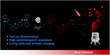
Fluorescence imaging in the near-infrared (NIR) window has great potential for clinical diagnosis and treatment because of its deep penetration and high contrast. Here, a NIR hemicyanine-based probe (CyP) was synthesized for selective detection and imaging of Hg2+ in living cells and animals. Using the rigid xanthene structure as the electron donor, trimethylindolenine as the electron acceptor and diphenylphosphinothioic chloride as the recognition element, the probe CyP could specifically respond to Hg2+ and transform into an activated donor–π-acceptor (D–π-A) motif with a NIR emission maximum at 710 nm. Cell and animal imaging experiments showed that the probe CyP could be activated by Hg2+ and had good concentration dependence in imaging. Meanwhile, animal imaging experiments showed that the activated CyP probe exhibited a higher tissue penetration depth and spatiotemporal resolution. Thus, the probe CyP could be a useful tool for monitoring and visually evaluating Hg2+ in living systems.


 Please wait while we load your content...