Design and fabrication of carbon fiber/carbonyl iron core–shell structure composites as high-performance microwave absorbers

Liu Yuan*, Liu Xiangxuan*, Li Rong, Wen Wu and Wang Xuanjun
Xi'an Research Institute of High Technology, Xi'an 710025, China. E-mail: xiangxuanstudy@sina.cn; liuyuanbixue@163.com

Received 2nd December 2014 , Accepted 11th December 2014

First published on 11th December 2014


Abstract

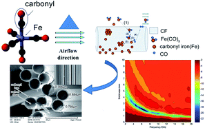
Microwave absorbing composites with carbon fiber (CF) and carbonyl iron (CI) particles as absorbers were prepared by a metal organic chemical vapor deposition (MOCVD) process. The structure and morphology analyses demonstrate that the composites have a complete core–shell structure with CF as core and CI layers as shell. The electromagnetic parameters can be adjusted by changing the CI content in the composites. Compared with CF, the composites have higher complex permeability and permittivity, which gradually increase with increasing CI content. When CF–CI = 1[thin space (1/6-em)]:[thin space (1/6-em)]8.8 (labeled as S2), the reflection loss (RL) of S2 exceeds −10 dB from 2 to 18 GHz for the absorber thickness between 0.9 and 3.9 mm, and a minimum RL value of −21.5 dB was observed at 6.6 GHz corresponding to a matching thickness of 2.0 mm. CI-doped CF by MOCVD can significantly improve the electromagnetic properties of CF and CF–CI composites could be used as an effective microwave absorption material.


1. Introduction

The research for electromagnetic wave (EMW) absorbing materials is becoming an important issue due to increase in electromagnetic pollution and in stealth technology for military platforms.1–6 A high-performance absorbent is the core in basic research of absorbing materials. Composites with core–shell structures are becoming promising EMW absorbing material because such structure can exhibit magnetic and dielectric characteristics through the proper selection of core and shell materials.7,8 Achieving a strong EMW absorption in the 2 to 18 GHz range for a thin absorber layer with low mass density is desired for the application of the core–shell structure.

Carbon fiber (CF) has broad application prospects in the EMW absorption fields because of its excellent performance properties, such as low density, high specific strength, high modulus and so on. However, CF is a resistive loss material with no magnetic property, and it has poor absorption properties when used alone. Thus, CF is always used with other materials, such as Ni,9 SiC,10 and Fe3O4 (ref. 11) to improve their combined absorbing performance. Carbonyl iron (CI) powders are widely applied in absorbing materials with high saturation magnetization and a relative permeability at microwave frequencies.12–14 According to related studies, CI powders can effectively improve high-frequency dielectric properties by adjusting the content of iron on the samples.15 However, CI has limited applications because of its narrow absorption bandwidth and heavy weight. Recently, microwave absorbing properties of several composites, such as CF–CI,16,17 CI/carbon black (CB)/CF18 have been investigated. Although aforementioned absorbers (CF–CI and CI/CB/CF) have exhibited favorable EMW absorption, both of them have not been reported to exhibit a reflection loss (RL) that exceeds −10 dB (90% EMW absorption) in the whole 2–18 GHz frequency range. Moreover, the CI and CF were simply mixed in the aforementioned research. There are no reported experimental results on the electromagnetic properties of the composites with CI as shell and CF as core. This special structure and interface interactions between the core and shell is in favor of EMW absorption.19

Thus, we designed a process to deposit CI on the CF surface to fabricate a new CF–CI composite with core–shell structures that possesses the advantages of these two materials by using the metal organic chemical vapor deposition (MOCVD) method, which has low deposition temperature, fast and flexible deposition rate, and controllable synthetic material composition.20 This new composite might obtain remarkable EMW absorption through thin and low-mass density layer absorbers in the entire 2 to 18 GHz frequency range. The morphology, complex permittivity and permeability, and EMW absorption of the composites are investigated.

2. Experimental

2.1 Sample preparation

2.1.1 In situ synthesis of CF–CI core–shell composite. The schematic diagram of the formation of CF–CI composites is illustrated in Fig. 1. Fe(CO)5 vapor is blown into the reactor and decomposed carbonyl iron (CI) and CO at high temperature. This reversible chemical reaction is Fe(CO)5 ↔ Fe + 5CO. The CF, having a large specific surface area, can effectively adsorb Fe(CO)5 gas, resulting in chemical reaction on the CF surface and generating a completely core–shell structure with the deposition time rise. The vapor deposition of Fe(CO)5 can be summarized into five main phases:21 (1) diffusion of the Fe(CO)5 vapor to the CF surface; (2) adsorption of the Fe(CO)5 vapor on the CF surface; (3) chemical reaction on the CF surface; (4) the surface of the material by-product gas from the CF surface; and (5) the generation of a reaction product leaving the clad layer.
image file: c4ra15654d-f1.tif
Fig. 1 Schematic illustration for the preparation process of CF–CI composites.

The pretreatment of CF (TORAY T300-1k) involved the following: (1) the CF was immersed into acetone for 2 h and then washed with distilled water; (2) the CF was treated with nitric acid [HNO3] for 5 h and then washed with distilled water.22 After the pretreatment, 1 g CF was placed on a stand and then put into the reactor. 15 mL of iron pentacarbonyl {Fe(CO)5, 99.9%; Shaanxi XingHua Chemical Co. Ltd.} was decanted into the evaporator. N2 was passed through the system to ensure that the air in the tube was blown out. The N2 supply was then turned off while closing the valve between the reactor and evaporator. When CF and Fe(CO)5 were heated to 240 and 80 °C, the valve between the reactor and the evaporator was opened. Fe(CO)5 vapor was blown into the reactor by N2 at a rate of 30 mL min−1 for 10, 30, 50, 70 min, respectively. The N2 flow was controlled by a gas flow meter. HH-SA digital thermostat oil bath pot was used to heat Fe(CO)5. Pipings were coated with a layer of insulation sleeve to prevent Fe(CO)5 gas condensation at low temperatures before Fe(CO)5 gas entered the reactor. The final sample was cooled to room temperature under N2 protection. The weight content of iron in the composites was determined by comparing the weights of specimens before and after the experiment. The mass ratios of CF to CI were approximately1: 5, 1[thin space (1/6-em)]:[thin space (1/6-em)]8.8, 1[thin space (1/6-em)]:[thin space (1/6-em)]11, and 1[thin space (1/6-em)]:[thin space (1/6-em)]14.3, and the composites were labeled as S1, S2, S3 and S4, respectively.

2.2 Measurement of properties

The resulting crystalline phases were characterized using X-ray diffraction (XRD; D/max-IIB, Japan). Data were recorded using Cu Kα radiation at 40.0 kV and 100.0 mA in the 2θ region, from 20° to 80°, with a scanning speed of 15° min−1. A VEGA II XMU INCA scanning electron microscope (SEM) was employed for morphological analysis, and using INCA 7718 spectroscopy (EDS) analysis of the distribution of elements in the coating. The electromagnetic (EM) parameters were measured and determined by using HP-8720ES vector network analyzer (2–18 GHz) and Agilent coaxial transmission airline. The frequency dependencies of complex permeability and permittivity for each composite specimen are then obtained using the Agilent 85071E material measurement software, which is based on Weir's reflection/transmission formulation. Samples used for EM parameter measurements were prepared by dispersing powders into paraffin wax at a mass fraction of 4% and then pressing the mixtures into a compact toroidal shape with outer and inner diameters of 7.0 and 3.0 mm, respectively.23 The RL of the prepared absorbers versus frequency was studied using an HP 8510B vector network analyzer and standard horn antennas in an anechoic chamber. The Absorbing agent powders (S2, 4 wt%) were dispersed into the epoxide resin via adding solvent and a high energy ultrasonic treatment for 30 min. Afterwards, a hardener was added into the mixtures, followed by stirring at 1000 rpm for 10 min. Finally, the composite materials are fabricated on an aluminum substrate with a standard size (180 mm × 180 mm × 3 mm).23 The RL was calculated by the transmission line theory,24,25 which was expressed by the following equations:
 
image file: c4ra15654d-t1.tif(1)
 
image file: c4ra15654d-t2.tif(2)
where Zin is the normalized input impedance of a metal-backed microwave absorption layer, Z0 = 377 Ω is the intrinsic impedance of free space, εr and μr are the relative complex permittivity and permeability, f is the frequency of the EMW, d is the thickness of an absorber, and c is the velocity of light in vacuum.

3. Results and discussion

3.1 SEM analysis

The CF surface is smooth with little grooves after the pretreatment, as shown in Fig. 2a. The surface SEM images of CF–CI composites are illustrated in Fig. 1b–e (the insets are the cross section SEM images of composites). When a small amount of Fe(CO)5 vapor was added, the CI was dispersed fine particles and less of generation result in the CF surface cannot be completely covered by CI. The CF–CI micro-structured composites have a full core–shell structure with the amount of Fe(CO)5 vapor increasing, as illustrated in Fig. 2c–e. CF coated with CI has smooth surfaces, as shown in Fig. 1c. From the cross section of SEM images of composites, an increase in the CI content of the CF–CI composites led to several small spherical particles on the surface, as shown in Fig. 2d and e. From the average shell thickness in Fig. 2c–e, it can be seen that the CI shell thickness increases linearly as y = 0.0866x − 1.7329 (R2 = 0.9963, x – deposition time, y – shell thickness), indicating that the CI content of composites can be controlled by deposition time. Fig. 2f shows the microstructure of the coating filled with 4% S2, the fillers are well dispersed in the epoxy resins (EP) matrix and no significant porosity is noticed.
image file: c4ra15654d-f2.tif
Fig. 2 SEM images of CF (a), CF–CI composites (b-S1, c-S2, d-S3, and e-S4) and SEM images of the microstructure of S2 composites coating surface (f).

3.2 XRD and EDS analysis

The XRD spectra of CF–CI composites are illustrated in Fig. 3. The patterns show the XRD intensity at a range of 20–30° for C diffracts. At 2θ = 44.8° and 65°correspond to the (110) and (200) planes of α-Fe, respectively. The growth speed of α-Fe (110) was 1.15 times faster than that of (200), which explains the preferred orientation of the film (110) for the different samples.26 As the amount of CI increased, the C peak gradually disappeared, and the intensity of the Fe peak obviously increased, indicating that the amount of CI in the composites can be adjusted through changing the reaction times.
image file: c4ra15654d-f3.tif
Fig. 3 XRD pattern of CF–CI composites.

The EDS line scan pattern through the sample surface and cross section (spots A and B in Fig. 2c and d) are shown in Fig. 4. The Fe, C distribution cladding layer confirmed that elemental iron existed, and the shell material was CI.


image file: c4ra15654d-f4.tif
Fig. 4 EDS line scan pattern of CF–CI composites.

3.3 Complex permittivity and permeability of samples

The complex permittivity of CF and CF–CI composites are shown in Fig. 5a and b, respectively. The ε′ and ε′′ values of the CF–CI composites were almost larger than that of the CF (except the ε′′ of CF at 10 GHz), and the values with fluctuations over the entire frequency range and the increase with increasing weight ratio of CI in the composites indicate that the dielectric loss of CF–CI composites has been improved. Complex permittivity of CF–CI showed multiple resonance phenomena, and resonance frequencies of 8.2, 12.6, and 16.8 GHz. The experiments showed that the CI shell, which forms a surface layer, greatly increases the conductivity of the samples and contributes to the enhancement of permittivity.27 It is likely to cause the composites to attain the permittivity necessary for EMW absorbing materials by optimizing the weight ratio of CI in the CF–CI composites.
image file: c4ra15654d-f5.tif
Fig. 5 Complex permittivity (a and b) and permeability (c and d) of CF and CF–CI composites at 2–18 GHz and Cole–Cole plots (e) and f−1(μ′)−2μ′′ (f) values of S2.

The resonance behavior of the material complex permittivity is typically sourced from the space charge, dipole, ionic, and electron polarizations.28 However, dipole polarization has a dominating function in the metal matrix composite and can operate at higher frequencies compared with that of the space charge polarization. Ionic and electronic polarizations generally operate at THz and PHz.28 Thus, the complex permittivity should be derived from the dipole polarization in CF–CI composites. According to the Debye dipolar relaxation expression: (ε′ − ε)2 + (ε′′)2 = (εsε)2, the plot of εversus ε′′ would be a single semicircle, which is usually defined as the Cole–Cole semicircle.29 Fig. 5e presents the Cole–Cole plots of S2 composite with circle radius continuously varying. Trajectory equation is no longer a circle or semi-circle, but a spiral curve precession, suggesting that the dipole polarization is a Lorentz-type resonance other than a Debye dipolar relaxation. It is reasonable that the higher complex permittivity (both the ε′ and ε′′) can be obtained when the composites are filled with higher content of CI, due to the higher Lorentz-type resonance in the CF–CI composites.

The complex permeability curves of CF and CF–CI composites versus frequency are shown in Fig. 5c and d. The complex permeability still showed a multiple resonance phenomenon and higher values were found for CF–CI composites with higher CI contents prior to this intersecting point. From the permeability real part of view, the value declined as the frequency increased. The real permittivity value showed fluctuation at about 11 and 16 GHz while the imaginary permittivity showed fluctuation about 9 and 14 GHz. A general loss of microwave magnetic material is primarily from eddy current losses, magnetization vector rotation, natural resonance, and magnetic domain wall resonance.30 Magnetization vector rotation only occurs in a strong magnetic field, and the magnetic domain wall resonance contribution at microwave frequency is very small and can be neglected.31 Therefore, the EMW loss of S2 was primarily caused by eddy current losses or natural resonance. If S2 magnetic loss is only from the eddy current loss, then f−1(μ′)2μ′′ should be a constant value.32 The values of f−1(μ′)−2μ′′ versus the frequency of S2 are shown in Fig. 5f. The value showed a downward trend as the frequency increased. Hence, the eddy current loss could be ruled out. Hence, S2 magnetic loss was primarily dominated by natural resonance.

3.4 Microwave absorption properties

In order to study the microwave absorption performance of S1 to S4, the relationship between RL and frequency at different thicknesses was calculated and shown in Fig. 6. It is can be seen that S2 has the best absorbing properties. Absorbing performance of CF–CI did not increase with the increase in the quality of CI. There is an optimum amount of CI. Studies have shown that this maybe because when the CI amount and shell thickness increased, the reflected EMW absorption performance is deteriorated due to the “skin depth” effects.33 The skin depth δ of the particle is expressed as image file: c4ra15654d-t3.tif, where ρ and μ0 are resistivity of the particle and permeability of free space, respectively.34 The average skin depth δ as a function of the frequency for the iron base ferromagnetic alloy is about 2 μm.33 For core–shell structure composites, the shell thickness of S3 and S4 exceed 2 μm, so the absorbing performances of composites turn bad. On the other hand, the real part of the complex permittivity, which increase (as shown in Fig. 5a) too larger with the CI mass increase, will impair the impedance matching between the absorb coating and free space.35
image file: c4ra15654d-f6.tif
Fig. 6 Microwave absorption diagrams of the S1 to S4 (a-S1, b-S2, c-S3, and d-S4).

The microwave absorbing properties of the S2 with different thicknesses is shown in Fig. 7a. It can be seen that the reflectance peak gradually moved to a lower frequency as the thickness increased. The absorption property increases with the thickness, and the minimum value was −21.5 dB at 6.6 GHz with a thickness of 2.0 mm, whereas the RL dramatically decreases when the thickness of coating exceeds 2.0 mm. The RL of S2 (0.9 to 3.9 mm in thickness) was less than −10 dB in the whole 2 to 18 GHz frequency range. It is worth noting that the EMW absorption of the coating can be tuned easily by simply changing the coating thickness. A comparison of the measured RL of the coating at thickness d = 0.9 mm and the calculated results is illustrated in Fig. 7b. Although the measured results and calculated values slightly differ, both measured and theoretically calculated results have similar curve patterns and absolute values.


image file: c4ra15654d-f7.tif
Fig. 7 Microwave absorbing properties of the S2 with different thicknesses (a) and comparison (b) of measured and calculated RL for S2 (d = 0.9 mm).

3.5 The comparison between the core–shell CF–CI composites and physical mixture

The EM parameters and EMW absorption of the core–shell CF–CI composites (S2) has been compared with the physical mixture (the mass ratios of CF to CI was 1[thin space (1/6-em)]:[thin space (1/6-em)]8.8, labeled as P1) between CF and CI to clarify the effect of the core–shell structure. The SEM image of P1 show that the CI particles and CF were uniformly mixed, which is illustrated in Fig. 8a. The complex permittivity of S2 and P1 composites are shown in Fig. 8b, respectively. The real part complex permittivity of P1 has the same phenomenon compared to S2, both with multiple resonances. The real part the complex permittivity of S2 is higher than that of P1, which indicates that the storage capability of electric energy of S2 is larger than P1. The imaginary part complex permittivity of P1 decreases as the frequency increases, and it is lower than S2 at most of the frequency. This indicates that the dielectric loss of the S2 is different from P1 and the core–shell structure increases the dielectric loss. Various studies comprising many types of core–shell absorbing materials draw the similar conclusion that the core–shell interface occurs at the micro dielectric relaxation, which causes the loss of EM energy.2,35–39 After the CI film grew on the CF surface, the conductive phase CI distributed on the surface of lower conductivity CF. The free charge gathered the two-phase interface because of the differences in the electrical properties of the two-phases. These aggregate charges induced polarization under the action of EM fields, thereby increased the dielectric constant. The composite strength of the free charge polarization size has a direct effect on the strength of relaxation, and polarization depends primarily on the size of the charge density and the size of the two-phase interface. For this study, the increase of the dielectric loss of S2 is also likely to be due to interfacial polarization phenomenon at the core–shell interface under the action of EMW.
image file: c4ra15654d-f8.tif
Fig. 8 SEM image(a) of P1, complex permittivity and permeability (b and c) of S2 and P1, and EMW spread schemes (d) of S2 composites.

The complex permeability curves of S2 and P1 versus frequency are shown in Fig. 8c. The permeability of S2 is almost higher than the peak of the P1, which shows significant multiple resonance phenomena. The results indicate that the magnetic energy storage capacity and magnetic loss of S2 are stronger than that of the mixed powder P1.40 Moreover, the core–shell structure effectively increased the propagation of EMW in the course of its interior for CF/Fe core–shell composites (Fig. 8d). By multiple reflections between the core–shell interfaces, the EMW attenuation is effectively improved.41,42

Microwave RL curves are shown in Fig. 9 with different thicknesses of P1. It is clear to see that the microwave absorbing properties of the composite powder S2 (Fig. 6b) is much better than that of the mixed powder P1. These results fully indicate that core–shell structure plays a dominant role in the EM properties of the samples.


image file: c4ra15654d-f9.tif
Fig. 9 Microwave absorbing properties of the P1 with different thicknesses.

Absorbing materials with a good effect must meet two conditions: one is EMW must penetrate into the interior of material as much as possible, that is a good wave impedance matching characteristic, the other is the material must be able to consume the incoming EMW, which should possess a large attenuation constant.43 The characteristic impedance of the material can be calculated according to image file: c4ra15654d-t4.tif.44 As can be seen from Fig. 10a, impedance characteristics of S2 was better than P1 over 2–18 GHz. The attenuation constant can be expressed as image file: c4ra15654d-t5.tif.45 EMW entering into the materials should be almost entirely attenuated (attenuation characteristic) for achieving low reflection. The attenuation constants of the samples are illustrated in Fig. 10b. The highest attenuation constant was obtained with S2. In summary, all the results mentioned in section 3.5 can explain why the dielectric properties of core–shell structure S2 have increased compared to the simple CF–CI mixture P1 and why S2 has the best EMW absorption properties.


image file: c4ra15654d-f10.tif
Fig. 10 Impedance (a) and attenuation constant (b) of P1 and S2 composites at 2–18 GHz.

We compare S2 with some similar reported composites for EMW absorption properties shown in Table 1. It can be seen that the S2 has stronger RL absorption and broader absorption bandwidth at a lower mass density and a thinner absorber layer.

Table 1 EMW absorption parameters of some reported composites and of as-prepared S2
Sample Weight ratio (wt%) RLmin (dB) Bandwidth (RL < −10 dB) (GHz) d (mm) Ref.
CF–CI 65% CI + 2% CF −11.2 8–18 1 16
CF–CI/CB 70% −25 9.0–13.5 1.6 18
CF–CI 5% CI + 0.6% CF −11.9 13.2–16.3 10 17
CF/Fe3O4 50% −35 3.52–10.01 2.90–5.12 22
CF/Ni–Fe 40% −20 8.3–12 2.4 39
S2 4% −21.5 2–18 0.9–3.9 Our work


4. Conclusions

CF-coated CI composites with core–shell structure were successfully prepared by a MOVCD process. Observations by SEM and XRD showed uniform coating of CI over CF, and EDS indicated CI plating layers over 90 wt%. Optimizing the weight ratio of CI would likely cause the composites to attain the EM parameter necessary in microwave absorbing materials. When the weight ratio of CF[thin space (1/6-em)]:[thin space (1/6-em)]CI was 1[thin space (1/6-em)]:[thin space (1/6-em)]8.8, a minimum RL value of −21.5 dB at 6.6 GHz on a coating with a matching thickness of 2.0 mm was found. An RL of composites (0.9–3.9 mm in thickness) is less than −10 dB over the range of 2–18 GHz.

This study fabricates an effective MOCVD method for the manufacturing of CI-coated CF as new composite fillers for EM wave absorption with good absorption properties, a simple process and low costs. The thicknesses and/or the compositions of the deposition layers can be modified for other applications through the control of catalytic processes and plating conditions.

References

  1. H. L. Lv, G. J. Ji, M. Wang, C. M. Shang, H. Q. Zhang and Y. W. Du, RSC Adv., 2014, 4, 57529 RSC.
  2. P. Bhattacharya, S. Dhibar, G. Hatui, A. Mandal, T. Das and C. K. Das, RSC Adv., 2014, 4, 17039 RSC.
  3. S. Hazra, B. K. Ghosh, H. R. a. Joshi, M. K. Patra, R. K. Jani, S. R. Vadera and N. N. Ghosh, RSC Adv., 2014, 4, 45715 RSC.
  4. Y. F. Zhu, Q. Q. Ni and Y. Q. Fu, RSC Adv., 2015, 5, 3748–3756 RSC.
  5. J. W. Liu, J. Cheng, R. C. Che, J. J. Xu, M. M. Liu and Z. W. Liu, J. Phys. Chem. C, 2013, 117, 489–495 CAS.
  6. S. Xie, X. N. Guo, G. Q. Jin and X. Y. Guo, Phys. Chem. Chem. Phys., 2013, 15, 16104 RSC.
  7. W. Wei, X. G. Yue, Y. Zhou, Z. Chen, J. Y. Fang, C. Gao and Z. H. Jiang, Phys. Chem. Chem. Phys., 2013, 15, 21043 RSC.
  8. L. G. Yan, J. B. Wang, X. H. Han, Y. Ren, Q. F. Liu and F. S. Li, Nanotechnology, 2010, 21(9), 095708 CrossRef PubMed.
  9. D. R. Igor Maria, D. Adrian, S. Fabrizio, S. S. Maria and T. Alessio, Compos. Sci. Technol., 2010, 70(1), 102–109 CrossRef PubMed.
  10. M. S. Cao, W. L. Song, Z. L. Hou, B. Wen and J. Yuan, Carbon, 2010, 48(3), 788–796 CrossRef CAS PubMed.
  11. X. G. Meng, Y. Z. Wan, Q. Y. Li, J. Wang and H. L. Luo, Appl. Surf. Sci., 2011, 257(24), 10808–10814 CrossRef CAS PubMed.
  12. Z. T. Zhu, X. Sun, H. R. Xue, H. Guo, X. LFan, X. C. Pan and J. P. He, J. Mater. Chem. C, 2014, 2, 6582–6591 RSC.
  13. M. S. Kim, Y. D. Liu, B. J. Park, C. Y. You and H. J. Choi, J. Ind. Eng. Chem., 2012, 18(2), 664–667 CrossRef CAS PubMed.
  14. X. M. Ni, Z. Zheng, K. Xiao, L. Huang and L. He, Mater. Chem. Phys., 2010, 120(1), 206–212 CrossRef CAS PubMed.
  15. Y. L. Cheng, J. M. Dai, D. J. Wu and Y. P. Sun, J. Magn. Magn. Mater., 2010, 322(1), 97–101 CrossRef CAS PubMed.
  16. Y. C. Qing, W. C. Zhou, S. Jia, F. Luo and D. M. Zhu, Appl. Phys. A, 2010, 100, 1177–1181 CrossRef CAS.
  17. Z. J. Wang, K. Z. Li, C. Wang and J. Xie, J. Chin. Ceram. Soc., 2011, 39(1), 69–73 CAS.
  18. Y. P. Duan, L. Wang, Z. Liu and S. C. Gu, Plast., Rubber Compos., 2013, 42(2), 82–87 CrossRef CAS PubMed.
  19. H. Wang, Y. Y. Dai, W. J. Gong, D. Y. Geng, S. Ma, D. Li, W. Liu and Z. D. Zhang, Appl. Phys. Lett., 2013, 102, 223113 CrossRef PubMed.
  20. Z. M. Li, J. P. Li, H. Y. Jiang, Y. B. Han, J. Q. Yin, Y. J. Xia, Y. M. Chang, J. C. Zhang and Y. Hao, J. Cryst. Growth, 2013, 368, 29–34 CrossRef CAS PubMed.
  21. Y. Li, J. Liu, Y. Wang and Z. L. Wang, Chem. Mater., 2001, 13(3), 1008–1014 CrossRef CAS.
  22. C. W. Qiang, J. C. Xu, Z. Q. Zhang, L. L. Tian, S. T. Xiao, Y. Liu and P. Xu, J. Alloys Compd., 2010, 506(1), 93–97 CrossRef CAS PubMed.
  23. Y. Liu, X. X. Liu and X. J. Wang, J. Alloys Compd., 2014, 584, 249–253 CrossRef CAS PubMed.
  24. R. C. Che, L. M. Peng, X. F. Duan, Q. Chen and X. L. Liang, Adv. Mater., 2004, 16, 401–405 CrossRef CAS.
  25. M. R. Meshram, N. K. Agrawal, B. Sinha and P. S. Misra, J. Magn. Magn. Mater., 2004, 271, 207–214 CrossRef CAS PubMed.
  26. P. Smereka, X. Q. Li and G. Russo, Acta Mater., 2005, 53(4), 1191–1204 CrossRef CAS PubMed.
  27. S. Ramo, J. R. Whinnery and T. V. Duzer, Fields and Waves in Communication Electronics, Wiley, New York, 1984 Search PubMed.
  28. T. Lacrevaz, B. Flechet, A. Farcy, J. Torres, M. Gros-Jean, C. Bermond, T. T. Vo, O. Cueto, B. Blampey, G. Angenieux, J. Piquet and F. deCrecyc, Microelectron. Eng., 2006, 83(11–12), 2184–2188 CrossRef CAS PubMed.
  29. X. L. Dong, X. F. Zhang, H. Huang and F. Zuo, Appl. Phys. Lett., 2008, 92(1), 013127–013129 CrossRef PubMed.
  30. S. Y. Zhang and Q. X. Cao, J. Mater. Sci. Eng. B, 2012, 177(8), 678–684 CrossRef CAS PubMed.
  31. X. F. Zhang, X. L. Dong, H. Huang, Y. Y. Liu, W. N. Wang, X. G. Zhu, B. Lv, J. P. Lei and C. G. Lee, Appl. Phys. Lett., 2006, 89(5), 053115 CrossRef PubMed.
  32. Y. Yang, C. L. Xu, Y. X. Xia, T. Wang and F. S. Li, J. Alloys Compd., 2010, 493(1–2), 549–552 CrossRef CAS PubMed.
  33. M. Zeng, X. X. Zhang, R. H. Yu, D. L. Zhao, Y. H. Zhang and X. L. Wang, J. Mater. Sci. Eng. B, 2014, 185, 21–25 CrossRef CAS PubMed.
  34. L. Z. Wu, J. Ding, H. B. Jiang, L. F. Chen and C. K. Ong, J. Magn. Magn. Mater., 2005, 285, 233 CrossRef CAS PubMed.
  35. Y. C. Du, W. W. Liu, R. Qiang, Y. Wang, X. J. Han, J. Ma and P. Xu, ACS Appl. Mater. Interfaces, 2014, 6, 12997–13006 CAS.
  36. K. Y. Park, J. H. Han, S. B. Lee and J. W. Yi, Composites, Part A, 2011, 42(5), 573–578 CrossRef PubMed.
  37. H. L. Luo, G. Y. Xiong, Z. W. Yang, Q. P. Li, C. Y. Ma, D. Y. Li, X. B. Wu, Z. R. Wang and Y. Z. Wan, Mater. Res. Bull., 2014, 53, 123–131 CrossRef CAS PubMed.
  38. Y. Liu, X. X. Liu, X. J. Wang and X. Chen, Acta Metall. Sin., 2014, 50(9), 1095–1101 Search PubMed.
  39. G. Q. Wang, Y. F. Chang, L. F. Wang and C. Liu, Adv. Powder Technol., 2012, 23, 861–865 CrossRef CAS PubMed.
  40. X. F. Pan, H. G. Shen, J. X. Qiu and M. Y. Gu, Mater. Chem. Phys., 2007, 101(2–3), 505–508 CrossRef CAS PubMed.
  41. Y. S. Chen, W. J. Feng, C. S. Li, R. S. Li, R. Fan and H. Yang, Acta Mater. Compositae Sin., 2012, 29(1), 111–116 CAS.
  42. W. Zhou, P. Xiao, Y. Li, H. Luo and W. Hong, J. Inorg. Mater., 2014, 29(10), 1093–1098 CrossRef CAS.
  43. B. Lu, H. Huang, X. L. Dong, X. F. Zhang, J. P. Lei, J. P. Sun and C. Dong, J. Appl. Phys., 2008, 104(11), 114313 CrossRef PubMed.
  44. X. Zhang, X. Dong, H. Huang, B. Lv, J. Lei and C. Choi, J. Phys. D: Appl. Phys., 2007, 40, 5383–5387 CrossRef CAS.
  45. M. C. Duan, L. M. Yu, L. M. Sheng, K. An, W. Ren and X. L. Zhao, J. Appl. Phys., 2014, 115(17), 174101 CrossRef PubMed.

This journal is © The Royal Society of Chemistry 2015
Click here to see how this site uses Cookies. View our privacy policy here.