Rigid three-dimensional Ni3S4 nanosheet frames: controlled synthesis and their enhanced electrochemical performance

Lina Wanga, Jiajia Liu*a, Li Li Zhangb, Baosong Daia, Meng Xua, Muwei Jia, X. S. Zhaoc, Chuanbao Caoa, Jiatao Zhang*a and Hesun Zhua
aSchool of Materials Science & Engineering, Beijing Institute of Technology, Beijing 100081, P. R. China. E-mail: liujiajia@bit.edu.cn; zhangjt@bit.edu.cn; Fax: +86-10-68918065; Tel: +86-10-68918065
bInstitute of Chemical and Engineering Sciences, A*STAR, 1 Pesek Road, Jurong Island 627833, Singapore
cSchool of Chemical Engineering, The University of Queensland, St Lucia, Brisbane, QLD 4072, Australia

Received 2nd December 2014 , Accepted 15th December 2014

First published on 15th December 2014


Abstract

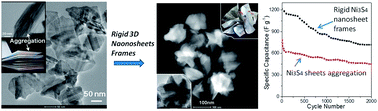
Rigid three-dimensional (3D) Ni3S4 nanosheet frames assembled from ultrathin nanosheets are synthesized via a facile solvothermal method. Compared to flat Ni3S4 sheets, 3D Ni3S4 nanosheet frames have both a high free volume and high compressive strength. They can deliver a very high specific capacitance of 1213 F g−1 with good rate performance. In addition, these 3D Ni3S4 nanosheet frames are stabilized by plastically deformed ridges. The stabilized nanosheet frames did not unfold or collapse during electrochemical tests, and thus showed enhanced cycling ability.


Nowadays, metal chalcogenide materials have been attracting significant attention in the energy field, due to their high theoretical capacitance and low cost.1 Key to their further development in these areas has been their improved surface area and the rigidity in their structure.2 To date, two-dimensional (2D) metal chalcogenide nanocrystals have provided a much higher specific surface area compared with their bulk counterparts, which is beneficial to energy devices, because the reaction/interaction between the devices and the interacting media can be significantly enhanced.3 However, during the electrode fabrication process, the nanosheet-like materials, such as graphene, tend to aggregate or restack due to strong inter-sheet van der Waals attraction.4 Consequently, many of the unique properties that individual sheets possess, such as high specific surface area and peculiar electron transport behaviours, are significantly compromised or are even unavailable during assembly. An efficient strategy to prevent nanosheet aggregation and thus to improve the energy storage performance of sheet-like materials is to synthesize three-dimensional (3D) architectures assembled with nanosheets, such as by fabricating sandwich-type structures by introducing “spacer” (e.g. carbon nanotubes, nanoparticles)5 and forming 3D macroporous structures.6 For instance, Luo et al.7 crumpled graphene sheets into paper ball-like, fractal-dimensional particles to make them aggregation-resistant in both solvents and the solid state, even after mechanical compression. Therefore, the preparation of large-area, aligned nanoporous nanostructured films in a highly crystalline state with well-oriented frameworks, excellent accessibility, and controlled porosity is an effective way to prepare metal chalcogenide-based electrode materials.

As an important class of transition metal chalcogenides, nickel sulphides with a controlled morphology is of great interest, due to their potential applications in hydrogenation catalysts and as electrode materials.8 The electrochemical performances of the electrode materials are strongly dependent on their sizes, morphologies and structures, and up to now, some progress has been made on nickel sulphide-based electrodes. For example, Hou et al.9 first investigated the potential application of NiS nanoparticles as a supercapacitor electrode and proposed an electrochemical reaction mechanism. Subsequently, Zhu et al.10 reported the synthesis of hierarchical NiS hollow spheres by a template-engaged conversion method. Due to the unique structural features and high surface area, these NiS hollow spheres showed a high specific capacitance of 583–927 F g−1 at various currents densities of 4.08–10.2 A g−1. Later, Yang et al.11 synthesized a hierarchical flower-like β-NiS electrode with a high specific discharge capacitance of 857.76 F g−1. Recently, Pang et al.12 reported the synthesis of uniform NiS2 nanocubes, and these NiS2 nanocube electrodes showed a large specific capacitance (695 F g−1 at 1.25 A g−1) and excellent cycling performance. However, Ni3S4 has attracted much less attention compared to other binary nickel sulfides, such as NiS, Ni3S2, and NiS2, due to the synthetic challenges in obtaining the single phase Ni3S4.13 Ni3S4 has a cubic spinel structure and is found in nature as the mineral polydymite.14 Much effort has been devoted to obtain single-phase Ni3S4 efficiently in laboratories. Through a selective control of the reaction conditions, such as the Ni precursor or capping agents, it is possible to obtain single phase Ni3S4 triangular nanoprisms and nanopyramids.15 Despite the success in the synthesis of various morphologies, the preparation of uniform 3D Ni3S4 nanosheet structure in a highly crystalline state still remains a significant challenge.

Herein, we present a facile, one-pot solvothermal route to prepare rigid 3D single-crystalline Ni3S4 nanosheet frames. To the best of our knowledge, the controlled synthesis of rigid 3D Ni3S4 nanosheet frames has not been reported. As a demonstration, the obtained 3D Ni3S4 nanosheet frames were used as the electrode of a supercapacitor and demonstrated excellent electrochemical performance, even outperforming most of the nickel sulfide-based electrodes from previous reports. We anticipate that the unique 3D Ni3S4 structure originating from the ultrathin Ni3S4 nanosheet is worth further exploring for its intrinsic properties in the fields of catalysis, cathode materials in lithium batteries, and so on.

In a typical reaction, a mixture of nickel stearate, oleic acid, n-dodecanethiol and toluene was added in to a Teflon-lined stainless autoclave. After continuous stirring for 5 min, the reaction solution was heated up to 200 °C for 2.5 h. Once the reaction was finished and cooled down to room temperature, the black products precipitated at the bottom of the autoclave were purified with excess ethanol by centrifugation, and were then easily dispersed in ethanol for further characterization.

The typical rigid 3D Ni3S4 nanosheet frames are displayed in Fig. 1. The size and morphology of the as-prepared frames were characterized by transmission electron microscopy (TEM). A panoramic view of the 3D frames (see Fig. 1A) reveals that the sample consists entirely of a 3D frame structure, with many ridges and vertices. The scanning transmission electron microscopy (STEM) (see Fig. 1B) and the high resolution transmission electron microscope (HRTEM) images (see Fig. 1C and D) show that the average size of the 3D nickel sulfide frames is ∼160 nm, and that they are assembled from very thin Ni3S4 sheets. The crystallographic phase of the nickel sulfide nanostructures was investigated by X-ray diffraction (XRD) (see Fig. 1F). The position of the diffraction peaks is in good accordance with the standard pattern of the cubic Ni3S4 (ICDD-JCPDS card no. 76-1813), indicating that the sample consists of pure single crystalline Ni3S4. It is worth noting that the 3D structures result in an apparent enhancement in peak (311) and a decrease in peak (111) of the Ni3S4 diffraction patterns. The single-crystalline feature of the Ni3S4 frames was further verified by HRTEM (see Fig. 1E). The lattice spacing of ∼0.54 and ∼0.33 nm matches well with the interplanar spacing of the (111) and (220) planes of the cubic Ni3S4, respectively. In addition, the selected area electron diffraction pattern from the edges shows strong Ni3S4 (111) and (220) diffraction spots, corresponding to the d-spacing of 0.54 nm and 0.33 nm, respectively. These results clearly demonstrate that the 3D Ni3S4 frame is composed of pure and single crystalline cubic Ni3S4 ultrathin nanosheets. It is notable that the obtained 3D Ni3S4 frames are very open, but are also rigid. No morphology change was observed after 30 min ultrasonication, further implying their good structural integrity and stability (see Fig. S1).


image file: c4ra15607b-f1.tif
Fig. 1 TEM image (A), STEM image (B), HRTEM images (C–E), inset FFT and XRD pattern (F) of the 3D Ni3S4 nanosheet frames.

When the ratio of n-dodecanethiol/oleic acid (DDT/OA) in the reaction system was increased, while keeping all the other parameters unchanged, relative flat nickel sulfide sheets were obtained. Fig. 2A shows a typical TEM image of the as-prepared nickel sulfide products, most of which exhibit a sheet-like morphology. The XRD pattern of the nickel sulfide sheets is similar to that of the Ni3S4 3D frames (see Fig. 2B), except there is no decrease in the intensity of the (111) peak of the Ni3S4 diffraction patterns. The HRTEM image (see Fig. 2C) reveals that the Ni3S4 sheets have near rectangular shape, with an average lateral size of ∼130 nm. The thickness of the sheet was determined with an atomic force microscope (AFM, see Fig. 2D). Prominently, the thickness was measured to be only 1.0 nm.


image file: c4ra15607b-f2.tif
Fig. 2 TEM image (A), XRD pattern (B), HRTEM images (C), AFM image with height information data (D) of the flat Ni3S4 sheets.

Our experimental results imply that the ratio of DDT/OA plays an important role in tuning the structure of Ni3S4, since the only varying parameter in preparing the 3D Ni3S4 frames and flat Ni3S4 sheets is the ratio of DDT/OA; n-dodecanethiol is also the source of sulfur for the formation of Ni3S4. Controlled experiments were carried out to investigate the evolution of the Ni3S4 sheets and 3D frames by varying the ratio of DDT/OA and the reaction time. The evolution of the morphology was examined by TEM, as shown in Fig. S2–S4. At a medium ratio of DDT/OA (e.g. 0.6 ml/6 ml or 0.8 ml/6 ml), the final product is composed of both sheets and 3D frames (see Fig. S2). The sample prepared at a low ratio (e.g. 0.5 ml/6 ml) and collected in the early stages (0.5 h, 1.0 h, 1.5 h) exhibit an immature 3D assembly structure, and the size and the sharpness of the 3D structure increases with the extension of time (see Fig. S3). The sample prepared at a high ratio (e.g. 1 ml/6 ml) and collected in the early stages (0.5 h, 1.0 h, 1.5 h) (see Fig. S4) exhibits a sheet structure, and the size of sheet increases with the extension of time (see Fig. S4). From the HRTEM image of a single relative small Ni3S4 3D frame (see Fig. S5), the clear lattice fringes at the extended end are visible and the interplanar distance is measured as ∼0.54 nm and ∼0.29 nm, corresponding to the (111) and (311) planes. This indicates that the growth direction is along the [311] direction. The growth rate of the {111} crystallographic facet was lower than that of the {311} facet, which is consistent with the intensity ratio for the (111) vs. the (311) peak (see Fig. 1F), in a qualitative manner. According to these observations and the previous literature,16 we propose that both the Ni3S4 sheets and the 3D frames could be obtained from the coalescence of the Ni3S4 nucleus (see Scheme 1a) in an oriented way. In the initial stages, Ni3S4 nanopyramids (see Scheme 1f) are formed in the reaction system. When the ratio of DDT/OA is low, the Ni3S4 nanopyramids tend to aggregate to minimize the total surface energy, and thus small Ni3S4 nano-aggregates are formed through a self-assembly process (see Scheme 1b). As the reaction continues, the growth rate of the {111} facets was lower than that of the {311} facets, which results in growth along the [311] direction, and finally leads to the formation of Ni3S4 3D frames assembled from the thin Ni3S4 nanosheets (see Scheme 1c). On the other hand, the higher DDT/OA ratio results in well-dispersed Ni3S4 nanopyramids initially (see Scheme 1f). As the reaction goes on, the reaction system tends to minimize the total energy by forming relatively flat Ni3S4 sheets through the coordination of n-dodecanethiol and oleic acid (see Scheme 1g). When the DDT/OA ratio is at a medium level, both 3D frames and relatively flat sheets are formed in the reaction system (see Scheme 1e).


image file: c4ra15607b-s1.tif
Scheme 1 Schematic illustration of the proposed mechanism for the formation of the 3D Ni3S4 nanosheet frames and Ni3S4 sheets.

A good electrode material for a pseudocapacitor should have a large specific surface area and a suitable pore-size distribution which allows efficient contact between the electroactive sites and the electrolyte ions for fast faradic energy storage.17 Since the Ni3S4 3D frames are a highly open structure, they should have a higher specific surface area than the stacked flat Ni3S4 sheets. Indeed, nitrogen adsorption/desorption analysis reveals that Ni3S4 3D frames have a higher BET surface area (122.6 m2 g−1) than the flat Ni3S4 sheets (66.7 m2 g−1). The value is also higher than some other nickel sulfide nanomaterials reported in the literature, such as NiS hierarchical hollow microspheres (34.3 m2 g−1)18 and hierarchical flower-like β-NiS nanostructures (24.0615 m2 g−1).19 The nitrogen adsorption/desorption isotherm of the Ni3S4 3D frames (see Fig. 3A) displays a type-IV isotherm with a type-H3 hysteresis loop in the relative pressure range of 0.6–1.0 p/p0, indicating the presence of a mesoporous structure. SEM images of the working electrode are shown in Fig. S6. As expected, the flat Ni3S4 nanosheets are packed like a stack of papers, with a smooth featureless surface. For the 3D Ni3S4 nanosheet frames, the surface has essentially indistinguishable microstructures. It should be noted that there is free space inside each individual 3D nanosheet frame, as well as between them.


image file: c4ra15607b-f3.tif
Fig. 3 N2-sorption isotherms and pore size distribution (inset) of the Ni3S4 3D frames (A) and flat Ni3S4 sheets (B).

The electrochemical properties of the Ni3S4 3D frames and flat Ni3S4 sheets were studied in 3 M KOH aqueous solution using a three-electrode system. Their typical cyclic voltammetry (CV) curves at a scan rate of 5 mV s−1 are shown in Fig. 4A. There can be seen a pair of redox peaks in the potential range of −0.15–0.55 V (vs. SCE). For nickel sulfide-based electrodes, it is widely accepted that the storage mechanism is mainly based on the surface redox reaction between Ni2+ and Ni3+.9,11,19 It is observed from the CV curves that the Ni3S4 3D frame electrode has a higher electrochemical activity. The rate capability of the Ni3S4 3D frame-based electrode is illustrated in Fig. S7A at different scan rates. With the increasing scan rate, the shape of the curves is maintained, suggesting a relatively good rate performance. Moreover, the peak current increases and the oxidation peak shifts to a more positive position, while the reduction peak shifts to a more negative position. This is due to the increased internal resistance within the pseudoactive material with the increase in scan rate.


image file: c4ra15607b-f4.tif
Fig. 4 The cyclic voltammogram curves at a scan rate of 5 mV s−1 (A), the galvanostatic charge–discharge curves at a current density of 2 A g−1 (B), the specific capacitance as a function of current density (C), and the dependence of the discharge specific capacitance on the charge–discharge cycle numbers at a current density of 2.0 A g−1 (D) of the Ni3S4 3D frame electrode and the flat Ni3S4 sheet electrode, respectively.

Consistent with the CV results, the plateaus in the charge–discharge curves (see Fig. 4B) indicate the existence of faradic processes. The ability for a high-rate discharge is crucial for a pseudocapacitor. Galvanostatic charge–discharge curves of the Ni3S4 3D frame-based electrode at different current densities are shown in Fig. S7B, and the corresponding rate dependent specific capacitances as a function of current density for Ni3S4 3D frame and Ni3S4 sheet-based electrodes are shown in Fig. 4C. The Ni3S4 3D frame electrode displays a high capacitance of 1213 F g−1 and 587 F g−1 at the current densities of 2 A g−1 and 12 A g−1, respectively; while, the capacitance of the Ni3S4 sheet electrode is only 789 F g−1 and 324 F g−1, respectively. The specific capacitance of our Ni3S4 3D frame electrode is significantly larger than some nickel sulfide-based electrodes from previous studies, such as NiS hollow spheres (927 F g−1 at 4.08 A g−1),10 flower-like β-NiS (857.76 F g−1 at 2 A g−1),11 and NiS2 nanocubes (695 F g−1 at 1.25 A g−1).12 The good pseudocapacitive performance of the Ni3S4 3D frame electrode could be attributed to its unique structural features. Specifically, the high surface area and open structure composed of ultrathin nanosheets can provide not only a high interfacial area between the electrode material and the electrolyte, but can also allow fast ionic diffusion.10 Fig. 4D shows the cycling performance of the as-prepared electrodes at a current density of 2 A g−1 in a voltage window of 0–0.4 V. The initial capacitance of the Ni3S4 3D frame electrode is 1213 F g−1, and around 60% of the initial capacitance was retained after 2000 cycles, indicating a relatively high cycling stability; while, the capacitance of the Ni3S4 sheet electrode dropped to 449 F g−1 after 2000 cycles, and around 56% of the initial capacitance was retained. The cycling performance of our Ni3S4 3D frame electrode is better than some nickel sulfide-based electrodes from literature, such as NiS hollow spheres (52% retention after 2000 cycles)10 and flower-like β-NiS (44% retention after 1000 cycles).11 This improved capacitance retention may be attributed to the enhanced structural integrity of these rigid 3D Ni3S4 nanosheet frames, which are stabilized by plastically deformed ridges.

Conclusions

In summary, a facile one-pot solvothermal method has been developed to prepare rigid 3D Ni3S4 nanosheet frames. The as-prepared 3D Ni3S4 nanosheet frames are shown to be very uniform in size, with an open and rigid structure. The DDT/OA ratio is shown to play an important role in altering the morphology of the Ni3S4 nanostructure; for instance, relatively flat Ni3S4 sheets were obtained with a high DDT/OA ratio. In virtue of its unique structure, the 3D Ni3S4 nanosheet frame electrode exhibits a high specific capacitance of 1213 F g−1 at the current density of 2 A g−1. Moreover, over 60% of the initial capacitances can be retained after 2000 cycles. This suggests its promising application as an electrode material for high-performance pseudocapacitors. Furthermore, we anticipate that this simple solvothermal method can be extended to the synthesis of other 3D metal sulfide nanostructures.

Acknowledgements

We acknowledge funding by the NNSF (91123001, 21322105, and 51372025), the Research Fund for the Doctoral Program of Higher Education of China (2011101120016), Beijing Excellent Talent Project (2012D009011000007) and Program for New Century Excellent Talents in University (NCET-11-0793).

Notes and references

  1. J. H. Han, S. Lee and J. Cheon, Chem. Soc. Rev., 2013, 42, 2581 RSC; M. Gao, Y. Xu, J. Jiang and S. Yu, Chem. Soc. Rev., 2013, 42, 2986 RSC.
  2. J. L. Mohanan, I. U. Arachchige and S. L. Brock, Science, 2005, 307, 397 Search PubMed; J. B. Rivest, R. Buonsanti, T. E. Pick, L. Zhu, E. Lim, C. Clavero, E. Schaible, B. A. Helms and D. J. Milliron, J. Am. Chem. Soc., 2013, 135, 7446 CrossRef CAS PubMed.
  3. J. Feng, X. Sun, C. Wu, L. Peng, C. Lin, S. Hu, J. Yang and Y. Xie, J. Am. Chem. Soc., 2011, 133, 17832 CrossRef CAS PubMed; Y. Du, Z. Yin, J. Zhu, X. Huang, X. WU, Z. Zeng, Q. Yan and H. Zhang, Nat. Commun., 2012, 3, 1 Search PubMed; Z. Zeng, T. Sun, J. Zhu, X. Huang, Z. Yin, G. Lu, Z. Fan, Q. Yan, H. H. Hng and H. Zhang, Angew. Chem., Int. Ed., 2012, 51, 9052 CrossRef PubMed; X. Zhang, J. Zhang, J. Zhao, B. Pan, M. Kong, J. Chen and Y. Xie, J. Am. Chem. Soc., 2012, 134, 11908 CrossRef PubMed; J. S. Son, X. Wen, J. Joo, J. Chae, S. Bake, K. Park, J. H. Kim, K. An, J. H. Yu, S. G. Kwon, S. Choi, Z. Wang, Y. Kim, Y. Kuk, R. Hoffmann and T. Hyeon, Angew. Chem., Int. Ed., 2009, 48, 6861 CrossRef PubMed.
  4. J. Luo, H. D. Jang, T. Sun, L. Xiao, Z. He, A. P. Katsoulidis, M. G. Kanatzides, J. M. Gibson and J. Huang, ACS Nano, 2011, 5, 8943 CrossRef CAS PubMed.
  5. Y. Li, Z. Li and P. K. Shen, Adv. Mater., 2013, 25, 2474 CrossRef CAS PubMed; L. Zhang, S. Zhao, X. N. Tian and X. S. Zhao, Langmuir, 2010, 26, 17624 CrossRef PubMed; L. Zhang, Z. Xiong and X. S. Zhao, J. Power Sources, 2013, 222, 326 CrossRef PubMed.
  6. S. Zhang, Y. Li and N. Pan, J. Power Sources, 2012, 206, 476 CrossRef CAS PubMed; J. Chen, K. Sheng, P. Luo, C. Li and G. Shi, Adv. Mater., 2012, 24, 4569 CrossRef PubMed; S. Yin, Z. Niu and X. Chen, Small, 2012, 8, 2458 CrossRef PubMed.
  7. J. Luo, J. Kim and J. Huang, Acc. Chem. Res., 2013, 46, 2225 CrossRef CAS PubMed; J. Luo, H. Jang and J. Huang, ACS Nano, 2013, 7, 1464 CrossRef PubMed.
  8. W. Wei, L. Mi, Y. Gao, Z. Zheng, W. Chen and X. Guan, Chem. Mater., 2000, 35, 3523,  DOI:10.1021/cm5006482; J. Yang, C. Bao, K. Zhu, T. Yu, F. Li, J. Liu, Z. Li and Z. Zou, Chem. Commun., 2014, 50, 4832 Search PubMed; C. Wei, Q. Lu, J. Sun and F. Gao, Nanoscale, 2013, 5, 12224 RSC.
  9. L. Hou, C. Yuan, D. Li, L. Yang, L. Shen and F. Zhang, Electrochim. Acta, 2011, 56, 7254 Search PubMed.
  10. T. Zhu, Z. Wang, S. Ding, J. S. Chen and X. W. Lou, RSC Adv., 2011, 1, 397 RSC.
  11. J. Yang, X. Duan, Q. Qin and W. Zheng, J. Mater. Chem., 2013, 1, 7880 RSC.
  12. H. Pang, C. Wei, X. Li, G. Li, Y. Ma, S. Li, J. Chen and J. Zhang, Sci. Rep., 2013, 4, 3577 Search PubMed.
  13. N. Mahmood, C. Zhang and Y. Hou, Small, 2013, 9, 1321 CrossRef CAS PubMed; X. Jiang, Y. Xie, J. Lu, L. Zhu, W. He and Y. Qian, Adv. Mater., 2001, 13, 1278 CrossRef; Y. Hu, J. Chen, W. Chen, X. Lin and X. Li, Adv. Mater., 2003, 15, 726 CrossRef.
  14. G. Kullerud and R. A. Yund, J. Petrol., 1962, 3, 126 CrossRef CAS PubMed.
  15. A. L. Abdelhady, M. A. Malik, P. O'Brien and F. Tuan, J. Phys. Chem. C, 2012, 116, 2253 Search PubMed; Q. Liu, A. Diaz, A. Prosvirin, Z. Luo and J. D. Batteas, Nanoscale, 2014, 6, 8935 RSC.
  16. C. Schliehe, B. H. Juareze, M. Pelletier, S. Jander, D. Greshnykh, M. Nagel, A. Merer, S. Foerster, A. Kornowski, C. Klinke and H. Weller, Science, 2010, 329, 550 CrossRef CAS PubMed; J. Li, Z. Chen, Y. Hu, X. Wang, T. Zhang, W. Chen and Q. Wang, J. Am. Chem. Soc., 2013, 125, 1213 CrossRef PubMed.
  17. X. Zhang, W. Shi, J. Zhu, W. Zhao, J. Ma, S. Mhaisalkar, T. L. Maria, Y. Yang, H. Zhang, H. H. Hng and Q. Yan, Nano Res., 2010, 3, 643 CrossRef CAS PubMed; H. Wan, J. Jiang, J. Yu, K. Xu, L. Miao, L. Zhang, H. Chen and Y. Ruan, CrystEngComm, 2013, 15, 7649 RSC.
  18. Y. Wang, Q. Zhu, L. Tao and X. Su, J. Mater. Chem., 2011, 21, 9248 RSC.
  19. W. We, L. Mi, Y. Gao, Z. Zheng, W. Chen and X. Guan, Chem. Mater., 2014, 26, 3418 CrossRef.

Footnote

Electronic supplementary information (ESI) available. See DOI: 10.1039/c4ra15607b

This journal is © The Royal Society of Chemistry 2015
Click here to see how this site uses Cookies. View our privacy policy here.