DOI:
10.1039/C4RA13764G
(Paper)
RSC Adv., 2014,
4, 64484-64493
In situ synthesis of hierarchical In2S3–graphene nanocomposite photocatalyst for selective oxidation†
Received
30th September 2014
, Accepted 14th November 2014
First published on 14th November 2014
Abstract
We report a simple, in situ hydrothermal way to fabricate In2S3–graphene (GR) nanocomposites in which hierarchical In2S3 “petals” spread over the surface of GR sheets in virtue of the “structure directing” role of graphene oxide (GO) as the precursor of GR in a solution phase. With the addition of an appropriate amount of GO, the hierarchical petal-like In2S3 structures have been successfully grown on the two-dimensional (2D) GR “mat”. The as-synthesized In2S3–GR nanocomposites featuring good interfacial contact exhibit much higher photocatalytic activity toward selective oxidation of alcohols under visible light irradiation than blank In2S3. A series of characterization results disclose that the significantly enhanced photocatalytic performance of In2S3–GR nanocomposites can be ascribed to the integrative effect of the increased separation and transfer efficiency of photogenerated electron–hole pairs and the larger surface area. This work highlights the wide scope of fabricating GR-based semiconductor nanocomposites with specific architectural morphology by rationally utilizing the “structure directing” property of GO and extending their applications toward photocatalytic selective transformations.
Introduction
In recent years, with the advance of two-dimensional (2D) graphene (GR) as an electrically conductive platform,1 there has been increasing interest in designing GR–semiconductor composites as efficient photocatalysts for converting solar energy into chemical energy.2–10 Through taking advantage of the brilliant physical and chemical properties of GR,11–16 diverse GR–semiconductor nanocomposites have been extensively reported to show enhanced photocatalytic performance toward a myriad of applications, including photodegradation of pollutants (dyes, volatile organic pollutant, and bacteria), hydrogen production from water splitting, and photocatalytic selective organic transformations.4,10,17–35
Generally, the synthetic method for fabrication of GR–semiconductor nanocomposites can be summarized as two categories.9,12,34 One is the hard integration of GR with solid semiconductor components and the other is the soft integration of graphene oxide (GO), as the precursor of GR, with soluble semiconductor precursors.10,21 For the hard integration method, the GR–semiconductor composites are obtained by the simple integration of GR with solid semiconductor, and the interfacial contact is often poor.21,35,36 In contrast, for the soft integration method, the “structure directing” role of GO in an aqueous phase can be readily utilized, which could promote the in situ anchoring of semiconductor particles onto the surface of reduced graphene oxide (RGO, also often called GR) mat in a solution phase. In this way, the as-obtained GR–semiconductor nanocomposites often have good interfacial contact, which is advantageous to the efficient separation of photogenerated charge carriers across the interfacial domain, thereby contributing to the enhanced photocatalytic performance of the GR–semiconductor nanocomposites.10,32,33
Indium sulfide (In2S3), as one of the common studied semiconductors, is well crystallized into three polymorphic forms, including α-In2S3, β-In2S3 and γ-In2S3. Among these three forms, β-In2S3 with a narrow band gap, has been widely used as a kind of potential photocatalyst under visible light irradiation due to its excellent photosensitivity, photoconductivity, stable chemical and physical properties, and low toxicity.37–42 Thus far, in order to make better use of the properties for β-In2S3 in photocatalysis, much effort has been devoted to constructing In2S3 based materials with different morphologies,37,38,43,44 such as one-dimensional (1D) In2S3 nanotubes and nanorods,37,45 2D In2S3 nanoplates and nanosheets,41,46 three-dimensional (3D) flowerlike In2S3 and hierarchical architectures.38,40,47 Among them, hierarchical architectures, with modified structural, optical, and surface properties, are potentially useful for the application of photocatalysis.48,49
Herein, we report a facile, in situ wet chemistry way to fabricate hierarchical In2S3–GR nanocomposites to boost the photoactivity of 3D In2S3. To the best of our knowledge, this is the first report on the application of In2S3–GR nanocomposites as visible light-driven photocatalyst for selective oxidation process. The higher photoactivity achieved on In2S3–GR than blank In2S3 toward selective oxidation of alcohols under visible light is mainly ascribed to the enhanced photogenerated electron–hole pairs separation and transfer as well as the larger surface area, resulting from the hierarchical architectures which are oriented by the “structure directing” role of GO. It is hoped that this work could provide useful information for careful design of GR–semiconductor nanocomposites with controllable architectures by making better use of the “structure directing” property of GO and extending their applications toward various photocatalytic selective transformations.
Experimental section
Materials
Indium(III) nitrate hydrate (In(NO3)3·4.5H2O), L-cysteine (C3H7NO2S), graphite powder, sulfuric acid (H2SO4), nitric acid (HNO3), hydrochloric acid (HCl), sodium hydroxide (NaOH), potassium persulfate (K2S2O8), phosphorus pentoxide (P2O5), potassium permanganate (KMnO4), hydrogen peroxide 30% (H2O2), ethanol (C2H6O), benzyl alcohol (C7H8O), ammonium oxalate (N2H8C2O4, AO), benzoquinone (C6H4O2, BQ), and tert-butyl alcohol (C4H10O, TBA) were supplied by Sinopharm chemical reagent Co., Ltd. (Shanghai, China). Benzotrifluoride (C7H5F3, BTF), 4-methylbenzyl alcohol (C8H10O), 4-methoxybenzyl alcohol (C8H10O2), 4-nitrobenzyl alcohol (C7H7NO3), 4-chlorobenzyl alcohol (C7H7OCl), 4-fluorobenzyl alcohol (C7H7OF), cinnamyl alcohol (C9H10O), and 3-methyl-2-buten-1-ol (C5H10O) were purchased from Alfa Aesar Co., Ltd. (Tianjin, China). All the reagents were analytical grade and used without further purification. The deionized water was from local sources.
Synthesis
(a) Synthesis of graphene oxide (GO). GO was synthesized by a modified Hummers method,50–55 which was also expatiated in our previous works.21,33,56,57 The detail of the preparation procedure is presented in the ESI.†
(b) Fabrication of In2S3–GR nanocomposites with different weight addition ratios of GR. In a typical procedure, the given amount of as-prepared GO was ultrasonically dispersed in 40 mL distilled water and mixed with 0.5 mmol In(NO3)3·4.5H2O. After stirring for 0.5 h, 2 mmol L-cysteine was added into the above mixture under vigorous stirring. After that, the pH was adjusted to 8 by adding 1 M NaOH dropwise. After stirring for 1 h, the mixture was transferred into a 50 mL Teflon-lined stainless steel autoclave, which was sealed and treated at 180 °C for 16 h. Subsequently, the as-synthesized products were separated by centrifugation and washed with distilled water and absolute ethanol for three times and one time, respectively. After drying at 60 °C overnight, the target products were obtained.
(c) Preparation of blank flowerlike In2S3 structures. Blank flowerlike In2S3 structures were fabricated through the same procedure as above except for the addition of GO.
Characterization
The crystal-phase information of the samples was measured on a Bruker D8 Advance X-ray diffractometer with Cu Kα radiation at 40 kV and 40 mA in the 2θ ranging from 10° to 80° with a scan rate of 0.02° s−1. The UV-vis diffuse reflectance spectroscopy (DRS) was used to characterize the optical properties of the samples using UV-vis spectrophotometer (Cary 500, Varian) in which BaSO4 was employed as a reference. Field-emission scanning electron microscopy (FESEM) was employed to detect the morphology of the samples on a FEI Nova NANOSEM 230 spectrophotometer. Transmission electron microscopy (TEM) images and high-resolution transmission electron microscopy (HRTEM) images were obtained using a JEOL model JEM 2010 EX instrument at an accelerating voltage of 200 kV. The Fourier transformed infrared spectroscopy (FTIR) was measured on a Nicolet Nexus 670 FTIR spectrophotometer at a resolution of 4 cm−1. The photoluminescence (PL) spectra were obtained using an Edinburgh FL/FS900 spectrophotometer with an excitation wavelength of 420 nm. The nitrogen adsorption–desorption isotherms and Brunauer–Emmett–Teller (BET) specific surface areas were obtained on Micromeritics ASAP2010 equipment. The electron spin resonance (ESR) signal of the radicals spin-trapped by 5,5-dimethyl-1-pyrroline-N-oxide (DMPO) was measured on a Bruker EPR A300 spectrometer. The sample (5 mg) was dispersed in 0.5 mL purified benzotrifluoride into which 25 μL of DMPO–benzyl alcohol solution (1![[thin space (1/6-em)]](https://www.rsc.org/images/entities/char_2009.gif) :10, v/v) was added. The mixture was oscillated to obtain well-blending suspension. The irradiation source (λ > 420 nm) was a 300 W Xe arc lamp system, the very light source for our photocatalytic selective oxidation of alcohols. The settings for the ESR spectrometer were as follows: center field = 3512 G, microwave frequency = 9.86 GHz, and power = 6.35 mW. The photoelectrochemical analysis was conducted in a homemade three electrode cell. A Pt plate was employed as the counter electrode and an Ag/AgCl electrode was used as the reference electrode. The electrolyte was 0.2 M Na2SO4 aqueous solution without additive (pH 6.8). The working electrode was prepared on fluorine doped tin oxide (FTO) glass that was sonicated in ethanol and dried at 80 °C before use. The boundary of FTO glass was protected using Scotch tape. The 2 mg sample was adequately dispersed in 0.5 mL of N,N-dimethylformamide with sonication. The as-obtained slurry was spread onto the pre-treated FTO glass. After air drying, the working electrode was further dried at 120 °C for 2 h to improve adhesion. Then, the Scotch tape was unstuck, and the uncoated part of the electrode was isolated with epoxy resin. The visible light irradiation source was a 300 W Xe arc lamp system equipped with a UV-CUT filter (λ > 420 nm). The electrochemical impedance spectroscopy (EIS) experiments and cyclic voltammograms were conducted on a CHI660D workstation (CH instrument, USA) in the electrolyte of 0.5 M KCl aqueous solution containing 0.01 M K3[Fe(CN)6]–K4[Fe(CN)6] (1:1) under open circuit potential conditions.
:10, v/v) was added. The mixture was oscillated to obtain well-blending suspension. The irradiation source (λ > 420 nm) was a 300 W Xe arc lamp system, the very light source for our photocatalytic selective oxidation of alcohols. The settings for the ESR spectrometer were as follows: center field = 3512 G, microwave frequency = 9.86 GHz, and power = 6.35 mW. The photoelectrochemical analysis was conducted in a homemade three electrode cell. A Pt plate was employed as the counter electrode and an Ag/AgCl electrode was used as the reference electrode. The electrolyte was 0.2 M Na2SO4 aqueous solution without additive (pH 6.8). The working electrode was prepared on fluorine doped tin oxide (FTO) glass that was sonicated in ethanol and dried at 80 °C before use. The boundary of FTO glass was protected using Scotch tape. The 2 mg sample was adequately dispersed in 0.5 mL of N,N-dimethylformamide with sonication. The as-obtained slurry was spread onto the pre-treated FTO glass. After air drying, the working electrode was further dried at 120 °C for 2 h to improve adhesion. Then, the Scotch tape was unstuck, and the uncoated part of the electrode was isolated with epoxy resin. The visible light irradiation source was a 300 W Xe arc lamp system equipped with a UV-CUT filter (λ > 420 nm). The electrochemical impedance spectroscopy (EIS) experiments and cyclic voltammograms were conducted on a CHI660D workstation (CH instrument, USA) in the electrolyte of 0.5 M KCl aqueous solution containing 0.01 M K3[Fe(CN)6]–K4[Fe(CN)6] (1:1) under open circuit potential conditions.
Catalytic activity
Photocatalytic selective oxidation of a range of alcohols was conducted according to the previous works.10,32,33,58–60 Typically, 0.1 mmol of alcohol and 8 mg of the as-synthesized catalyst were suspended in 1.5 mL of benzotrifluoride (BTF), which was saturated with molecular oxygen. Subsequently, the above mixture was transferred into a 10 mL Pyrex glass bottle, which was filled with molecular oxygen at a pressure of 0.1 MPa and stirred for 20 min to establish the adsorption–desorption equilibrium. The mixture was irradiated by a 300 W Xe arc lamp (PLS-SXE 300, Beijing Perfect light Co., Ltd.) with a UV-CUT filter to cut off light of wavelength less than 420 nm. After that, the mixture was centrifuged at 12000 rpm for 10 min to remove the catalyst particles. The remaining solution was analyzed using an Agilent gas chromatograph (GC-7820). Conversion of alcohol, yield and selectivity for aldehyde were defined as follows:
| Conversion (%) = [(C0 − Cr)/C0] × 100 |
| Selectivity (%) = [Cp/(C0 − Cr)] × 100 |
where C0 is the initial concentration of alcohol and Cr and Cp are the concentrations of the substrate alcohol and the corresponding product aldehyde, respectively, at a certain time after the photocatalytic reaction.
Results and discussion
The synthesis of blank In2S3 and In2S3–GR nanocomposites with different weight addition ratios of GR is graphically portrayed in Scheme 1. For blank In2S3, the functional groups in L-cysteine molecule, such as –NH2, –COOH, and –SH (Fig. S1†), are crucial for the formation of flowerlike In2S3 structure. Firstly, a complex is formed via the coordination between In3+ ions and L-cysteine due to their strong interactions.40,49 During the hydrothermal process, the complex is decomposed to form In2S3 nuclei, followed by crystal growth at the expense of the small crystals. With the evolution of the hydrothermal reaction, the flowerlike In2S3 structures are gradually assembled using L-cysteine as a sulfide source and grafting molecular.49 As for the fabrication of In2S3–GR nanocomposites, In(NO3)3 aqueous solution is firstly mixed with a certain amount of GO, during which In3+ ions primarily anchor on the surface of GO mat through electrostatic attractive interaction between them. In the process of the subsequent hydrothermal treatment, L-cysteine is decomposed to provide S2− and simultaneously hierarchical petal-like In2S3 architectures grow on conductive GR sheets formed by the reduction of nonconductive GO. The efficient reduction of GO to GR after the hydrothermal process can be evidenced by the Fourier transformed infrared spectroscopy (FT-IR),35,61 as displayed in Fig. S2 (ESI†). According to the FT-IR spectra of the pristine GO, the stretching vibrations including O–H (3426 cm−1), C![[double bond, length as m-dash]](https://www.rsc.org/images/entities/char_e001.gif) O (1720 cm−1), CC (1622 cm−1), C–O–H (1408 cm−1), C–OH (1224 cm−1), and C–O–C (1057 cm−1) are observed, indicating the existence of various oxygen-containing functional groups on the GO surface.35,57,61 For In2S3–1%GR, the intensity of various oxygen-containing functional groups is significantly decreased, which manifests the efficient reduction of GO to GR after the hydrothermal treatment.
O (1720 cm−1), CC (1622 cm−1), C–O–H (1408 cm−1), C–OH (1224 cm−1), and C–O–C (1057 cm−1) are observed, indicating the existence of various oxygen-containing functional groups on the GO surface.35,57,61 For In2S3–1%GR, the intensity of various oxygen-containing functional groups is significantly decreased, which manifests the efficient reduction of GO to GR after the hydrothermal treatment.
 |
| | Scheme 1 The schematic illustration for the preparation of blank flowerlike In2S3 structure and In2S3–GR nanocomposites via a facile, in situ hydrothermal method. | |
Typical SEM analysis is used to observe the morphology of blank In2S3 and In2S3–GR nanocomposites. As shown in Fig. 1A, without addition of GO, flowerlike structures for blank In2S3 with the average diameter of 1 μm are obtained. Once GO is introduced, the morphology of flowerlike In2S3 structure changes dramatically. As displayed in Fig. 1B, when the weight addition ratio of GR is 0.1%, the flowerlike morphology of blank In2S3 has been damaged apparently and petal-like In2S3 architectures start to grow along the 2D GR platform. With the further increase of GR to 1%, the compact hierarchical petal-like architectures of In2S3 grown onto the GR mat become more obvious (Fig. 1C). When the weight addition ratio of GR is increased to 2% (Fig. 1D), the petal-like architectures of In2S3 disappear, and the evident agglomeration of In2S3 particles occurs. The SEM results reveal that the addition of GO has a great influence on the morphology of the as-obtained In2S3–GR nanocomposites, and the synthesis of In2S3–GR with an appropriate amount of GO is a key factor for achieving different morphology of In2S3 spreading onto the 2D GR scaffold.
 |
| | Fig. 1 Typical FESEM images of (A) blank In2S3, (B) In2S3–0.1%GR, (C) In2S3–1%GR, and (D) In2S3–2%GR. | |
To further obtain the microscopic structure information of blank In2S3 and In2S3–GR, transmission electron microscopy (TEM) analysis has been conducted as displayed in Fig. 2. It can be seen from Fig. 2A that without the addition of GO, flowerlike structure of blank In2S3 has been obtained. In comparison, with the addition of an appropriate amount of GO such as 1%, as shown in Fig. 2B and C, it is obvious to see that the hierarchical petal-like architectures of In2S3 are growing onto the GR mat compactly which ensures the good interfacial contact between In2S3 and GR. The results are in good accordance with the SEM analysis. The image of high resolution TEM (HRTEM) in Fig. 2D shows the distinct lattice fringe and the spacing is measured to be 0.324 nm, which is corresponding to the (311) planes of cubic β-In2S3 phase. The selected area electron diffraction (SAED) pattern indicates that the In2S3–1%GR nanocomposite possesses polycrystalline structure, which is consistent with the following XRD results.
 |
| | Fig. 2 TEM images of (A) blank In2S3, (B) and (C) In2S3–1%GR, and high-resolution TEM (HR-TEM) image of (D) In2S3–1%GR; the inset of (D) is the image of SAED pattern of In2S3–1%GR. | |
The XRD patterns in Fig. 3 exhibit the crystallographic structure and phase purity of the In2S3–GR nanocomposites and blank In2S3. It can be seen that the In2S3–GR nanocomposites with different weight ratios of GR display similar XRD patterns to that of blank In2S3. The peaks located at ca. 14.2°, 20.9°, 27.4°, 28.7°, 33.2°, and 47.7° can be indexed to the (111), (211), (311), (222), (400), and (440) crystal planes for the cubic β-In2S3 phase (JCPDS no. 32-0456), respectively. No diffraction peaks of other crystal phases are observed, manifesting that the pure cubic β-In2S3 is formed during the current synthesis conditions. Notably, as compared to blank In2S3, there are no apparent peaks for the separate GR in the In2S3–GR nanocomposites to be detected, which can be probably ascribed to the relatively low addition amount of GR in the In2S3–GR nanocomposites and the overlapping of the main characteristic peak of GR at 26.0° with the (311) peak at 27.4° for cubic β-In2S3.10,32,33,58,62
 |
| | Fig. 3 The XRD spectra of the as-synthesized blank In2S3 and In2S3–GR nanocomposites with different weight addition ratios of GR. | |
Fig. 4 displays the UV-vis diffuse reflectance spectra (DRS) for characterization of the optical properties of In2S3–GR nanocomposites and blank In2S3. It can be found that the addition of different amounts of GR has a great influence on the visible light absorption property of In2S3–GR nanocomposites. With the increase of the weight addition ratios of GR, there is gradually enhanced absorption intensity in the visible light region of 560–800 nm, which should be ascribed to the intrinsic absorption of black colored GR. From the plot for the samples via the transformation based on the Kubelka–Munk function versus the energy of light, as shown in Fig. S3 (ESI†), the band gap values of the samples can be estimated to be ca. 2.54, 2.53, 2.49, and 2.46 eV, corresponding to blank In2S3, In2S3–0.1%GR, In2S3–1%GR, and In2S3–2%GR, respectively, indicating a band gap narrowing of the semiconductor In2S3 due to the introduction of GR into the matrix of In2S3–GR nanocomposites. Such an extended optical absorption has also been observed in previous works regarding GR–semiconductor photocatalysts for solar-driven catalytic applications.21,27,28,63,64
 |
| | Fig. 4 The UV-vis DRS spectra of blank In2S3 and In2S3–GR nanocomposites with different weight addition ratios of GR. | |
The photocatalytic performance of the In2S3–GR nanocomposites is initially estimated by selective oxidation of benzyl alcohol to benzaldehyde under visible light irradiation (λ > 420 nm). It can be seen from Fig. 5 that the In2S3–GR nanocomposites with different weight addition ratios of GR exhibit higher photoactivity than blank In2S3 toward selective oxidation of benzyl alcohol. Meanwhile, In2S3–1%GR exhibits the highest photocatalytic activity, over which the conversion of benzyl alcohol is about 55% with high selectivity (ca. 99%) after the visible light irradiation for 2 h. When the weight addition ratio of GR reaches 2%, the photoactivity and selectivity are both reduced, which could be ascribed to the lowered contact surface of semiconductor In2S3 with the light irradiation in In2S3–2%GR.21,33 Another reason for the decline in the selectivity is that the target product aldehyde may not be easily desorbed from the surface of photocatalyst due to the higher adsorption ability of carbon material GR with relatively higher addition in In2S3–2%GR nanocomposite. Under this circumstance, further deep oxidation of aldehyde may occur, therefore resulting in the drop in selectivity.33 As shown in Fig. 6, time-online profiles of selective oxidation of benzyl alcohol over blank In2S3 and the optimal In2S3–1%GR indicate that the conversion of benzyl alcohol and yield of benzaldehyde increase gradually with the irradiation time and the superior photoactivity of In2S3–1%GR over blank In2S3 is obvious.
 |
| | Fig. 5 Photocatalytic selective oxidation of benzyl alcohol to benzaldehyde over blank In2S3 and In2S3–GR nanocomposites with different weight addition ratios of GR under visible light irradiation (λ > 420 nm) for 2 h. | |
 |
| | Fig. 6 Time-online photocatalytic selective oxidation of benzyl alcohol to benzaldehyde over the as-prepared blank In2S3 and In2S3–1%GR under visible light irradiation (λ > 420 nm). | |
To further confirm the higher photoactivity of In2S3–1%GR than blank In2S3, we have also conducted the activity testing on oxidation of other benzylic alcohols and allylic alcohols under visible light irradiation. As demonstrated in Table 1, the In2S3–1%GR nanocomposite exhibits higher photocatalytic activity than blank In2S3 in all selected reaction systems. Hence, it is clear that with the addition of appropriate amount of GR, the as-prepared In2S3–1%GR shows remarkably enhanced photoactivity toward selective oxidation of a range of alcohols under visible light irradiation as compared to blank In2S3.
Table 1 The photocatalytic performance of blank In2S3 and In2S3–1%GR for selective oxidation of a range of alcohols under visible light irradiation (λ > 420 nm) for 2 h
| Entry |
Substrate |
Product |
Time (h) |
Blank In2S3 |
In2S3–1%GR |
| Conv. (%) |
Yield (%) |
Conv. (%) |
Yield (%) |
| 1 |
 |
 |
2 |
14 |
14 |
55 |
54 |
| 2 |
 |
 |
2 |
18 |
15 |
32 |
30 |
| 3 |
 |
 |
2 |
43 |
39 |
56 |
56 |
| 4 |
 |
 |
2 |
10 |
9 |
20 |
20 |
| 5 |
 |
 |
2 |
24 |
23 |
50 |
49 |
| 6 |
 |
 |
2 |
14 |
14 |
31 |
29 |
| 7 |
 |
 |
2 |
10 |
7 |
18 |
15 |
| 8 |
 |
 |
2 |
14 |
12 |
25 |
25 |
To understand and explore the origins of the higher photoactivity of In2S3–1%GR for selective oxidation of alcohols than blank In2S3, a series of characterizations have been carried out. The transient photocurrent density of In2S3–1%GR and blank In2S3 electrodes under visible light irradiation has shown in Fig. 7. It is clear to see that the photocurrent density of In2S3–1%GR under visible light illumination is much higher than that of blank In2S3. It is well known that the photocurrent is generated mainly by the diffusion of photo-induced electrons to the back contact, and at the same time, the photogenerated holes are taken up by the hole acceptor in the electrolyte.33,65 Therefore, the enhanced photocurrent over In2S3–1%GR implies more efficient separation of photo-induced charge carriers and longer lifetime of the photogenerated electron–hole pairs as compared with blank In2S3, which can be ascribed to the introduction of conductive GR and the good interfacial contact between In2S3 and GR. This can be further confirmed by the photoluminescence (PL) spectra measurements. The PL spectra give the information about photoexcited energy/electron transfer and recombination processes.33,36,66–68 As displayed in Fig. 8, the PL intensity observed for In2S3–1%GR is weaker than that of blank In2S3 with the excitation wavelength of 420 nm, thus indicating the more effective inhibition of the recombination of photogenerated charge carriers over In2S3–1%GR than blank In2S3.
 |
| | Fig. 7 Transient photocurrent density of as-prepared blank In2S3 and In2S3–1%GR electrodes in the electrolyte of 0.2 M Na2SO4 aqueous solution without bias versus Ag/AgCl under visible light irradiation (λ > 420 nm). | |
 |
| | Fig. 8 Photoluminescence (PL) spectra of blank In2S3 and In2S3–1%GR with an excitation wavelength of 420 nm. | |
Additionally, electrochemical impedance spectroscopy (EIS) has been performed to study the migration of charge carriers in the electrode materials.69 From the EIS Nyquist plots of blank In2S3 and In2S3–1%GR electrode materials cycled in 0.5 M KCl aqueous solution containing 0.01 M K3[Fe(CN)6]–K4[Fe(CN)6] (1:1) electrolyte solution (Fig. 9), we can see that both of the samples show semicycles at high frequencies. Under the similar preparation of the electrodes and electrolyte, the high-frequency arc corresponds to the charge transfer limiting process and can be attributed to the charge transfer resistance at the contact interface between the electrode and electrolyte solution.33,35,58,61 The smaller arc of In2S3–1%GR than that of blank In2S3 indicates that the In2S3–1%GR photocatalyst has faster interfacial electron transfer than blank In2S3.
 |
| | Fig. 9 Nyquist impedance plots of blank In2S3 and In2S3–1%GR electrodes in 0.5 M KCl aqueous solution containing 0.01 M K3[Fe(CN)6]–K4[Fe(CN)6] (1:1). | |
Furthermore, to understand the role of GR on affecting the surface area and pore structure of the samples, the Brunauer–Emmett–Teller (BET) surface area and porosity for In2S3–1%GR and blank In2S3 have also been investigated. It can be found from Fig. 10 that both of the In2S3–1%GR and blank In2S3 exhibit type IV isotherm with a typical H3 hysteresis loop characteristic of mesoporous solids according to the IUPAC classification.62,70 As illustrated in Table S1 (ESI†), the specific surface area and pore volume for In2S3–1%GR are measured to be ca. 37 m2 g−1 and 0.23 cm3 g−1, respectively, which are larger than those for blank In2S3 (ca. 18 m2 g−1 and 0.11 cm3 g−1). Besides, adsorption experiments in the dark for various alcohols have been conducted (Fig. S4, ESI†). The results indicate that the In2S3–1%GR with larger surface area shows enhanced adsorptivity toward various alcohols than blank In2S3, which is favorable to promote the photoactivity.10–12
 |
| | Fig. 10 Nitrogen adsorption–desorption isotherms of blank In2S3 and In2S3–1%GR. | |
To investigate the possible mechanism for the photocatalytic performances over the samples, controlled experiments with addition of various radical scavengers for the photocatalytic selective oxidation of benzyl alcohol over blank In2S3 and In2S3–1%GR in the BTF solvent have been carried out.32,71,72 The results displayed in Fig. 11 suggest that the primary active radical species are photogenerated holes, electrons and activated oxygen (e.g., O2˙−), which can be also evidenced by the ESR spectra displayed in Fig. S5 (ESI†), as reflected by the decreased photoactivity obtained with the addition of AO, BQ, and K2S2O8 as scavenger of photogenerated holes, superoxide radicals, and photogenerated electrons, respectively. Notably, although the photogenerated electrons can not directly participate in the oxidation reaction, they can activate molecular oxygen. Thus, we can observe the decrease of conversion when the scavenger K2S2O8 for photogenerated electrons is added into the reaction system. The addition of TBA as scavenger for hydroxyl radicals has almost no influence on the photoactivity of blank In2S3 and In2S3–1%GR. This is reasonable because there are no strong and nonselective hydroxyl radicals present in the BTF solvent system.10,32,33,73
 |
| | Fig. 11 Controlled experiments using different radical scavengers for the photocatalytic selective oxidation of benzyl alcohol over blank In2S3 and In2S3–1%GR in the BTF solvent under visible light irradiation (λ > 420 nm) for 2 h; ammonium oxalate (AO) is scavenger for photogenerated holes, K2S2O8 for photogenerated electrons, benzoquinone (BQ) for superoxide radicals and tert-butyl alcohol (TBA) for hydroxyl radicals. | |
The photostability of In2S3–1%GR has also been investigated, as shown in Fig. 12. In2S3–1%GR has the excellent recycled performance in four successive recycling tests toward photocatalytic selective oxidation of benzyl alcohol in the organic solvent of BTF under visible light. After four recycles, the In2S3–1%GR nanocomposite shows almost no deactivation, suggesting that In2S3–1%GR can serve as a stable visible light responsive photocatalyst toward selective oxidation of alcohols in our reaction conditions.
 |
| | Fig. 12 Recycled testing of In2S3–1%GR toward selective oxidation of benzyl alcohol under visible light irradiation (λ > 420 nm) for 2 h. | |
Based on the above discussions, the remarkable enhancement of photoactivity for In2S3–GR nanocomposites over blank In2S3 can be attributed to the efficient separation and transfer efficiency of photogenerated electron–hole pairs and the larger surface area.
Scheme 2 shows the possible mechanism in which the electrons are firstly excited from the valence band (VB) to the conduction band (CB) (−0.8 V vs. NHE) of In2S3 in the In2S3–GR nanocomposites, leaving holes in the VB. Then, photogenerated electrons can transfer to GR sheets (its work function is −0.08 V vs. NHE). Due to the good interfacial contact between In2S3 and GR, the photoexcited electrons can readily transfer to GR, which efficiently hampers the recombination of electrons and holes. The adsorbed alcohol can be oxidized by the hole to form alcohol radical cation, reacting with molecular oxygen or superoxide radicals to produce target aldehyde.74
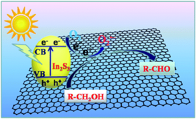 |
| | Scheme 2 Schematic diagram of the charge carriers transfer and proposed mechanism for selective oxidation of alcohols to corresponding aldehydes over the In2S3–GR nanocomposites under visible light irradiation (λ > 420 nm). | |
Conclusions
In summary, we have successfully fabricated In2S3–graphene (GR) nanocomposites with hierarchical petal-like In2S3 overspreading the GR sheets. In virtue of the “structure directing” role of graphene oxide (GO) as the precursor of GR, the as-synthesized In2S3–GR featuring the good interfacial contact between In2S3 and GR exhibit much higher photocatalytic activity toward selective oxidation of alcohols to aldehydes under visible light irradiation than blank flowerlike In2S3. It is revealed that the significantly enhanced photocatalytic performance of In2S3–GR is ascribed to the integrative effect of the boosted separation and transfer efficiency of photogenerated electron–hole pairs and the larger surface area. Our work highlights the promising scope of fabricating GR-based semiconductor nanocomposites with controllable architectural nanostructures by rationally utilizing the “structure directing” property of GO and extending their applications toward photocatalytic selective transformations.
Acknowledgements
The support by the National Natural Science Foundation of China (NSFC) (20903023, 21173045), the Award Program for Minjiang Scholar Professorship, the Natural Science Foundation (NSF) of Fujian Province for Distinguished Young Investigator Grant (2012J06003), and Program for Returned High-Level Overseas Chinese Scholars of Fujian province is gratefully acknowledged.
Notes and references
- K. S. Novoselov, A. K. Geim, S. V. Morozov, D. Jiang, Y. Zhang, S. V. Dubonos, I. V. Grigorieva and A. A. Firsov, Science, 2004, 306, 666–669 CrossRef CAS PubMed.
- N. Zhang, Y. Zhang and Y.-J. Xu, Nanoscale, 2012, 4, 5792–5813 RSC.
- G. Xie, K. Zhang, B. Guo, Q. Liu, L. Fang and J. R. Gong, Adv. Mater., 2013, 25, 3820–3839 CrossRef CAS PubMed.
- C. Han, M.-Q. Yang, N. Zhang and Y.-J. Xu, J. Mater. Chem. A, 2014, 2, 19156–19166 CAS.
- M. Zhu, P. Chen and M. Liu, ACS Nano, 2011, 5, 4529–4536 CrossRef CAS PubMed.
- Q. Zhang, C. Tian, A. Wu, T. Tan, L. Sun, L. Wang and H. Fu, J. Mater. Chem., 2012, 22, 11778–11784 RSC.
- J. Zhang, J. Yu, M. Jaroniec and J. R. Gong, Nano Lett., 2012, 12, 4584–4589 CrossRef CAS PubMed.
- W. Tu, Y. Zhou, Q. Liu, Z. Tian, J. Gao, X. Chen, H. Zhang, J. Liu and Z. Zou, Adv. Funct. Mater., 2012, 22, 1215–1221 CrossRef CAS.
- A. Iwase, Y. H. Ng, Y. Ishiguro, A. Kudo and R. Amal, J. Am. Chem. Soc., 2011, 133, 11054–11057 CrossRef CAS PubMed.
- Y. Zhang, Z.-R. Tang, X. Fu and Y.-J. Xu, ACS Nano, 2011, 5, 7426–7435 CrossRef CAS PubMed.
- C. Han, M.-Q. Yang, B. Weng and Y.-J. Xu, Phys. Chem. Chem. Phys., 2014, 16, 16891–16903 RSC.
- M.-Q. Yang, N. Zhang, M. Pagliaro and Y.-J. Xu, Chem. Soc. Rev., 2014, 43, 8240–8254 RSC.
- Q. Xiang, J. Yu and M. Jaroniec, Chem. Soc. Rev., 2012, 41, 782–796 RSC.
- I. V. Lightcap and P. V. Kamat, Acc. Chem. Res., 2012, 46, 2235–2243 CrossRef PubMed.
- L. Han, P. Wang and S. Dong, Nanoscale, 2012, 4, 5814–5825 RSC.
- W. Fan, Q. Zhang and Y. Wang, Phys. Chem. Chem. Phys., 2013, 15, 2632–2649 RSC.
- O. Akhavan and E. Ghaderi, J. Phys. Chem. C, 2009, 113, 20214–20220 CAS.
- P. Gao, J. Liu, S. Lee, T. Zhang and D. D. Sun, J. Mater. Chem., 2012, 22, 2292–2298 RSC.
- W. Fan, Q. Lai, Q. Zhang and Y. Wang, J. Phys. Chem. C, 2011, 115, 10694–10701 CAS.
- J. Zhou, G. Tian, Y. Chen, X. Meng, Y. Shi, X. Cao, K. Pan and H. Fu, Chem. Commun., 2013, 49, 2237–2239 RSC.
- Y. Zhang, Z.-R. Tang, X. Fu and Y.-J. Xu, ACS Nano, 2010, 4, 7303–7314 CrossRef CAS PubMed.
- J. Zhang, Z. Xiong and X. Zhao, J. Mater. Chem., 2011, 21, 3634–3640 RSC.
- Y. H. Ng, A. Iwase, A. Kudo and R. Amal, J. Phys. Chem. Lett., 2010, 1, 2607–2612 CrossRef CAS.
- I. Y. Kim, J. M. Lee, T. W. Kim, H. N. Kim, H. i. Kim, W. Choi and S. J. Hwang, Small, 2012, 8, 1038–1048 CrossRef CAS PubMed.
- A. Mukherji, B. Seger, G. Q. Lu and L. Wang, ACS Nano, 2011, 5, 3483–3492 CrossRef CAS PubMed.
- J. Du, X. Lai, N. Yang, J. Zhai, D. Kisailus, F. Su, D. Wang and L. Jiang, ACS Nano, 2010, 5, 590–596 CrossRef PubMed.
- H. Zhang, X. Lv, Y. Li, Y. Wang and J. Li, ACS Nano, 2009, 4, 380–386 CrossRef PubMed.
- C. Chen, W. Cai, M. Long, B. Zhou, Y. Wu, D. Wu and Y. Feng, ACS Nano, 2010, 4, 6425–6432 CrossRef CAS PubMed.
- E. Gao, W. Wang, M. Shang and J. Xu, Phys. Chem. Chem. Phys., 2011, 13, 2887–2893 RSC.
- Y. Fu, P. Xiong, H. Chen, X. Sun and X. Wang, Ind. Eng. Chem. Res., 2012, 51, 725–731 CrossRef.
- X. An, X. Yu, C. Y. Jimmy and G. Zhang, J. Mater. Chem. A, 2013, 1, 5158–5164 CAS.
- N. Zhang, M.-Q. Yang, Z.-R. Tang and Y.-J. Xu, ACS Nano, 2014, 8, 623–633 CrossRef CAS PubMed.
- N. Zhang, Y. Zhang, X. Pan, X. Fu, S. Liu and Y.-J. Xu, J. Phys. Chem. C, 2011, 115, 23501–23511 CAS.
- M.-Q. Yang and Y.-J. Xu, Phys. Chem. Chem. Phys., 2013, 15, 19102–19118 RSC.
- S. Liu, Z. Chen, N. Zhang, Z.-R. Tang and Y.-J. Xu, J. Phys. Chem. C, 2013, 117, 8251–8261 CAS.
- M.-Q. Yang, B. Weng and Y.-J. Xu, Langmuir, 2013, 29, 10549–10558 CrossRef CAS PubMed.
- Y. HeeáKim, J. HakáLee, S. MináPark, J. SooáMoon and J. GyuáNam, Chem. Commun., 2010, 46, 2292–2294 RSC.
- S. Rengaraj, S. Venkataraj, C.-w. Tai, Y. Kim, E. Repo and M. Sillanpää, Langmuir, 2011, 27, 5534–5541 CrossRef CAS PubMed.
- W. Qiu, M. Xu, X. Yang, F. Chen, Y. Nan, J. Zhang, H. Iwai and H. Chen, J. Mater. Chem., 2011, 21, 13327–13333 RSC.
- L.-Y. Chen, Z.-D. Zhang and W.-Z. Wang, J. Phys. Chem. C, 2008, 112, 4117–4123 CAS.
- K. H. Park, K. Jang and S. U. Son, Angew. Chem., Int. Ed., 2006, 45, 4608–4612 CrossRef CAS PubMed.
- B. Pejova and I. Bineva, J. Phys. Chem. C, 2013, 117, 7303–7314 CAS.
- Y. Gu and Y. Wang, RSC Adv., 2014, 4, 8582–8589 RSC.
- F. Ye, G. Du, Z. Jiang, Y. Zhong, X. Wang, Q. Cao and J. Z. Jiang, Nanoscale, 2012, 4, 7354–7357 RSC.
- W. Du, J. Zhu, S. Li and X. Qian, Cryst. Growth Des., 2008, 8, 2130–2136 CAS.
- F. Ye, C. Wang, G. Du, X. Chen, Y. Zhong and J. Jiang, J. Mater. Chem., 2011, 21, 17063–17065 RSC.
- L. Liu, H. Liu, H.-Z. Kou, Y. Wang, Z. Zhou, M. Ren, M. Ge and X. He, Cryst. Growth Des., 2008, 9, 113–117 Search PubMed.
- Y. He, D. Li, G. Xiao, W. Chen, Y. Chen, M. Sun, H. Huang and X. Fu, J. Phys. Chem. C, 2009, 113, 5254–5262 CAS.
- X. An, J. C. Yu, F. Wang, C. Li and Y. Li, Appl. Catal., B, 2013, 129, 80–88 CrossRef CAS PubMed.
- W. S. Hummers and R. E. Offeman, J. Am. Chem. Soc., 1958, 80, 1339 CrossRef CAS.
- N. I. Kovtyukhova, P. J. Ollivier, B. R. Martin, T. E. Mallouk, S. A. Chizhik, E. V. Buzaneva and A. D. Gorchinskiy, Chem. Mater., 1999, 11, 771–778 CrossRef CAS.
- L. J. Cote, F. Kim and J. Huang, J. Am. Chem. Soc., 2008, 131, 1043–1049 CrossRef PubMed.
- D. Pan, S. Wang, B. Zhao, M. Wu, H. Zhang, Y. Wang and Z. Jiao, Chem. Mater., 2009, 21, 3136–3142 CrossRef CAS.
- D. Li, M. B. Müller, S. Gilje, R. B. Kaner and G. G. Wallace, Nat. Nanotechnol., 2008, 3, 101–105 CrossRef CAS PubMed.
- L. J. Cote, F. Kim and J. Huang, J. Am. Chem. Soc., 2008, 131, 1043–1049 CrossRef PubMed.
- Y. Zhang, N. Zhang, Z.-R. Tang and Y.-J. Xu, J. Phys. Chem. C, 2014, 118, 5299–5308 CAS.
- Y. Zhang, N. Zhang, Z.-R. Tang and Y.-J. Xu, ACS Nano, 2012, 6, 9777–9789 CrossRef CAS PubMed.
- N. Zhang, Y. Zhang, X. Pan, M.-Q. Yang and Y.-J. Xu, J. Phys. Chem. C, 2012, 116, 18023–18031 CAS.
- M. Zhang, C. Chen, W. Ma and J. Zhao, Angew. Chem., Int. Ed., 2008, 47, 9730–9733 CrossRef CAS PubMed.
- M. Zhang, Q. Wang, C. Chen, L. Zang, W. Ma and J. Zhao, Angew. Chem., Int. Ed., 2009, 48, 6081–6084 CrossRef CAS PubMed.
- Z. Chen, S. Liu, M.-Q. Yang and Y.-J. Xu, ACS Appl. Mater. Interfaces, 2013, 5, 4309–4319 CAS.
- N. Zhang, Y. Zhang, M.-Q. Yang, Z.-R. Tang and Y.-J. Xu, J. Catal., 2013, 299, 210–221 CrossRef CAS PubMed.
- Y.-J. Xu, Y. Zhuang and X. Fu, J. Phys. Chem. C, 2010, 114, 2669–2676 CAS.
- X.-Y. Zhang, H.-P. Li, X.-L. Cui and Y. Lin, J. Mater. Chem., 2010, 20, 2801–2806 RSC.
- S. Soedergren, A. Hagfeldt, J. Olsson and S.-E. Lindquist, J. Phys. Chem., 1994, 98, 5552–5556 CrossRef CAS.
- J. Yang, H. Yan, X. Wang, F. Wen, Z. Wang, D. Fan, J. Shi and C. Li, J. Catal., 2012, 290, 151–157 CrossRef CAS PubMed.
- A. Cao, Z. Liu, S. Chu, M. Wu, Z. Ye, Z. Cai, Y. Chang, S. Wang, Q. Gong and Y. Liu, Adv. Mater., 2010, 22, 103–106 CrossRef CAS PubMed.
- M.-Q. Yang, B. Weng and Y.-J. Xu, J. Mater. Chem. A, 2014, 2, 1710–1720 CAS.
- T. Lu, Y. Zhang, H. Li, L. Pan, Y. Li and Z. Sun, Electrochim. Acta, 2010, 55, 4170–4173 CrossRef CAS PubMed.
- R. Pierotti and J. Rouquerol, Pure Appl. Chem., 1985, 57, 603–619 Search PubMed.
- Y. Zhang, N. Zhang, Z.-R. Tang and Y.-J. Xu, Chem. Sci., 2013, 4, 1820–1824 RSC.
- Y. Zhang, N. Zhang, Z.-R. Tang and Y.-J. Xu, Chem. Sci., 2012, 3, 2812–2822 RSC.
- Q. Wang, M. Zhang, C. Chen, W. Ma and J. Zhao, Angew. Chem., Int. Ed., 2010, 49, 7976–7979 CrossRef CAS PubMed.
- N. Zhang, X. Fu and Y.-J. Xu, J. Mater. Chem., 2011, 21, 8152–8158 RSC.
Footnote |
| † Electronic supplementary information (ESI) available: Experimental section for preparation of graphene oxide; the chemical structure of L-cysteine; the Fourier transformed infrared spectra (FTIR) of In2S3–1%GR and the original GO; the plot of transformed Kubelka–Munk function versus the energy of light for blank In2S3 and In2S3–GR nanocomposites with different weight addition ratios of GR; remaining fraction of various alcohols after the adsorption–desorption equilibrium is achieved over blank In2S3 and In2S3–1%GR; ESR spectra of radical adduct trapped by DMPO (DMPO–O2˙−) over In2S3–1%GR suspension in the BTF solution without or with the visible light irradiation; summary of BET surface area and pore volume of blank In2S3 and In2S3–1%GR. See DOI: 10.1039/c4ra13764g |
|
| This journal is © The Royal Society of Chemistry 2014 |
Click here to see how this site uses Cookies. View our privacy policy here. 


